

PDF Version - Glidewell Dental Labs
PDF Version - Glidewell Dental Labs
PDF Version - Glidewell Dental Labs

Create successful ePaper yourself
Turn your PDF publications into a flip-book with our unique Google optimized e-Paper software.
Dr. Melker and I use an exacting protocol for approaching<br />
these kinds of cases. It requires a team approach with<br />
everyone paying attention to the smallest of details. Our<br />
objectives are to create an environment that facilitates the<br />
control and prevention of microbial infection so the restorative<br />
dentistry is easier, more predictable and profitable.<br />
It makes no sense to avoid sending a patient to the<br />
periodontist when it makes the case easier, more predictable<br />
and more profitable.<br />
Provisionals are required, then removed at the time of<br />
surgery to allow vertical access to the prepared tooth. The<br />
old restorations and caries are removed and core buildups<br />
are done so the extent of compromised tooth structure<br />
in an apical direction can be assessed relative to the<br />
biologic width.<br />
Rita’s crowns were removed along with the gross caries,<br />
stain and old filling material. Core buildups were done<br />
using a specific bonding protocol for self-cure resin composite.<br />
The provisionals were cemented with polycarboxylate<br />
cement, which is antimicrobial and sticks to the<br />
teeth when the provisionals are removed. The teeth were<br />
sealed with SuperSeal before cementation to protect the<br />
pulp in #29. Because the cement sticks to the tooth, when<br />
microleakage occurs under the provisionals, microbial<br />
growth is restricted between the cement and the intaglio<br />
of the provisional, thus denying microbes access to dentinal<br />
tubules.<br />
Surgical provisionals are easily removed, using mosquito<br />
hemostats in a gently rocking motion several times from<br />
the incisal/occlusal. Aggressive forces should be avoided<br />
to prevent tooth fracture. Surgical provisionals must be<br />
made of methylmethacrylate, not bis-acryl, because the<br />
provisional must be able to flex for its removal. Bis-acryl<br />
does not flex, it fractures and is, therefore, unacceptable<br />
as a surgical provisional material.<br />
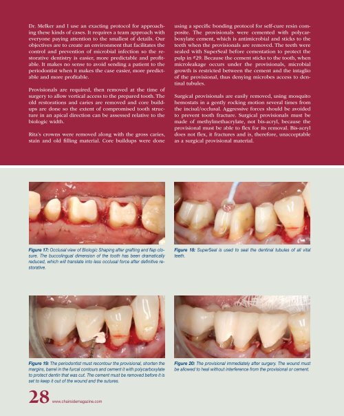
Figure 17: Occlusal view of Biologic Shaping after grafting and flap closure.<br />
The buccolingual dimension of the tooth has been dramatically<br />
reduced, which will translate into less occlusal force after definitive restorative.<br />
Figure 18: SuperSeal is used to seal the dentinal tubules of all vital<br />
teeth.<br />
Figure 19: The periodontist must recontour the provisional, shorten the<br />
margins, barrel in the furcal contours and cement it with polycarboxylate<br />
to protect dentin that was cut. The cement must be removed before it is<br />
set to keep it out of the wound and the sutures.<br />
Figure 20: The provisional immediately after surgery. The wound must<br />
be allowed to heal without interference from the provisional or cement.<br />
28 www.chairsidemagazine.com


