World Congress of Brachytherapy 10-12 May, 2012 - Estro-events.org
World Congress of Brachytherapy 10-12 May, 2012 - Estro-events.org
World Congress of Brachytherapy 10-12 May, 2012 - Estro-events.org

Create successful ePaper yourself
Turn your PDF publications into a flip-book with our unique Google optimized e-Paper software.
S134 <strong>World</strong> <strong>Congress</strong> <strong>of</strong> <strong>Brachytherapy</strong> 20<strong>12</strong><br />
errors was simulated by rotating 3D dose cloud manually using the 3D<br />
imaging registration system (VelocityAI), as shown in figure. The<br />
rotational errors <strong>of</strong> ± 1°, ± 3°, ± 5°, ± 7° , ± <strong>10</strong>° , ± 30°,± 45° and ±<br />
90 ° along each axis were simulated. Dosimetric effects <strong>of</strong> the rotated<br />
plans were compared with the original plans. In previous study, the<br />
balloon to skin/chest wall distance, (BtoS Dist.) was the most critical<br />
factor to affect the treatment results. Results for two groups <strong>of</strong><br />
patients were analyzed: (1) BtoS Dist. ≤ 7mm (the small BtoS group) ;(<br />
2) BtoS Dist.>7mm (the large BtoS group).<br />
: A comparison <strong>of</strong> various dosimetric parameters is presented<br />
in the Table. The mean volume <strong>of</strong> PTV_Eval (1cm rind <strong>of</strong> breast tissue<br />
outside <strong>of</strong> the balloon) that received the prescription dose decreased,<br />
for the small BtoS group, from 1.7% ± 2.2% (less than <strong>10</strong>°) to 28.1% ±<br />
25.9% (up to 90°); for the large BtoS group, from 0.7% ± 0.5% (less<br />
than <strong>10</strong>°) to 3.3% ± 3.4% (up to 90°). In general, the increase in the<br />
volumes <strong>of</strong> the <strong>org</strong>ans at risk (OARs) receiving the tolerance doses was<br />
small and did not exceed the tolerance, except for cases where the<br />
OARs were in close proximity to the balloon (3% tumor coverage loss and >36%<br />
maximal dose increase to the OARs, for the small and large BtoS<br />
groups, respectively. However, for small BtoS Dist. ≤ 3mm targets<br />
with the elliptical shape <strong>of</strong> dose cloud, the target coverage and dose<br />
<strong>of</strong> OARs decreased significantly as rotational errors <strong>of</strong> 3° or more<br />
were present.<br />
: Rotational errors should be evaluated carefully for<br />
clinical cases involving small balloon to skin/chest wall distance and<br />
for dose cloud with elliptical shape. For the best dosimetric results,<br />
APBI treatments using Contura should have rotational errors ≤ 3° (BtoS<br />
Dist. ≤ 3mm), ≤ <strong>10</strong>° (BtoS Dist.≤ 7mm) and ≤ 45° (BtoS Dist.> 7mm)<br />
PO335<br />
INVERSE PLANNING OF CONFORMAL HDRPROSTATE BRACHYTHERAPY<br />
BOOST TECHNIQUE USING TWO CTVS<br />
S. Wolf 1 , G. Bockelmann 1 , F.A. Siebert 1<br />
1 University Hospital SH Campus Kiel, Radiotherapy, Kiel, Germany<br />
: Ultrasoundbased intraoperative HDRprostate<br />
brachytherapy (BT) as boost technique is a well established method in<br />
our clinic. To date the treatment planning is performed manually as<br />
conventional planning technique (CP). Despite the success <strong>of</strong> this<br />
method, the introduction <strong>of</strong> inverse planning techniques (IP) is hoped<br />
to reduce planning time and to decrease user dependency <strong>of</strong> the<br />
planning process itself. In this study the clinical implementation and<br />
evaluation <strong>of</strong> inverse planning for HDR prostate BT is presented. In<br />
contrast to other HDR prostate techniques the target structure is<br />
segmented into two parts: the peripheral zone <strong>of</strong> the prostate (CTV2)<br />
and the whole prostate gland (CTV2).<br />
: For the patient cohort considered in mean <strong>12</strong><br />
needles were implanted in the typical ushape form. Using 38<br />
different clinical treatment plans appropriate dose constraints for the<br />
IP algorithm (BrachyVision v8.8, Varian Medical Systems, Palo Alto,<br />
CA) were identified. Prescription doses for CTV1/CTV2 were 15/8.5<br />
Gy. Doses in urethra and rectum should be lower than <strong>10</strong> and 8 Gy,<br />
resp. Because the CTV1 is <strong>of</strong>ten badly visible with ultrasound after<br />
insertion <strong>of</strong> the implant needles, the CTV1 was geometrically defined<br />
by a threedimensional 0.5 cm hull around the implanted needles.<br />
After establishing a general template <strong>of</strong> dose constraints and their<br />
appropriate weightings, applicable to all patient anatomies, all<br />
treatment plans were calculated according the TG43 formalism using<br />
identical start conditions as well: three seconds dwell times in all<br />
dwell positions, calculation time for the IP algorithm in the treatment<br />
planning system was one minute, a maximum dwell time <strong>of</strong> <strong>10</strong> seconds<br />
was allowed, and dwell time smooth function was set to maximum<br />
<strong>10</strong>0%. IP results were compared against the existing clinical CP by<br />
using D90CTV1/2, V200CTV1/2, D2ccrectum, D0.1ccurethra, and COIN index.<br />
Significant statistical differences (pvalue < 0.05) were analyzed by<br />
the MannWhitney rank sum test.<br />
: The inverse planning results in this study were available after<br />
one minute. In contrast to this an experienced medical physicist needs<br />
about <strong>10</strong> to 15 minutes per treatment plan. The results <strong>of</strong> this study<br />
are summarized in table 1. Dosimetric indices are very similar<br />
between CP and IP techniques. Only V200CTV1 is significantly lower for<br />
CP. Moreover the IP technique fairly reduced (not statistical<br />
significant) the D0.1ccurethra.<br />
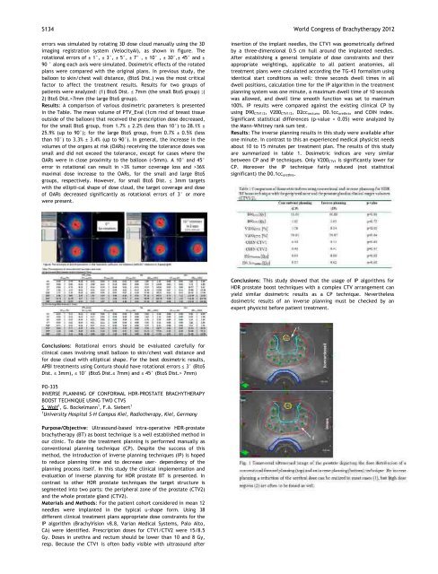
: This study showed that the usage <strong>of</strong> IP algorithms for<br />
HDR prostate boost techniques with a complex CTV arrangement can<br />
yield similar dosimetric results as a CP technique. Nevertheless<br />
dosimetric results <strong>of</strong> an inverse planning must be checked by an<br />
expert physicist before patient treatment.<br />
<strong>World</strong> <strong>Congress</strong> <strong>of</strong> <strong>Brachytherapy</strong> 20<strong>12</strong> S 135<br />
PO336<br />
BRACHYTHERAPY ACTIVITIES IN THE FRAMEWORK OF THE EUROPEAN<br />
METROLOGY RESEARCH PROGRAMME<br />
T. Schneider 1 , U. Ankerhold 1 , J. Solc 2<br />
1<br />
Phys. Techn. Bundesanstalt (PTB), Dosimetry for Radiation Therapy,<br />
Braunschweig, Germany<br />
2<br />
Czech Metrology Institute, Inspectorate for Ionizing Radiation,<br />
Praha, Czech Republic<br />
: The European Metrology Research Programme<br />
(EMRP) enables European metrology institutes to collaborate with<br />
industrial <strong>org</strong>anisations and academia on joint research projects<br />
within specified fields. The EMRP is implemented by EURAMET e.V.,<br />
<strong>org</strong>anised by 22 European National Metrology Institutes (NMIs),<br />
supported by the European Union and has a budget <strong>of</strong> 400 M€ over an<br />
approximately seven year period. In 2011 a new Joint Research<br />
Project (JRP) in the field dosimetry <strong>of</strong> ionizing radiation was initiated<br />
and approved after a review conference. A part <strong>of</strong> this project covers<br />
the recent developments in brachytherapy. In this presentation an<br />
outline <strong>of</strong> the planned activities for dosimetry in brachytherapy will<br />
be given.<br />
: The work within the new JRP addresses the<br />
establishing <strong>of</strong> miniature xray tubes for the treatment with low<br />
energy photons as well as the work in the community <strong>of</strong> medical<br />
physicists to bring up the treatment modality and treatment planning<br />
to the level already achieved in External Beam Radiation Therapy,<br />
manifested in the work <strong>of</strong> the AAPM TG 186 task group members in<br />
close cooperation with the European society ESTRO.<br />
: The dosimetry system <strong>of</strong> two miniature xray tubes will be<br />
investigated: one from the vendor Carl Zeiss called 'Intrabeam ® ' and<br />
another one from the vendor X<strong>of</strong>t called 'Axxent ® ' tube. The<br />
calibration procedures <strong>of</strong>fered by the vendors are totally different.<br />
The Axxent system uses a well type chamber calibrated in terms <strong>of</strong> Air<br />
Kerma Strength (AKS) for the radiation field <strong>of</strong> I<strong>12</strong>5. Calibration<br />
factor in AKS for the Axxent field are under development (NIST,<br />
Univ. Wisconsin). For the Intrabeam device the calibration is based on<br />
measurements in a water phantom in terms <strong>of</strong> absorbed dose to water<br />
with a s<strong>of</strong>t xray ionization chamber. As no NMI <strong>of</strong>fers calibrations in<br />
terms <strong>of</strong> absorbed dose to water DW in a certain depth in a water<br />
phantom in the respective radiation field, this calibration factor is<br />
derived from Air Kerma calibrations and a conversion into DW<br />
according to ICRU 17. The goal <strong>of</strong> the first part <strong>of</strong> the brachytherapy<br />
work is to characterize the sources, to establish a primary standard in<br />
terms <strong>of</strong> absorbed dose to water and to give recommendations on a<br />
calibration chain.<br />
: In most publications the results <strong>of</strong> treatment planning<br />
are compared only to MonteCarlo calculations and not to<br />
experimental data. It is the topic <strong>of</strong> the second part <strong>of</strong> the work to<br />
overcome this problem by developing measuring techniques to canvass<br />
the results <strong>of</strong> treatment planning systems in the presence <strong>of</strong> clinical<br />
applicators. For NMIs this means a step apart from the standard fields<br />
usually applied to a complex field close to clinical conditions. The<br />
applied measuring devices will be characterized to achieve an<br />
absolute uncertainty below 2% (k=1) in this irregular, nonstandard<br />
field and the uncertainty <strong>of</strong> the positioning will be below 30 m.<br />
PO337<br />
MAPPING OF RELATIVE DOSE RATE DISTRIBUTION UNCERTAINTY OWING<br />
TO SOURCE CONSTRUCTION<br />
K. Zourari 1 , E. Pantelis 1 , L. Sakelliou 2 , E. Ge<strong>org</strong>iou 1 , P. Papagiannis 1<br />
1<br />
University <strong>of</strong> Athens, Medical School Medical Physics Laboratory,<br />
Athens, Greece<br />
2<br />
University <strong>of</strong> Athens, Physics Department Nuclear and Particle<br />
Physics Section, Athens, Greece<br />
: A joint AAPM/GECESTRO report 1 recommends<br />
that brachytherapy source dosimetry investigators using Monte Carlo<br />
simulation (MC) should determine the effect <strong>of</strong> type B uncertainties<br />
on the quantities proposed for clinical use. This work presents results<br />
<strong>of</strong> a method to map the total uncertainty owing to source construction<br />
related type B uncertainties based on the ISO Guide to the Expression<br />
<strong>of</strong> Uncertainty in Measurement 2 and MC.<br />
: Calculations were performed for one<br />
commercially available <strong>12</strong>5 I LDR seed. 3 Information on the dimensions<br />
<strong>of</strong> the cylindrical silver marker, the thickness <strong>of</strong> its radioactive silver<br />
halide coating, and the dimensions <strong>of</strong> the titanium encapsulation,<br />
were available 3 as: xi±axi. Corresponding standard uncertainties were<br />
calculated assuming uniform probability distributions. Let be a<br />
point in the 2D matrix <strong>of</strong> dose rate per unit air kerma strength on a<br />
plane including the source long axis.<br />
is a function <strong>of</strong> xi (i.e. ) and, assuming f is<br />
approximately linear and no correlation exists between xi, its standard<br />
uncertainty can be calculated according to the law <strong>of</strong> uncertainty<br />
propagation 2 : . MC simulations were<br />
performed to obtain the 2D distribution <strong>of</strong> around the nominal<br />
source design, as well as source designs where each <strong>of</strong> the quantities<br />
xi was individually varied, assuming values <strong>of</strong> xiaxi or xi+axi. This<br />
allowed for the forward and backward finite difference<br />
approximations <strong>of</strong> the partial derivatives in the above equation, and<br />
hence the calculation <strong>of</strong> 2D maps <strong>of</strong> the total standard uncertainty<br />
owing to source construction and the partial contributions to it.<br />
: The figure presents the overall % relative standard<br />
uncertainty <strong>of</strong> around the source (left) compared to the<br />
corresponding type A MC uncertainty (right). As expected, radioactive<br />
coating thickness, followed by end weld radius, were the major<br />
uncertainty contributors affecting mainly points close to the source<br />
longitudinal axis. Uncertainty results at these points are comparable<br />
to the uncertainty <strong>of</strong> NIST traceable SK calibration <strong>of</strong> seeds delivered<br />
to the clinic. 1 The former however is position dependent and<br />
therefore subject to smoothing out when multiple sources are used.<br />
: Results support recommendations 1 that detailed<br />
information on the source should be openly provided by<br />
manufacturers and considered by dosimetry investigators. The<br />
proposed 2D mapping method facilitates the overview <strong>of</strong> total<br />
uncertainty while providing all the input necessary for the calculation<br />
<strong>of</strong> uncertainty <strong>of</strong> TG43 quantities. The brute force implementation <strong>of</strong><br />
MC could be replaced by methods <strong>of</strong> improved computational<br />
efficiency or physically intuitive models.<br />
Acknowledgments<br />
Kiveli Zourari was supported by Irakleitos II.<br />
References<br />
1. L. De Werd et al. Med. Phys. 38, 782801, 2011.<br />
2. Guide to the expression <strong>of</strong> uncertainty in measurement, ISO, JCGM<br />
<strong>10</strong>0, 2008.<br />
3. P. Karaiskos et al. Med. Phys. 28, 1753–1760, 2001.