World Congress of Brachytherapy 10-12 May, 2012 - Estro-events.org
World Congress of Brachytherapy 10-12 May, 2012 - Estro-events.org
World Congress of Brachytherapy 10-12 May, 2012 - Estro-events.org

Create successful ePaper yourself
Turn your PDF publications into a flip-book with our unique Google optimized e-Paper software.
S136 <strong>World</strong> <strong>Congress</strong> <strong>of</strong> <strong>Brachytherapy</strong> 20<strong>12</strong><br />
PO338<br />
VAGINAL CUFF BRACHYTHERAPY USING OVOIDS: COMPARISON OF<br />
POINT BASED AND TARGET BASED PRESCRIPTION<br />
S. Sharma 1 , D. Manigandan 1 , A.K. Gandhi 1 , D.N. Sharma 1 , V.<br />
Subramani 1 , P.K. Julka 1 , G.K. Rath 1<br />
1<br />
All India Institute <strong>of</strong> Medical Sciences (AIIMS), Radiation Oncology,<br />
Delhi, India<br />
: To compare the treatment plans with point based<br />
and target dose point based prescription methods for vaginal cuff<br />
brachytherapy with two ovoids.<br />
: Twenty previously treated sessions <strong>of</strong> post<br />
operative carcinoma uterine cervix patients were studied. For all the<br />
patients, application was done by using two Fletcher ovoids under<br />
sedation. After application, CT scan was done at 3mm slice thickness.<br />
The target volume was contoured and it assumed to be around the<br />
applicator, however, in some cases, modification was done according<br />
to the clinical and planning CT findings. Similarly, <strong>org</strong>ans at risk (OAR)<br />
i.e. bladder and rectum were contoured. The ICRU38 bladder and<br />
rectum points were also marked on CT images. Applicators were<br />
reconstructed using 2.5mm source step size and plans were generated<br />
for Nucletron HDRV2 machine using PLATO planning system. In both<br />
the ovoids, first four dwell positions were activated and dose<br />
prescription was done by using two different prescription methods.<br />
One is conventional point dose (PD) based and the second is target<br />
dose point (TDP) based. For conventional PD method, the dose<br />
prescription point was at 5mm above the superior surface <strong>of</strong> mid<br />
dwell position <strong>of</strong> two ovoids. For TDP based method, target dose<br />
points were created 5mm around the target. No optimization was<br />
done after prescription. For comparison purposes, dose was<br />
prescribed at <strong>10</strong>0cGy. For plan evaluation, following dose volume<br />
indices were used: Volume <strong>of</strong> target that receive <strong>10</strong>0% (V<strong>10</strong>0), 95%<br />
(V95), and 90% (V90) <strong>of</strong> prescription dose, coverage index<br />
(CI=V<strong>10</strong>0/Target volume) and overdose volume index (OI=V200/V<strong>10</strong>0). For<br />
bladder and rectum, doses received by 0.1cc (D0.1cc), 1cc (D1cc), 2cc<br />
(D2cc), 5cc (D5cc), and <strong>10</strong>cc (D<strong>10</strong>cc) <strong>of</strong> volumes were noted. The ICRU38<br />
bladder and rectum point doses were also noted to correlate with<br />
volumetric doses.<br />
: The plan comparison is summarized in Table.1. Volumetric<br />
evaluation shows that target coverage was lower in PD based plans<br />
than the TDP based plans. However, the OI index shows that high dose<br />
volume was more in TDP based plans compared to PD plans. In<br />
addition, the OAR doses were higher in TDP based method than the PD<br />
based. In the both TDP and PD methods, the bladder doses received<br />
by 0.1cc, 1cc, 2cc and 5cc volumes were higher compared to ICRU38<br />
bladder point doses and doses to <strong>10</strong>cc bladder volume were in closer<br />
agreement with the ICRU38 bladder points. Similarly, for rectum, the<br />
doses received by 0.1cc, and 1cc volumes were higher compared to<br />
ICRU38 rectum point doses and doses to 2–5cc volume <strong>of</strong> rectum were<br />
in closer agreement with the ICRU38 rectum points.<br />
: In the CT guided PD prescription method, lesser target<br />
coverage and lower OAR doses were observed. In TDP based<br />
prescription, target coverage was higher and resulted in higher OAR<br />
doses. Doses received by <strong>10</strong>cc <strong>of</strong> bladder and 2–5cc <strong>of</strong> rectum were<br />
closer to ICRU38 bladder and rectum points, respectively.<br />
PO339<br />
MRIDUMMY MARKERS OF MRIGUIDED HDR BRACHYTHERAPY FOR<br />
INTERSTITIAL PROSTATE AND INTRACAVITARY GYN CANCERS<br />
J. Schindel 1 , M. Muruganandham 1 , A. Eagle 2 , M. Hewitt 2 , T.<br />
Stockman 2 , C. Pigge 3 , Y. Kim 1<br />
1<br />
University <strong>of</strong> Iowa, Radiation Oncology, Iowa City, USA<br />
2<br />
University <strong>of</strong> Iowa, Electrical Computer Engineering, Iowa City, USA<br />
3<br />
University <strong>of</strong> Iowa, Chemistry, Iowa City, USA<br />
: Compare seven different MRIdummy markers for<br />
interstitial prostate and intracavitary GYN HDR brachytherapy.<br />
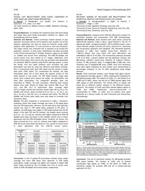
: Seven markers were used: Saline, Conray60<br />
(Mallinckrodt medical), CuSO4 (1.5g/L), liquid Vitamin E, fish oil, 1%<br />
Agarose gel (AGEL: 1g Agarose powder/ <strong>10</strong>0 ml distilled water), and a<br />
cobaltchloride complex contrast (C4) (CoCl2: Glycine=4:1). Interstitial<br />
and intracavitary phantoms were designed. The interstitial phantom<br />
consisted <strong>of</strong> eight flexi needles (outer/inner diameter <strong>of</strong><br />
1.98mm/1.45mm) that were filled with each MRImarker and<br />
embedded in 450 mL <strong>of</strong> 3% AGEL. The intracavitary phantom consisted<br />
<strong>of</strong> 4L <strong>of</strong> 3% AGEL, a plastic tandem and ring applicator (Varian), and<br />
MRIdummy catheters (outer/inner diameter <strong>of</strong> 2.66mm/1.87mm).<br />
Clinical 3T MRI protocols (both T1weightedMRI (T1MRI:1mm slice<br />
thickness) and T2weightedMRI scans (T2MRI: 3mm slice thickness))<br />
were used. Signal intensities for each marker were characterized as<br />
percentages compared to the 3% AGEL background using ImageJ<br />
s<strong>of</strong>tware.<br />
: Some interstitial markers, even though small agent volume,<br />
could generate favorable signals (> <strong>10</strong>0%), implying their feasibility for<br />
MRIguided prostate HDR. The markers <strong>of</strong> CuSO4, C4, and Vitamin E on<br />
T1MRI and 1% AGEL, Saline, and fish oil on T2MRI showed higher than<br />
<strong>10</strong>0% signals on both phantoms. The volume effect on signals was<br />
found due to different marker volumes in the different dummy<br />
catheters. The markers <strong>of</strong> CuSO4 and Saline showed highest signals on<br />
T1MRI and T2MRI, respectively. Sourcereconstruction is<br />
recommended on T1MRI, so CuSO4 and C4 markers have great<br />
potentials as a dummy marker for both interstitial and intracavitary<br />
brachytherapy.<br />
: The use <strong>of</strong> interstitial markers for MRIguided prostate<br />
HDR seems feasible. The markers <strong>of</strong> CuSO4 and C4 showed<br />
considerably high signals on T1MRI, as did Saline on T2MRI, for<br />
interstitial needles for prostate HDR and for a tandemandring<br />
applicator for GYN.<br />
<strong>World</strong> <strong>Congress</strong> <strong>of</strong> <strong>Brachytherapy</strong> 20<strong>12</strong> S 137<br />
PO340<br />
DOSIMETRIC IMPACT OF NOT CORRECTING FOR THE DISTAL SHIFT<br />
REPORTED IN VARIAN TANDEM AND RING (T&R) APPLICATORS<br />
O. Craciunescu 1 , J. Sánchez Mazón 2 , L. Lan 3 , J. Maurer 1 , B. Steffey 1 ,<br />
J. Cai 1 , J. Adamson 1 , J. Chino 1<br />
1<br />
Duke University Medical Center, Radiation Oncology, Durham NC,<br />
USA<br />
2<br />
Hospital Universitario Marqués de Valdecilla, Servicio de Radi<strong>of</strong>ísica<br />
y Protección Radiológica, Santander, Spain<br />
3<br />
Duke University Medical Center, Department <strong>of</strong> Biostatistics and<br />
Bioinformatics, Durham NC, USA<br />
: Studies have shown that source stop positions<br />
within Varian’s HDR CT/MR compatible ring applicators can deviate<br />
from the intended positions by several millimeters, and that a<br />
corrective shift has to be applied to limit the effect <strong>of</strong> these inherent<br />
<strong>of</strong>fsets. The purpose <strong>of</strong> this study was to investigate the dosimetric<br />
impact <strong>of</strong> NOT applying this correction.<br />
: Twentyseven HDR T&R clinical plans (cP)<br />
from six different patients treated for cervical cancer in our clinic<br />
were used in this study. Both CT and MR were acquired for each plan.<br />
The dose per fraction was 5.5 Gy. GECESTRO guidelines were<br />
followed for HRCTV and <strong>org</strong>ans at risk (OARs): bladder, rectum,<br />
sigmoid. ICRU 38 points A were also defined. The planning was done in<br />
BrachyVision with a hybrid volume optimization method based on<br />
HRCTV and ABS recommended reference lines. Depending on the<br />
shape <strong>of</strong> the HRCTV, the posterior ring dwell positions were allowed<br />
in the optimization for 5 patients. For 1 patient, a standard ring<br />
loading that mimicks ovoid placement was used. To show the<br />
dosimetric impact <strong>of</strong> NOT applying distal corrections, plans were<br />
generated by shifting the dwells in the ring in the opposite direction<br />
incrementally by 1 5 mm. The plans were calculated using the TG43<br />
formalism and ACUROS, a modelbased analytical solver that<br />
accounts for inhomogeneities. To establish clinical significance, the<br />
mean percent difference between all metrics in each <strong>of</strong> the shifted<br />
plans and the cP were calculated. The Wilcoxon signedrank test was<br />
used to determine if the differences have any statistical significance,<br />
for all cases together, but also separating patients depending on the<br />
ring dwells activated.<br />
: For brevity, only D2cc results for OARs for all 27 plans are<br />
presented. Ring loading strategy did not impact the dosmetric results.<br />
The mean percent differences between the largest shift (5mm) and<br />
the cP for the D2cc bladder, rectum and sigmoid were 1.6, 1.7, 2.5%<br />
for TG43, and 1.2, 1.8 and 1.4 % for ACUROS. The Wilcoxon rank sum<br />
pvalues were > 0.05 for all the OAR metrics, shift amounts, and<br />
method <strong>of</strong> calculation. For A_RT, A_LT and HRCTV D90, the mean<br />
percent differences were 0.8, 0.5 and 0.6 %, and 0.6, 0.8 and 0.9 %<br />
for TG43 and ACUROS, respectively. However, the Wilcoxon signed<br />
rank pvalues for Point A_RT were < 0.05 for both TG43 and ACUROS,<br />
for all shifts. Only when using ACUROS, the pvalues for HRCTV D90<br />
were < 0.05 for all shifts.<br />
: The dosimetric impact <strong>of</strong> NOT applying corrective shifts<br />