Download the uHead Surgical Technique - Small Bone Innovations
Download the uHead Surgical Technique - Small Bone Innovations
Download the uHead Surgical Technique - Small Bone Innovations

You also want an ePaper? Increase the reach of your titles
YUMPU automatically turns print PDFs into web optimized ePapers that Google loves.
4<br />
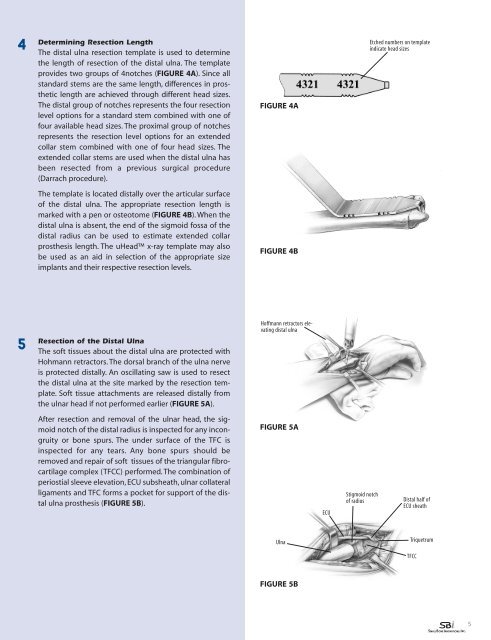
Determining Resection Length<br />
The distal ulna resection template is used to determine<br />
<strong>the</strong> length of resection of <strong>the</strong> distal ulna. The template<br />
provides two groups of 4notches (FIGURE 4A). Since all<br />
standard stems are <strong>the</strong> same length, differences in pros<strong>the</strong>tic<br />
length are achieved through different head sizes.<br />
The distal group of notches represents <strong>the</strong> four resection<br />
level options for a standard stem combined with one of<br />
four available head sizes. The proximal group of notches<br />
represents <strong>the</strong> resection level options for an extended<br />
collar stem combined with one of four head sizes. The<br />
extended collar stems are used when <strong>the</strong> distal ulna has<br />
been resected from a previous surgical procedure<br />
(Darrach procedure).<br />
FIGURE 4A<br />
Etched numbers on template<br />
indicate head sizes<br />
The template is located distally over <strong>the</strong> articular surface<br />
of <strong>the</strong> distal ulna. The appropriate resection length is<br />
marked with a pen or osteotome (FIGURE 4B). When <strong>the</strong><br />
distal ulna is absent, <strong>the</strong> end of <strong>the</strong> sigmoid fossa of <strong>the</strong><br />
distal radius can be used to estimate extended collar<br />
pros<strong>the</strong>sis length. The <strong>uHead</strong> x-ray template may also<br />
be used as an aid in selection of <strong>the</strong> appropriate size<br />
implants and <strong>the</strong>ir respective resection levels.<br />
FIGURE 4B<br />
5<br />
Resection of <strong>the</strong> Distal Ulna<br />
The soft tissues about <strong>the</strong> distal ulna are protected with<br />
Hohmann retractors. The dorsal branch of <strong>the</strong> ulna nerve<br />
is protected distally. An oscillating saw is used to resect<br />
<strong>the</strong> distal ulna at <strong>the</strong> site marked by <strong>the</strong> resection template.<br />
Soft tissue attachments are released distally from<br />
<strong>the</strong> ulnar head if not performed earlier (FIGURE 5A).<br />
Hoffmann retractors elevating<br />
distal ulna<br />
After resection and removal of <strong>the</strong> ulnar head, <strong>the</strong> sigmoid<br />
notch of <strong>the</strong> distal radius is inspected for any incongruity<br />
or bone spurs. The under surface of <strong>the</strong> TFC is<br />
inspected for any tears. Any bone spurs should be<br />
removed and repair of soft tissues of <strong>the</strong> triangular fibrocartilage<br />
complex (TFCC) performed. The combination of<br />
periostial sleeve elevation, ECU subsheath, ulnar collateral<br />
ligaments and TFC forms a pocket for support of <strong>the</strong> distal<br />
ulna pros<strong>the</strong>sis (FIGURE 5B).<br />
FIGURE 5A<br />
ECU<br />
Stigmoid notch<br />
of radius<br />
Distal half of<br />
ECU sheath<br />
Ulna<br />
Triquetrum<br />
TFCC<br />
FIGURE 5B<br />
5