Download the rHead⢠RECON Surgical Technique - Small Bone ...
Download the rHead⢠RECON Surgical Technique - Small Bone ...
Download the rHead⢠RECON Surgical Technique - Small Bone ...

You also want an ePaper? Increase the reach of your titles
YUMPU automatically turns print PDFs into web optimized ePapers that Google loves.
2<br />
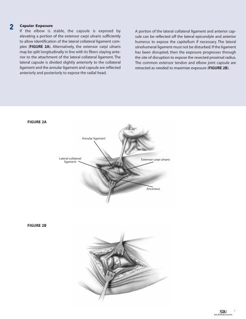
Capular Exposure<br />
If <strong>the</strong> elbow is stable, <strong>the</strong> capsule is exposed by<br />
elevating a portion of <strong>the</strong> extensor carpi ulnaris sufficiently<br />
to allow identification of <strong>the</strong> lateral collateral ligament complex<br />
(FIGURE 2A). Alternatively, <strong>the</strong> extensor carpi ulnaris<br />
may be split longitudinally in line with its fibers staying anterior<br />
to <strong>the</strong> attachment of <strong>the</strong> lateral collateral ligament. The<br />
lateral capsule is divided slightly anteriorly to <strong>the</strong> collateral<br />
ligament and <strong>the</strong> annular ligament and capsule are reflected<br />
anteriorly and posteriorly to expose <strong>the</strong> radial head.<br />
A portion of <strong>the</strong> lateral collateral ligament and anterior capsule<br />
can be reflected off <strong>the</strong> lateral epicondyle and anterior<br />
humerus to expose <strong>the</strong> capitellum if necessary. The lateral<br />
ulnohumeral ligament must not be disturbed. If <strong>the</strong> ligament<br />
has been disrupted, <strong>the</strong>n <strong>the</strong> exposure progresses through<br />
<strong>the</strong> site of disruption to expose <strong>the</strong> resected proximal radius.<br />
The common extensor tendon and elbow joint capsule are<br />
retracted as needed to maximize exposure (FIGURE 2B).<br />
FIGURE 2A<br />
Annular ligament<br />
Lateral collateral<br />
ligament<br />
Extensor carpi ulnaris<br />
Anconeus<br />
FIGURE 2B<br />
7