American Journalof Orthopedics® - Small Bone Innovations
American Journalof Orthopedics® - Small Bone Innovations
American Journalof Orthopedics® - Small Bone Innovations

You also want an ePaper? Increase the reach of your titles
YUMPU automatically turns print PDFs into web optimized ePapers that Google loves.
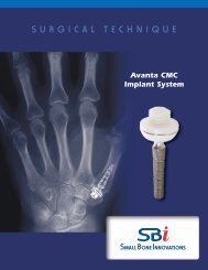
Total Joint Arthroplasty for the Arthritic Thumb CMC Joint<br />
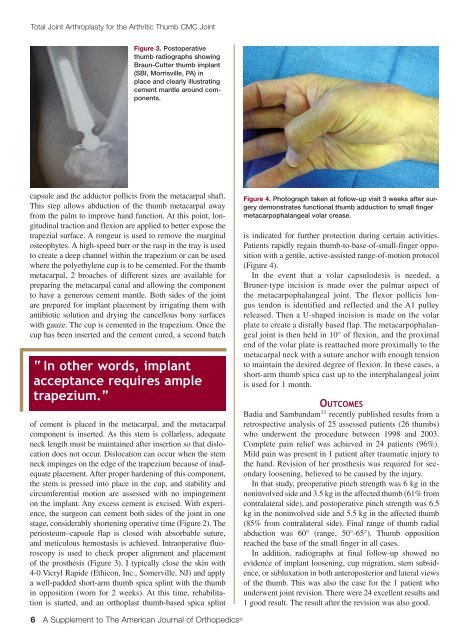
Figure 3. Postoperative<br />
thumb radiographs showing<br />
Braun-Cutter thumb implant<br />
(SBI, Morrisville, PA) in<br />
place and clearly illustrating<br />
cement mantle around components.<br />
capsule and the adductor pollicis from the metacarpal shaft.<br />
This step allows abduction of the thumb metacarpal away<br />
from the palm to improve hand function. At this point, longitudinal<br />
traction and flexion are applied to better expose the<br />
trapezial surface. A rongeur is used to remove the marginal<br />
osteophytes. A high-speed burr or the rasp in the tray is used<br />
to create a deep channel within the trapezium or can be used<br />
where the polyethylene cup is to be cemented. For the thumb<br />
metacarpal, 2 broaches of different sizes are available for<br />
preparing the metacarpal canal and allowing the component<br />
to have a generous cement mantle. Both sides of the joint<br />
are prepared for implant placement by irrigating them with<br />
antibiotic solution and drying the cancellous bony surfaces<br />
with gauze. The cup is cemented in the trapezium. Once the<br />
cup has been inserted and the cement cured, a second batch<br />
“ In other words, implant<br />
acceptance requires ample<br />
trapezium.”<br />
of cement is placed in the metacarpal, and the metacarpal<br />
component is inserted. As this stem is collarless, adequate<br />
neck length must be maintained after insertion so that dislocation<br />
does not occur. Dislocation can occur when the stem<br />
neck impinges on the edge of the trapezium because of inadequate<br />
placement. After proper hardening of this component,<br />
the stem is pressed into place in the cup, and stability and<br />
circumferential motion are assessed with no impingement<br />
on the implant. Any excess cement is excised. With experience,<br />
the surgeon can cement both sides of the joint in one<br />
stage, considerably shortening operative time (Figure 2). The<br />
periosteum–capsule flap is closed with absorbable suture,<br />
and meticulous hemostasis is achieved. Intraoperative fluoroscopy<br />
is used to check proper alignment and placement<br />
of the prosthesis (Figure 3). I typically close the skin with<br />
4-0 Vicryl Rapide (Ethicon, Inc., Somerville, NJ) and apply<br />
a well-padded short-arm thumb spica splint with the thumb<br />
in opposition (worn for 2 weeks). At this time, rehabilitation<br />
is started, and an orthoplast thumb-based spica splint<br />
Figure 4. Photograph taken at follow-up visit 3 weeks after surgery<br />
demonstrates functional thumb adduction to small finger<br />
metacarpophalangeal volar crease.<br />
is indicated for further protection during certain activities.<br />
Patients rapidly regain thumb-to-base-of-small-finger opposition<br />
with a gentle, active-assisted range-of-motion protocol<br />
(Figure 4).<br />
In the event that a volar capsulodesis is needed, a<br />
Bruner-type incision is made over the palmar aspect of<br />
the metacarpophalangeal joint. The flexor pollicis longus<br />
tendon is identified and reflected and the A1 pulley<br />
released. Then a U-shaped incision is made on the volar<br />
plate to create a distally based flap. The metacarpophalangeal<br />
joint is then held in 10° of flexion, and the proximal<br />
end of the volar plate is reattached more proximally to the<br />
metacarpal neck with a suture anchor with enough tension<br />
to maintain the desired degree of flexion. In these cases, a<br />
short-arm thumb spica cast up to the interphalangeal joint<br />
is used for 1 month.<br />
Outcomes<br />
Badia and Sambandam 11 recently published results from a<br />
retrospective analysis of 25 assessed patients (26 thumbs)<br />
who underwent the procedure between 1998 and 2003.<br />
Complete pain relief was achieved in 24 patients (96%).<br />
Mild pain was present in 1 patient after traumatic injury to<br />
the hand. Revision of her prosthesis was required for secondary<br />
loosening, believed to be caused by the injury.<br />
In that study, preoperative pinch strength was 6 kg in the<br />
noninvolved side and 3.5 kg in the affected thumb (61% from<br />
contralateral side), and postoperative pinch strength was 6.5<br />
kg in the noninvolved side and 5.5 kg in the affected thumb<br />
(85% from contralateral side). Final range of thumb radial<br />
abduction was 60° (range, 50°-65°). Thumb opposition<br />
reached the base of the small finger in all cases.<br />
In addition, radiographs at final follow-up showed no<br />
evidence of implant loosening, cup migration, stem subsidence,<br />
or subluxation in both anteroposterior and lateral views<br />
of the thumb. This was also the case for the 1 patient who<br />
underwent joint revision. There were 24 excellent results and<br />
1 good result. The result after the revision was also good.<br />
6 A Supplement to The <strong>American</strong> Journal of Orthopedics ®