Comparative dental development and microstructure of ... - UCL
Comparative dental development and microstructure of ... - UCL
Comparative dental development and microstructure of ... - UCL

Create successful ePaper yourself
Turn your PDF publications into a flip-book with our unique Google optimized e-Paper software.
A. D. Beynon<br />
Department <strong>of</strong> Oral Biology,<br />
The Dental School, University<br />
<strong>of</strong> Newcastle upon Tyne,<br />
NE2 4BW, U.K. E-mail:<br />
a.d.beynon@newcastle.ac.uk<br />
M. C. Dean*<br />
Evolutionary Anatomy Unit,<br />
Department <strong>of</strong> Anatomy &<br />
Developmental Biology,<br />
University College London,<br />
Gower Street, London<br />
WC1E 6BT, U.K.<br />
E-mail: ucgacrd@ucl.ac.uk<br />
M. G. Leakey<br />
Department <strong>of</strong> Palaeontology,<br />
Kenya National Museums,<br />
P.O. Box 40658, Nairobi,<br />
Kenya. E-mail:<br />
palaeo@swiftkenya.com<br />
D. J. Reid<br />
Department <strong>of</strong> Oral Biology,<br />
The Dental School, University<br />
<strong>of</strong> Newcastle upon Tyne,<br />
NE2 4BW, U.K. E-mail:<br />
d.j.reid@newcastle.ac.uk<br />
A. Walker<br />
Department <strong>of</strong> Anthropology,<br />
409 Carpenter Building,<br />
The Pennsylvania State<br />
University, University<br />
Park, PA 16802-3404,<br />
U.S.A. E-mail:<br />
axw8@psu.edu<br />
Received 10 July 1997<br />
Revision received<br />
1 March 1998<br />
Accepted 12 March 1998<br />
Keywords: Proconsul, Miocene<br />
hominoids, enamel<br />
thickness, Striae <strong>of</strong> Retzius,<br />
enamel, dentine.<br />
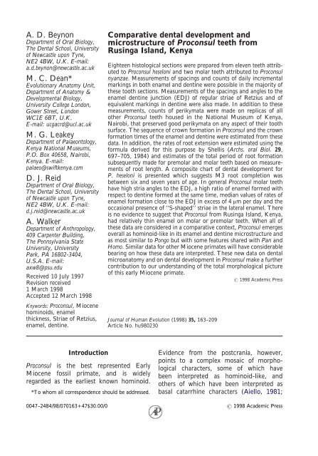
<strong>Comparative</strong> <strong>dental</strong> <strong>development</strong> <strong>and</strong><br />
<strong>microstructure</strong> <strong>of</strong> Proconsul teeth from<br />
Rusinga Isl<strong>and</strong>, Kenya<br />
Eighteen histological sections were prepared from eleven teeth attributed<br />
to Proconsul heseloni <strong>and</strong> two molar teeth attributed to Proconsul<br />
nyanzae. Measurements <strong>of</strong> spacings <strong>and</strong> counts <strong>of</strong> daily incremental<br />
markings in both enamel <strong>and</strong> dentine were possible in the majority <strong>of</strong><br />
these tooth sections. Measurements <strong>of</strong> the spacings <strong>and</strong> angles to the<br />
enamel dentine junction (EDJ) <strong>of</strong> regular striae <strong>of</strong> Retzius <strong>and</strong> <strong>of</strong><br />
equivalent markings in dentine were also made. In addition to these<br />
measurements, counts <strong>of</strong> perikymata were made on replicas <strong>of</strong> all<br />
other Proconsul teeth housed in the National Museum <strong>of</strong> Kenya,<br />
Nairobi, that preserved good perikymata on any aspect <strong>of</strong> their tooth<br />
surface. The sequence <strong>of</strong> crown formation in Proconsul <strong>and</strong> the crown<br />
formation times <strong>of</strong> the enamel <strong>and</strong> dentine were estimated from these<br />
data. In addition, the rates <strong>of</strong> root extension were estimated using the<br />
formula derived for this purpose by Shellis (Archs. oral Biol. 29,<br />
697–705, 1984) <strong>and</strong> estimates <strong>of</strong> the total period <strong>of</strong> root formation<br />
subsequently made for premolar <strong>and</strong> molar teeth based on measurements<br />
<strong>of</strong> root length. A composite chart <strong>of</strong> <strong>dental</strong> <strong>development</strong> for<br />
P. heseloni is presented which suggests M3 root completion was<br />
between six <strong>and</strong> seven years <strong>of</strong> age. In general Proconsul molar teeth<br />
have high stria angles to the EDJ, a high ratio <strong>of</strong> enamel formed with<br />
respect to dentine formed at the same time, median values <strong>of</strong> rates <strong>of</strong><br />
enamel formation close to the EDJ in excess <strong>of</strong> 4 μm per day <strong>and</strong> the<br />
occasional presence <strong>of</strong> ‘‘S-shaped’’ striae in the lateral enamel. There<br />
is no evidence to suggest that Proconsul from Rusinga Isl<strong>and</strong>, Kenya,<br />
had relatively thin enamel on molar or premolar teeth. When all <strong>of</strong><br />
these data are considered in a comparative context, Proconsul emerges<br />
overall as hominoid-like in its enamel <strong>and</strong> dentine <strong>microstructure</strong> <strong>and</strong><br />
as most similar to Pongo but with some features shared with Pan <strong>and</strong><br />
Homo. Similar data for other Miocene primates will have considerable<br />
bearing on how these data are interpreted. These new data on <strong>dental</strong><br />
microanatomy <strong>and</strong> on <strong>dental</strong> <strong>development</strong> in Proconsul make a further<br />
contribution to our underst<strong>and</strong>ing <strong>of</strong> the total morphological picture<br />
<strong>of</strong> this early Miocene primate.<br />
1998 Academic Press<br />
Journal <strong>of</strong> Human Evolution (1998) 35, 163–209<br />
Article No. hu980230<br />
Introduction<br />
Proconsul is the best represented Early<br />
Miocene fossil primate, <strong>and</strong> is widely<br />
regarded as the earliest known hominoid.<br />
*To whom all correspondence should be addressed.<br />
Evidence from the postcrania, however,<br />
points to a complex mosaic <strong>of</strong> morphological<br />
characters, some <strong>of</strong> which have<br />
been interpreted as hominoid-like, <strong>and</strong><br />
others <strong>of</strong> which have been interpreted as<br />
basal catarrhine characters (Aiello, 1981;<br />
0047–2484/98/070163+47$30.00/0 1998 Academic Press
164 A. D. BEYNON ET AL.<br />
Beard et al., 1986; Begun et al., 1993;<br />
Fleagle, 1983; Harrison, 1987, 1993; Lewis,<br />
1971; Napier & Davis, 1959; Rose, 1997;<br />
Ward et al., 1991, 1993). Overall, these<br />
postcranial characters suggest Proconsul was<br />
an arboreal quadruped with a varied positional<br />
repertoire that indulged in relatively<br />
slow climbing but which showed few signs <strong>of</strong><br />
forelimb suspensory behaviour (Walker,<br />
1997). Evidence from the skull <strong>and</strong> dentition<br />
includes characters that link Proconsul<br />
with later hominoids. The estimated degree<br />
<strong>of</strong> encephalization <strong>of</strong> P. heseloni, although<br />
based on one specimen (KNM-RU 7290),<br />
suggests that Proconsul had a bigger brain<br />
than modern cercopithecoids <strong>of</strong> a comparable<br />
body mass (Walker et al., 1983). Other<br />
cranio<strong>dental</strong> features such as the presence <strong>of</strong><br />
a frontal air sinus (Walker & Teaford,<br />
1989), a wide frontal bone at bregma, the<br />
<strong>development</strong> <strong>of</strong> a maxillary jugum, a low<br />
crowned P 3 <strong>and</strong> reduced cusp heteromorphy<br />
<strong>of</strong> the upper premolars are also each considered<br />
by some to be hominoid synapomorphies<br />
(Andrews, 1985). For a recent<br />
review see Walker (1997).<br />
Four species <strong>of</strong> Proconsul are now<br />
described (Walker et al., 1993; Teaford<br />
et al., 1993; Andrews, 1996). Proconsul<br />
africanus <strong>and</strong> Proconsul major are known<br />
from the type sites <strong>of</strong> Koru <strong>and</strong> Songhor in<br />
western Kenya <strong>and</strong> P. major also from<br />
Meswa Bridge in Kenya <strong>and</strong> Napak in<br />
Ug<strong>and</strong>a. P. heseloni <strong>and</strong> P. nyanzae are the<br />
two species that are represented at Rusinga<br />
<strong>and</strong> Mfangano Isl<strong>and</strong>s in Kenya (Walker<br />
et al., 1993). Ruff et al. (1989) used crosssectional<br />
measurements <strong>of</strong> the femoral<br />
diaphysis <strong>and</strong> articular dimensions to estimate<br />
the body weight <strong>of</strong> Proconsul specimens<br />
from Rusinga <strong>and</strong> Mfangano. Rafferty et al.<br />
(1995) subsequently made estimates from<br />
ankle joint surface areas. Both estimates are<br />
around 9–12 kg for the smaller P. heseloni<br />
specimens (about the same as a siamang, or<br />
twice that <strong>of</strong> smaller Hylobates species).<br />
Body weight estimates for P. nyanzae are<br />
closer to those <strong>of</strong> female chimpanzees <strong>of</strong><br />
the smallest subspecies averaging 35·6 kg<br />
(Rafferty et al., 1995).<br />
Reviewing the paleoecology <strong>and</strong> the<br />
hominoid paleoenvironments, Andrews<br />
(1996) presented evidence that Songhor <strong>and</strong><br />
Koru (dated at 19–20 Ma) have fossil faunas<br />
which suggest environments closest to<br />
tropical African, non-seasonal, wet, evergreen<br />
forest faunas today, whereas the<br />
slightly younger (17·5–17·9 Ma) Rusinga<br />
<strong>and</strong> Mfangano Isl<strong>and</strong> sites were most similar<br />
to dry seasonal forests <strong>and</strong> also had more<br />
open conditions. This evidence may turn<br />
out to be important in considering information<br />
about whether the diets <strong>of</strong> these<br />
different species <strong>of</strong> Proconsul were similar<br />
<strong>and</strong> in interpreting the effects <strong>of</strong> seasonality<br />
on developing tooth tissues among different<br />
species <strong>of</strong> Proconsul (Macho et al., 1996).<br />
The paleosols also provide useful environmental<br />
information. Retallack et al. (1995)<br />
have associated Proconsul from Rusinga<br />
Isl<strong>and</strong> with soils interpreted as having supported<br />
riparian woodl<strong>and</strong> early in the ecological<br />
succession <strong>of</strong> streamsides. These<br />
workers found no evidence <strong>of</strong> soils that<br />
would indicate extensive dry grassl<strong>and</strong>s or<br />
wet rain forest. Substantial paleobotanical<br />
remains are found on Rusinga <strong>and</strong><br />
Mfangano. The type <strong>of</strong> P. heseloni was<br />
deposited by a predator in a large hollow<br />
tree (Walker & Teaford, 1988). Fossilized<br />
fruits have been found in the same paleosol<br />
as the partial skeletons, some teeth <strong>of</strong> which<br />
form the biggest sample for this study, at the<br />
Kaswanga Primate Site (Walker et al., 1985)<br />
<strong>and</strong> again these paleobotanical finds point to<br />
potential differences in seasonality <strong>and</strong> diet<br />
in Proconsul from Rusinga Isl<strong>and</strong>.<br />
On the basis <strong>of</strong> histological sections <strong>of</strong><br />
nine molar teeth attributed to P. africanus,<br />
P. major <strong>and</strong> P. nyanzae, Gantt (1983,<br />
1986), has previously reported that linear<br />
measurements <strong>of</strong> enamel indicate thick<br />
enamel, relative to body size estimates.<br />
Gantt estimated enamel thickness in these
DENTAL DEVELOPMENT IN PROCONSUL<br />
165<br />
species <strong>of</strong> Proconsul as equivalent to that in<br />
Sivapithecus. However, Andrews & Martin<br />
(1991) defined enamel thickness in a different<br />
way, using the dentine cap area to correct<br />
for body size, <strong>and</strong> found specimens <strong>of</strong><br />
both P. africanus <strong>and</strong> P. major from Songhor<br />
<strong>and</strong> Koru to have thin enamel. These<br />
results are consistent with a predominantly<br />
frugivorous diet with limited degrees <strong>of</strong><br />
folivory, similar to extant forest-living,<br />
arboreal cercopithecine monkeys. Nothing<br />
further has been published about enamel<br />
thickness or about enamel <strong>and</strong> dentine<br />
<strong>microstructure</strong> in P. heseloni or P. nyanzae<br />
which might contribute to our underst<strong>and</strong>ing<br />
<strong>of</strong> the variation in enamel thickness<br />
between these species or indeed on the<br />
underlying processes <strong>of</strong> enamel growth in<br />
these early Miocene hominoids.<br />
Kelley (1992, 1993, 1997) has proposed<br />
that one way <strong>of</strong> distinguishing between Old<br />
World monkeys <strong>and</strong> apes would be to define<br />
their life history pr<strong>of</strong>iles more precisely <strong>and</strong><br />
that it would be important to learn more<br />
about life history pr<strong>of</strong>iles in early Miocene<br />
hominoids. Smith (1989, 1991, 1994) <strong>and</strong><br />
Smith et al. (1995) have demonstrated that<br />
many life history traits correlate with age <strong>of</strong><br />
first permanent molar emergence or brain<br />
weight, for example. Kelley (1997) has<br />
drawn on the apparently tight relationship<br />
between brain weight <strong>and</strong> M1 emergence<br />
(but see Smith et al., 1995) <strong>and</strong> used cranial<br />
capacity estimates available for P. heseloni to<br />
suggest that an approximate age <strong>of</strong> emergence<br />
for M1 in this taxon would have<br />
been 20·6 months. Kelley has cautiously<br />
argued that this result may point to a more<br />
prolonged set <strong>of</strong> life history traits in P.<br />
heseloni than would be expected for an<br />
early Miocene catarrhine <strong>of</strong> the same body<br />
size.<br />
A key aim <strong>of</strong> the present study is to<br />
reconstruct the sequence <strong>and</strong> timing <strong>of</strong><br />
<strong>dental</strong> <strong>development</strong> in P. heseloni using a<br />
variety <strong>of</strong> techniques. It is clear that there<br />
is much to learn about growth <strong>and</strong> <strong>development</strong><br />
<strong>and</strong> about life history in Proconsul.<br />
Thus, the present study attempts to establish<br />
a preliminary chronological schedule for<br />
<strong>dental</strong> <strong>development</strong> in Proconsul heseloni.<br />
There are now several juvenile partial skeletons<br />
associated with developing tooth<br />
germs from Rusinga Isl<strong>and</strong>. Some idea<br />
about a schedule <strong>of</strong> <strong>dental</strong> <strong>development</strong> in<br />
this one species <strong>of</strong> Proconsul would make it<br />
easier to associate these germs securely as<br />
different individuals. A time scale for <strong>dental</strong><br />
<strong>development</strong> would also provide a better<br />
comparative framework to describe juvenile<br />
postcranial material. A second aim <strong>of</strong> this<br />
study is to report further on enamel thickness<br />
in P. heseloni <strong>and</strong> P. nyanzae <strong>and</strong> on the<br />
processes through which enamel grows<br />
thicker or thinner. Thirdly, we aim to<br />
describe microanatomical features in the<br />
enamel <strong>and</strong> dentine <strong>of</strong> Proconsul that can be<br />
compared to other species <strong>of</strong> both extant<br />
<strong>and</strong> Miocene monkeys <strong>and</strong> hominoids.<br />
Describing growth processes that underlie<br />
morphological characters in both enamel<br />
<strong>and</strong> dentine between different species <strong>of</strong><br />
primate is a sound way to establish <strong>development</strong>al<br />
homologies which are useful for<br />
phylogenetic analyses.<br />
Materials<br />
This study combines information from<br />
ground sections <strong>of</strong> Proconsul teeth with data<br />
from perikymata counts made from surface<br />
replicas <strong>of</strong> other teeth. Four developing<br />
m<strong>and</strong>ibular permanent tooth germs (I 1 ,I 2 ,<br />
M 1 <strong>and</strong> M 2 ) <strong>and</strong> two deciduous m<strong>and</strong>ibular<br />
teeth (dm 1 <strong>and</strong> dm 2 ), attributed to P.<br />
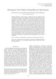
heseloni (Figure 1) were prepared for histological<br />
examination (Individual IV from the<br />
Kaswanga primate site on Rusinga Isl<strong>and</strong>).<br />
These teeth had well-preserved, unworn<br />
incisal or occlusal enamel, although in<br />
some <strong>of</strong> the germs the lateral enamel was<br />
incomplete or abraded post-mortem at the<br />
developing cervix. It should be noted that at<br />
least ten partial Proconsul skeletons were
166 A. D. BEYNON ET AL.<br />
Figure 1. P. heseloni teeth belonging to the juvenile specimen prior to sectioning. (All to the same scale<br />
with a mm scale bar at the foot <strong>of</strong> the plate.) Top row left to right: dm 1 buccal view, dm 2 lingual view, M 1<br />
occlusal view. Middle row left to right: dm 1 occlusal view, dm 2 occlusal view, M 1 , fractured base <strong>of</strong> crown.<br />
Bottom row left to right: I 2 germ, I 1 germ, M 2 germ occlusal view.<br />
comingled at the Kaswanga Primate Site. In<br />
nearly all cases, the maxillary <strong>and</strong> m<strong>and</strong>ibular<br />
bone had been broken up so that isolated<br />
teeth were collected from the deflation<br />
surface. Teeth were matched to individuals<br />
by size, degree <strong>of</strong> wear <strong>and</strong> interstitial facets,<br />
but this is a difficult undertaking <strong>and</strong> there<br />
may have been mistaken allocations. Five<br />
m<strong>and</strong>ibular permanent teeth attributed to<br />
a single adult specimen <strong>of</strong> P. heseloni
DENTAL DEVELOPMENT IN PROCONSUL<br />
167<br />
Figure 2. P. heseloni teeth belonging to the adult specimen prior to sectioning. (All to the same scale with<br />
a mm scale bar at the foot <strong>of</strong> the plate.) Top row left to right: canine, M 1 occlusal view, M 1 mesiobuccal<br />
view. Middle row: base <strong>and</strong> incomplete lingual aspect <strong>of</strong> canine. Bottom row: occlusal views <strong>of</strong> P 3 ,M 2 <strong>and</strong><br />
M 3 .<br />
(Individual III from the Kaswanga Primate<br />
Site on Rusinga Isl<strong>and</strong>) were also prepared<br />
for histological examination (Figure 2).<br />
These were a canine, P 4 ,M 1 ,M 2 <strong>and</strong> M 3<br />
from the lower right m<strong>and</strong>ibular quadrant.<br />
Although worn occlusally, each <strong>of</strong> these<br />
teeth preserves the lateral enamel on one or<br />
more aspects <strong>of</strong> the crown. In addition, two<br />
complete adult tooth crowns without roots<br />
preserved (KNM-RU 1721 <strong>and</strong> KNM-RU<br />
1695, both surface finds) attributed to P.<br />
nyanzae were also prepared for histological<br />
examination (Figure 3). These are an M 1<br />
<strong>and</strong> a right M 2 respectively. All <strong>of</strong> these<br />
teeth are housed in The Kenya National<br />
Museum, Nairobi. In addition all teeth<br />
attributed to Proconsul <strong>and</strong> housed in The<br />
Kenya National Museum, Nairobi were<br />
examined <strong>and</strong> many included as part <strong>of</strong> this<br />
study.<br />
Other ground sections <strong>of</strong> primate teeth<br />
including Pan troglodytes, Gorilla gorilla,<br />
Pongo pygmaeus, Hylobates moloch, Hylobates<br />
(Symphalangus) syndactylus, Theropithecus
168 A. D. BEYNON ET AL.<br />
Figure 3. P. nyanzae M 1 (RU 1721) <strong>and</strong> M 2 (RU 1695) crowns prior to sectioning. (All to the same scale<br />
with a mm scale bar at the foot <strong>of</strong> the plate.) The three views on the left h<strong>and</strong> side are <strong>of</strong> RU 1695 <strong>and</strong><br />
the three views on the right h<strong>and</strong> side are <strong>of</strong> RU 1721.<br />
gelada, <strong>and</strong> Cebus apella were also used for<br />
reference in this study. These sections form<br />
part <strong>of</strong> a large reference collection housed in<br />
the Department <strong>of</strong> Oral Biology, The<br />
Dental School, University <strong>of</strong> Newcastle<br />
upon Tyne. Many are from zoo animals<br />
(the great apes) but others are <strong>of</strong> unknown<br />
provenance.<br />
Methods<br />
Perikymata<br />
All teeth housed in the National Museum<br />
<strong>of</strong> Kenya, Nairobi, that are attributed to<br />
Proconsul were first examined using a<br />
Wild M8 binocular microscope. Those<br />
that preserve surface incremental markings
DENTAL DEVELOPMENT IN PROCONSUL<br />
169<br />
(perikymata) over some or all <strong>of</strong> their buccal<br />
or lingual enamel were cleaned with<br />
alcohol <strong>and</strong> cotton wool <strong>and</strong> impressions<br />
taken <strong>of</strong> the buccal <strong>and</strong> or lingual surfaces<br />
using the Coltene President putty Light<br />
Body wash system (Beynon, 1987). The<br />
moulds were then cast in Spurr Resin following<br />
the methods described by Beynon<br />
(1987). The resin replicas were sputter<br />
coated with gold to maximize surface<br />
reflectance. Counts <strong>of</strong> perikymata were<br />
then made with the replica illuminated in<br />
polarized incident light using a Wild M8<br />
binocular microscope at appropriate magnifications<br />
for each tooth. These magnifications<br />
ranged between 20 <strong>and</strong> 80 times.<br />
All counts were made with the tooth surface<br />
mounted perpendicular to the optical<br />
axis <strong>of</strong> the microscope <strong>and</strong> with the tooth<br />
continually tilted on a microscope stage to<br />
maintain this relationship. Counts were<br />
recorded as numbers <strong>of</strong> perikymata present<br />
per millimetre <strong>of</strong> the total tooth height<br />
along the buccal or lingual surfaces.<br />
Approximately 45 counts <strong>of</strong> perikymata on<br />
various aspects <strong>of</strong> 25 teeth were made. The<br />
counts were tabulated from the most occlusal<br />
or incisive part <strong>of</strong> the tooth to the<br />
cervix. Rarely was it possible to make complete<br />
counts <strong>of</strong> perikymata on a tooth.<br />
Where areas <strong>of</strong> tooth were abraded or<br />
worn, estimates <strong>of</strong> the numbers <strong>of</strong> missing<br />
perikymata within any one millimetre <strong>of</strong><br />
the tooth surface under study were made<br />
(i) on the basis <strong>of</strong> true counts made adjacent<br />
to these regions, or alternatively (ii)<br />
on the basis <strong>of</strong> actual counts made in<br />
contralateral teeth from the same specimen.<br />
In practice, the trends in packing<br />
patterns were obvious <strong>and</strong> facilitated reconstruction<br />
<strong>of</strong> sequential counts estimated as<br />
above. In this way a general pr<strong>of</strong>ile <strong>of</strong><br />
perikymata counts <strong>and</strong> <strong>of</strong> their packing<br />
patterns was recorded for most tooth types<br />
<strong>of</strong> Proconsul. Any portion <strong>of</strong> any count that<br />
was estimated appears in brackets in<br />
Appendix 1.<br />
Histological methods<br />
Each <strong>of</strong> the teeth to be sectioned was first<br />
cleaned under a dissecting microscope using<br />
<strong>dental</strong> instruments, alcohol, <strong>and</strong> cotton<br />
wool. The teeth were then photographed<br />
(Figures 1, 2, 3) <strong>and</strong> replicated using the<br />
Coltene President putty <strong>and</strong> Coltene Light<br />
Body wash silicone addition curing impression<br />
system (Beynon, 1987). Teeth were<br />
then dehydrated in alcohol <strong>and</strong> acetone <strong>and</strong><br />
included in Clear Cast Resin. In the case<br />
<strong>of</strong> the dm 1 ,dm 2 ,I 1 ,I 2 ,M 1 <strong>and</strong> M 2 <strong>of</strong> the<br />
juvenile specimen, just one section was cut<br />
buccolingually through the tooth using<br />
an annular diamond saw. Each <strong>of</strong> these<br />
sections was made either centrally through<br />
the incisal edge <strong>of</strong> incisors or, in the case <strong>of</strong><br />
the other teeth, mesially through the tallest<br />
buccal cusp <strong>and</strong> lingual cusp. In the case <strong>of</strong><br />
the permanent molar teeth <strong>of</strong> the adult<br />
specimens, one section was cut buccolingually<br />
through the mesial cusps <strong>and</strong> another<br />
through the distal cusps with the aim <strong>of</strong><br />
preserving the points <strong>of</strong> both dentine horns<br />
in each section. All sections were then<br />
lapped plane parallel with a PM2 Logitech<br />
lapping jig to a thickness <strong>of</strong> approximately<br />
100 μm (range 99 to 155 μm for all the<br />
sections cut) such that the point <strong>of</strong> the<br />
dentine horn was preserved within the section<br />
as truly axial as possible to the plane <strong>of</strong><br />
section through the cusps. Figure 4 illustrates<br />
in outline one section from each <strong>of</strong> the<br />
teeth used in this study. The remaining cut<br />
block faces <strong>of</strong> each tooth were then removed<br />
from the Clear Cast resin <strong>and</strong> replaced in<br />
the Coltene moulds in their correct positions.<br />
Composite resin light-curing restorative<br />
filling materials, previously colour<br />
matched to each tooth, were then placed<br />
into the moulds between the cut block<br />
faces to restore the teeth to their original<br />
dimensions <strong>and</strong> appearance. Light curing<br />
was done sequentially in layers <strong>of</strong> appropriate<br />
colour in the manner prescribed to<br />
restore their original appearance. In this<br />
way, a total <strong>of</strong> 18 ground sections were
170 A. D. BEYNON ET AL.<br />
Figure 4. Crown outlines, drawn from ground sections, <strong>of</strong> the teeth used in the histological part <strong>of</strong> this<br />
study. One section only from each tooth is represented even though several posterior teeth were sectioned<br />
more than once (see text). From top left to right through rows 1 <strong>and</strong> 2: dm 1 ,dm 2 ,I 1 ,I 2 ,M 1 ,M 2 (juvenile<br />
P. heseloni) <strong>and</strong>M 1 . Row 3: canine P 4 ,M 3 (adult Proconsul heseloni). Bottom row: M 1 <strong>and</strong> M 2 (P.<br />
nyanzae). Four tooth sections were reconstructed over the cusps in order to estimate enamel cap area <strong>and</strong><br />
EDJ length used for the calculations <strong>of</strong> relative enamel thickness. (None <strong>of</strong> the linear enamel thickness<br />
measurements that appear in Table 1 were made on reconstructed outlines.) Representations <strong>of</strong> the high<br />
power reconstructions are shown with dashed lines as appropriate.
DENTAL DEVELOPMENT IN PROCONSUL<br />
171<br />
prepared from 13 teeth. Ground sections<br />
were first examined in polarized transmitted<br />
light, then in reflectance mode with a Leica<br />
laser confocal microscope at key locations.<br />
Measurements in this study were made both<br />
from high power photomontages <strong>of</strong> the<br />
tooth sections <strong>and</strong> also directly using a Zeiss<br />
Filar micrometer eyepiece.<br />
Enamel thickness<br />
For several unworn anterior <strong>and</strong> posterior<br />
teeth, <strong>of</strong> both P. heseloni <strong>and</strong> P. nyanzae, it<br />
was possible to make linear measurements<br />
<strong>of</strong> enamel thickness. For some other teeth,<br />
as in previous studies on enamel thickness,<br />
minor reconstructions on tracings <strong>of</strong> crown<br />
outlines were possible, either at the cervix<br />
or at the cusp tips to correct for damage<br />
or wear. It was then possible to make<br />
additional estimates <strong>of</strong> the area <strong>of</strong> the<br />
enamel cap, the dentine cap <strong>and</strong> the length<br />
<strong>of</strong> the EDJ in four slightly worn or damaged<br />
teeth. Measurements <strong>of</strong> enamel thickness<br />
were made in several ways that reflect previous<br />
studies on enamel thickness <strong>and</strong> which<br />
therefore allow comparison with the results<br />
<strong>of</strong> these studies. Linear measurements <strong>of</strong><br />
enamel thickness were made on teeth where<br />
there was no occlusal wear, in the way<br />
detailed by Beynon & Wood (1986) <strong>and</strong> in<br />
Figure 1 <strong>of</strong> Macho & Berner (1994), but<br />
were made here on both m<strong>and</strong>ibular <strong>and</strong><br />
one maxillary tooth. Measurements 1 <strong>and</strong> 8<br />
were omitted as they are not directly comparable<br />
in upper <strong>and</strong> lower teeth. Andrews<br />
& Martin (1991) present data for enamel<br />
thickness in P. africanus <strong>and</strong> P. major. Two<br />
measurements <strong>of</strong> enamel thickness as<br />
defined by Martin (1983) were therefore<br />
included to facilitate comparisons that are<br />
derived from measurements <strong>of</strong> the enamel<br />
<strong>and</strong> dentine cap area <strong>and</strong> from the length <strong>of</strong><br />
the enamel dentine junction. These were:<br />
average enamel thickness (the area <strong>of</strong> the<br />
enamel cap ‘‘c’’ divided by the length <strong>of</strong> the<br />
enamel–dentine junction ‘‘e’’ as measured<br />
from longitudinal sections <strong>of</strong> teeth) <strong>and</strong><br />
relative enamel thickness (the average<br />
enamel thickness value, c/e, corrected as a<br />
dimensionless index relative to ‘‘b’’, the area<br />
<strong>of</strong> the dentine cap).<br />
Enamel cross striations <strong>and</strong> striae <strong>of</strong> Retzius<br />
Evidence supporting the fact that enamel<br />
cross striations represent circadian increments<br />
<strong>of</strong> growth has been reviewed previously<br />
(Bromage, 1991; Dean, 1987, 1989,<br />
1995a). Counts <strong>of</strong> cross striations can be<br />
used to estimate the time <strong>of</strong> cuspal enamel<br />
formation. Measurements <strong>of</strong> the distance<br />
(spacing) between cross striations provide<br />
an estimate <strong>of</strong> the daily rate <strong>of</strong> enamel<br />
secretion. It is also well established that<br />
counts <strong>of</strong> regular striae <strong>of</strong> Retzius in enamel<br />
or <strong>of</strong> surface perikymata can be used to<br />
calculate the time <strong>of</strong> lateral enamel formation<br />
when the number <strong>of</strong> cross striations,<br />
or days, between them is known (Bromage<br />
& Dean, 1985; Dean, 1987; Beynon & Dean<br />
1998). In many places in the sections <strong>of</strong> the<br />
permanent teeth <strong>of</strong> P. heseloni <strong>and</strong> P. nyanzae<br />
it was possible to see enamel cross<br />
striations <strong>and</strong> regular striae <strong>of</strong> Retzius. However,<br />
these were less clear in the ground<br />
sections <strong>of</strong> the deciduous teeth. In the cuspal<br />
enamel <strong>of</strong> the P. nyanzae M 2 , enamel<br />
cross striations were exceptionally well preserved<br />
<strong>and</strong> could be tracked continuously<br />
from the dentine horn to the outer surface <strong>of</strong><br />
the enamel along paths <strong>of</strong> groups <strong>of</strong> prism.<br />
This tooth was therefore chosen to make a<br />
more careful comparative study <strong>of</strong> cross<br />
striations in cuspal enamel in Proconsul <strong>and</strong><br />
other primates. Figures 5(a) <strong>and</strong> 5(b) are<br />
confocal reflected light images <strong>of</strong> cross<br />
striations at the EDJ <strong>and</strong> at the surface <strong>of</strong> the<br />
cuspal enamel in the M 2 <strong>of</strong> P. nyanzae.<br />
Cuspal cross striations<br />
Measurements <strong>of</strong> cross striations were made<br />
in zones, or b<strong>and</strong>s, <strong>of</strong> enamel spaced<br />
approximately 30 days apart (e.g., at roughly<br />
monthly intervals) through the cuspal<br />
enamel <strong>of</strong> the mesiobuccal cusp <strong>of</strong> second
DENTAL DEVELOPMENT IN PROCONSUL<br />
173<br />
molars <strong>of</strong> P. nyanzae, H. sapiens, Pan<br />
troglodytes, G. gorilla, Pongo pygmaeus, H.<br />
moloch <strong>and</strong> T. gelada. Data for a modern<br />
human dm 2 are also included. These data<br />
are presented as graphs for each taxon. Each<br />
box plot in each graph is equivalent to a<br />
monthly zone <strong>and</strong> represents between 50<br />
<strong>and</strong> 100 measurements <strong>of</strong> the distance<br />
between cross striations within that zone,<br />
depending on how many could be reliably<br />
measured. The median values <strong>of</strong> the<br />
measurements <strong>of</strong> cross striations for each<br />
monthly zone in KNM-RU 1695 were used<br />
to calculate median values for inner, middle<br />
<strong>and</strong> cuspal enamel. The enamel prism track<br />
used in the mesiobuccal cuspal enamel was<br />
divided into three equal linear portions<br />
between the dentine horn <strong>and</strong> the cusp tip.<br />
Zones one to four were contained in the<br />
inner portion, zones five to eight in the<br />
middle <strong>and</strong> zones nine to 11 in the outer<br />
portion. Other measurements <strong>of</strong> cuspal<br />
cross striations in the Proconsul sample were<br />
compared with these <strong>and</strong> found to match<br />
well. Therefore, an overall average cuspal<br />
enamel secretion rate was calculated in<br />
order to estimate the time taken to form<br />
known thicknesses <strong>of</strong> enamel in other<br />
unworn Proconsul teeth. The times for cuspal<br />
enamel estimated in this way for several<br />
teeth were subsequently used in one <strong>of</strong> the<br />
methods for calculating crown formation<br />
times in unworn teeth (see below).<br />
Form <strong>and</strong> periodicity <strong>of</strong> striae <strong>of</strong> Retzius<br />
Measurements <strong>of</strong> the spacings between<br />
adjacent striae in inner, middle <strong>and</strong> outer<br />
enamel <strong>and</strong> <strong>of</strong> the angle <strong>of</strong> the striae <strong>of</strong><br />
Retzius to the enamel dentine junction<br />
were made in as many <strong>of</strong> the Proconsul<br />
ground sections as possible. These data were<br />
collected in the same way as in previous<br />
studies <strong>of</strong> great ape enamel (Beynon & Reid,<br />
1995). Total counts <strong>of</strong> the striae <strong>of</strong> Retzius<br />
were made in as many <strong>of</strong> the Proconsul<br />
ground sections as possible. Counts were<br />
made between the estimated position <strong>of</strong> the<br />
first striae that appeared at the surface <strong>of</strong> the<br />
enamel (as a perikyma) to the last stria<br />
formed at the enamel cervix. This portion <strong>of</strong><br />
the enamel is referred to as the lateral<br />
enamel in this study (but is equivalent to<br />
that defined as the imbricational enamel <strong>of</strong><br />
some previous studies). The number <strong>of</strong><br />
days between adjacent striae <strong>of</strong> Retzius was<br />
determined in one <strong>of</strong> two ways. Direct<br />
counts <strong>of</strong> cross striations between Retzius<br />
lines were possible in some sections on<br />
photomontages. In other places two authors<br />
independently measured the average distance<br />
between cross striations <strong>and</strong> striae <strong>of</strong><br />
Retzius in the same field <strong>of</strong> view. The<br />
average number <strong>of</strong> days between striae was<br />
estimated in this way. The total number <strong>of</strong><br />
striae in the lateral enamel <strong>of</strong> a tooth multiplied<br />
by the number <strong>of</strong> days between two<br />
adjacent striae is equivalent to the total<br />
lateral enamel formation time.<br />
Incremental markings in dentine<br />
While cross striations in enamel are better<br />
described in primates than daily (von<br />
Ebner’s) lines are in dentine, the experimental<br />
evidence for these lines being daily in<br />
primates <strong>and</strong> in other animals is probably<br />
better than that for enamel cross striations<br />
(see Dean et al., 1993a; Dean, 1995a;<br />
Ohtsuka & Shinoda, 1995; <strong>and</strong> Erickson,<br />
1996 for reviews). Long-period incremental<br />
markings (Andresen lines) that match the<br />
periodicity <strong>of</strong> striae <strong>of</strong> Retzius in enamel,<br />
also exist in dentine (Dean, 1995a). Both<br />
long-period <strong>and</strong> daily lines are preserved in<br />
many <strong>of</strong> the sections <strong>of</strong> Proconsul <strong>and</strong> the<br />
spacings <strong>of</strong> both were measured. Measurements<br />
<strong>of</strong> the spacing between these lines<br />
Figure 5. (a) Confocal reflected (backscattered) light image <strong>of</strong> enamel cross striations at the EDJ in P.<br />
nyanzae. (b) Confocal reflected (backscattered) light image <strong>of</strong> enamel cross striations at the outer enamel<br />
surface in P. nyanzae. (Fieldwidth 220 μm in both micrographs.)
174 A. D. BEYNON ET AL.<br />
were made in the cuspal regions <strong>of</strong> teeth <strong>and</strong><br />
close to the enamel–dentine junction.<br />
The following eight criteria were carefully<br />
considered when identifying daily lines in<br />
extant primate material <strong>and</strong> in Proconsul<br />
dentine: (i) markings in dentine should<br />
show a calcospheritic pattern (Boyde &<br />
Jones, 1983) close to the granular layer <strong>of</strong><br />
Tomes in the root <strong>and</strong> gradually become<br />
more laminar in their contour, (ii) they<br />
should appear as a continuous series <strong>of</strong><br />
evenly spaced lines, (iii) they should follow<br />
the contours <strong>of</strong> the growing tooth crown <strong>and</strong><br />
root, (iv) they should be maximally spaced<br />
in the axial plane <strong>of</strong> the tallest cusp, (v) the<br />
spacing between daily lines in dentine close<br />
to the enamel–dentine junction should<br />
match that predicted from the geometry <strong>of</strong><br />
the enamel forming at the same time, (vi)<br />
the number <strong>of</strong> short-period daily increments<br />
in enamel <strong>and</strong> dentine growing at the same<br />
time (between accentuated markings that<br />
occur in both enamel <strong>and</strong> dentine) should<br />
be equal in number, (vii) when visible, the<br />
number <strong>of</strong> daily lines between long-period<br />
markings in dentine should be the same as<br />
that for cross striations counted between<br />
adjacent striae <strong>of</strong> Retzius in the same individual<br />
(Dean, 1995a), (viii) the spacing <strong>of</strong><br />
dentine increments in a given part <strong>of</strong> the<br />
tooth crown or root should be equal to or<br />
close to values for the rate <strong>of</strong> dentine formation<br />
determined in experimental studies<br />
<strong>of</strong> humans <strong>and</strong> nonhuman primates. Figure<br />
6 illustrates daily lines in Proconsul dentine.<br />
The mean value for the spacings <strong>of</strong> incremental<br />
lines in dentine was used to calculate<br />
the average daily rate <strong>of</strong> dentine formation<br />
in the Proconsul teeth as follows. A line<br />
equivalent to the last formed stria <strong>of</strong> Retzius,<br />
<strong>and</strong> therefore formed at the same time as<br />
enamel completion, was traced into the dentine<br />
from the enamel cervix up to the axial<br />
plane <strong>of</strong> the tallest cusp with the longest<br />
enamel formation time on each ground section.<br />
The distance between the dentine horn<br />
<strong>and</strong> the point at which this line crossed the<br />
axial plane <strong>of</strong> the cusp was measured. The<br />
total length <strong>of</strong> the line (in microns)<br />
measured along the path <strong>of</strong> dentine tubules<br />
was divided by the mean value <strong>of</strong> the incremental<br />
lines (in microns). This method <strong>of</strong><br />
calculating crown formation using dentine is<br />
described in more detail in Dean (1998).<br />
The spacing between daily lines in dentine<br />
were also measured close to the enamel–<br />
dentine junction <strong>and</strong> used to calculate the<br />
ratio <strong>of</strong> dentine to enamel formation in<br />
Proconsul. In addition, daily lines in the<br />
dentine <strong>of</strong> H. moloch, H. (Symphalangus)<br />
syndactylus, were measured for comparison<br />
with the results obtained for Proconsul.<br />
Estimates <strong>of</strong> crown formation times<br />
Estimating the total time to form enamel<br />
from histological sections is complicated.<br />
Different molar <strong>and</strong> premolar cusps differ in<br />
cuspal enamel thickness <strong>and</strong> striae counts on<br />
the lingual <strong>and</strong> buccal aspects <strong>of</strong> molar tooth<br />
sections also <strong>of</strong>ten differ. There is some<br />
relationship between the two variables since<br />
thicker cuspal enamel is associated with<br />
fewer striae in lateral enamel <strong>and</strong> conversely,<br />
thinner cuspal enamel with a greater<br />
number in the same tooth when enamel<br />
formation begins <strong>and</strong> ends in both cusps<br />
together (Ramirez Rozzi, 1993, 1995). If<br />
cusps were to begin to mineralize together,<br />
<strong>and</strong> if the buccal <strong>and</strong> lingual cervix were<br />
coincident, such that enamel formation ends<br />
at the same time on all aspects <strong>of</strong> the tooth,<br />
the sum <strong>of</strong> cuspal enamel formation times<br />
<strong>and</strong> lateral enamel formation times would be<br />
equal on both lingual <strong>and</strong> buccal aspects <strong>of</strong><br />
the same tooth. However, if as there is,<br />
disparity between the initial times <strong>of</strong> cusp<br />
mineralization <strong>and</strong>/or a cervical enamel<br />
margin that continues to form for longer on<br />
the buccal or lingual aspect, then estimates<br />
<strong>of</strong> total enamel formation times will differ<br />
when made on different aspects <strong>of</strong> the same<br />
tooth.<br />
In incisors, canines <strong>and</strong> premolars, estimating<br />
the total crown formation period
DENTAL DEVELOPMENT IN PROCONSUL<br />
175<br />
Figure 6. Daily lines in dentine in (a) the midline axial plane <strong>of</strong> the cusp <strong>of</strong> a juvenile M 1 <strong>of</strong> P. heseloni<br />
<strong>and</strong> (b) daily lines in the cervical dentine <strong>of</strong> the adult permanent M 1 . (Transmitted light. Original<br />
magnification 500. Fieldwidth 120 μm in both micrographs.)<br />
using cuspal enamel formation times <strong>and</strong> the<br />
total buccal stria or perikymata counts was<br />
straightforward in both species <strong>of</strong> Proconsul.<br />
One method <strong>of</strong> estimating total enamel formation<br />
times in molars <strong>of</strong> P. heseloni in<br />
unworn tooth sections was by summing the<br />
estimate for mesiobuccal cusp formation<br />
time with that for lateral enamel formation<br />
time estimated from the same mesiobuccal<br />
aspect. In order to be objective <strong>and</strong> consistent<br />
on all occasions the mesiobuccal cusp <strong>of</strong><br />
P. heseloni molars, which contains the first<br />
formed enamel, was used <strong>and</strong> the number<br />
<strong>of</strong> additional striae in the lateral enamel <strong>of</strong><br />
that same cusp to the end <strong>of</strong> enamel formation<br />
was counted. A second method <strong>of</strong><br />
estimating crown formation times was by<br />
using incremental markings in dentine when<br />
possible as described above. A third method<br />
<strong>of</strong> estimating crown formation times in P.<br />
heseloni was to combine the histological estimates<br />
for the cuspal enamel formation times
176 A. D. BEYNON ET AL.<br />
(mesiobuccal cuspal times in molars) with<br />
average perikymata counts made on the<br />
buccal (incisors, canines <strong>and</strong> premolars) or<br />
mesiobuccal (molars) cusps. In this way<br />
estimates for canines <strong>and</strong> P 3 s, for example,<br />
could be included, <strong>and</strong> a more realistic estimate<br />
<strong>of</strong> the average lateral enamel formation<br />
time for several teeth <strong>of</strong> each tooth type used<br />
in the composite reconstruction. In the two<br />
more complete sections <strong>of</strong> P. nyanzae it was<br />
possible to estimate enamel formation times<br />
in more than one cusp. It was also possible<br />
to use daily lines in dentine to estimate<br />
crown formation times for each tooth. These<br />
data are presented in full together with those<br />
for P. heseloni.<br />
Root extension rates<br />
Three things must be measured in order to<br />
estimate the rate at which the crowns <strong>and</strong><br />
roots <strong>of</strong> teeth grow in length. (i) The daily<br />
rate at which cells produce matrix. (ii) The<br />
direction <strong>of</strong> cell movement <strong>and</strong> (iii) the<br />
number <strong>of</strong> mature secretory cells active at<br />
any one time (their rate <strong>of</strong> differentiation).<br />
Shellis (1984) has expressed the ‘‘extension<br />
rate’’ <strong>of</strong> teeth at the enamel–dentine junction<br />
in the crown or at the cement–dentine<br />
junction (CEJ) in the root mathematically.<br />
In the equation c=d{sin I/tan D)cos I},<br />
‘‘c’’ is the extension rate, ‘‘d’’ the daily rate<br />
<strong>of</strong> dentine secretion, Angle ‘‘I’’ is the angle<br />
the dentine tubules make with the root surface<br />
<strong>and</strong> Angle ‘‘D’’ is the angle between an<br />
incremental or accentuated line <strong>and</strong> the root<br />
surface. These variables are illustrated with<br />
respect to the root dentine <strong>of</strong> the P. heseloni<br />
P 4 in Figure 7. The equation defines how<br />
each <strong>of</strong> these variables can be used to estimate<br />
the rate <strong>of</strong> tooth root extension. In<br />
order to calculate the rate <strong>of</strong> extension <strong>of</strong><br />
tooth roots in Proconsul, three things need to<br />
be measured from photomontages made<br />
using high power reflected or transmitted<br />
light images <strong>of</strong> tooth roots. These are: (i)<br />
The amount <strong>of</strong> tissue secreted in a day<br />
which is equivalent to the spacing between<br />
daily lines in dentine, (ii) the direction <strong>of</strong><br />
travel <strong>of</strong> the odontoblast relative to the EDJ<br />
or CEJ (which can be inferred from the<br />
alignment <strong>of</strong> a dentine tubule) <strong>and</strong> (iii) the<br />
angle that the active cell sheet subtends to<br />
the EDJ (which is a reflection <strong>of</strong> the number<br />
<strong>of</strong> active secretory cells). It was possible to<br />
measure each <strong>of</strong> these variables in the dm 2<br />
<strong>of</strong> the juvenile specimen <strong>and</strong> in the M 1 ,M 2<br />
<strong>and</strong> P 4 <strong>of</strong> the adult Proconsul specimen.<br />
Estimates <strong>of</strong> the rate at which roots<br />
extended (the extension rate) were therefore<br />
possible in these teeth, in more than one<br />
position in some teeth.<br />
Sequence <strong>of</strong> <strong>dental</strong> <strong>development</strong><br />
In order to reconstruct a chronology <strong>of</strong><br />
<strong>dental</strong> <strong>development</strong> in P. heseloni, the positions<br />
<strong>of</strong> homologous accentuated lines in<br />
each individual (that represent a single<br />
event) were identified in ground sections <strong>of</strong><br />
both the adult <strong>and</strong> juvenile specimens. This<br />
allowed the parts <strong>of</strong> teeth forming at the<br />
same time in each individual to be crossmatched.<br />
To provide additional evidence for<br />
a sequence <strong>of</strong> <strong>dental</strong> <strong>development</strong> in P.<br />
heseloni, linear hypoplastic markings, visible<br />
on the resin replicas <strong>of</strong> all <strong>of</strong> the permanent<br />
upper <strong>and</strong> lower teeth <strong>of</strong> the exquisitelypreserved<br />
specimen KNM-RU 7290 were<br />
studied across all teeth. On the basis <strong>of</strong> the<br />
combined evidence from accentuated lines<br />
in the ground sections <strong>and</strong> from the distribution<br />
<strong>of</strong> linear hypoplasia in KNM-RU<br />
7290, a sequence <strong>of</strong> tooth <strong>development</strong> was<br />
proposed. Details <strong>of</strong> the histological procedure<br />
for doing this in the ground sections<br />
are detailed here.<br />
Examination <strong>of</strong> the dm 2 ,M 1 ,I 1 <strong>and</strong> I 2<br />
germs <strong>of</strong> the juvenile specimen revealed neonatal<br />
lines in the dm 2 <strong>and</strong> M 1 that allowed<br />
their <strong>dental</strong> <strong>development</strong> to be registered to<br />
birth. An additional accentuated marking,<br />
with a constant number <strong>of</strong> cross striations<br />
between it <strong>and</strong> the neonatal line in the M 1 ,<br />
I 1 <strong>and</strong> I 2 also allowed these teeth to be<br />
securely registered with each other. Since
DENTAL DEVELOPMENT IN PROCONSUL<br />
177<br />
Figure 7. Incremental markings in the cervical dentine <strong>of</strong> the P. heseloni P 4 (polarized light). Over these,<br />
the tubule direction (Angle I) is indicated, the angulation <strong>of</strong> the incremental lines to the EDJ (Angle D)<br />
<strong>and</strong> the distance c–c’ over which the extension rate is calculated using the formula ‘‘c=d[(sin I/tan D)–cos<br />
I]’’ (Shellis, 1984) described in the text.<br />
the last dentine formation occurred at death,<br />
estimates <strong>of</strong> the length <strong>of</strong> time for dentine<br />
to form in these tooth germs subsequent<br />
to the occurrence <strong>of</strong> the accentuated line<br />
made it possible to check that all germs<br />
were compatible as belonging to one<br />
individual.<br />
Within the enamel <strong>of</strong> the M 1 ,P 4 <strong>and</strong> M 2<br />
<strong>of</strong> the adult specimen there were also several<br />
irregular accentuated markings. The time<br />
between each <strong>of</strong> these accentuated markings<br />
was estimated in these teeth using cross<br />
striations <strong>and</strong> striae <strong>of</strong> Retzius such that a<br />
matching chronological sequence <strong>of</strong> lines<br />
could be identified across the developing<br />
dentition. In this way a precise sequence <strong>of</strong><br />
tooth mineralization was established for<br />
these tooth types.<br />
Estimates <strong>of</strong> the average cuspal enamel<br />
formation times, the lateral enamel formation<br />
times for each tooth type <strong>and</strong> where<br />
possible, estimates <strong>of</strong> the times <strong>of</strong> root
178 A. D. BEYNON ET AL.<br />
growth (derived from the formula to estimate<br />
extension rates as defined by Shellis,<br />
1984) were then used to construct a composite<br />
chart <strong>of</strong> <strong>dental</strong> <strong>development</strong> in P.<br />
heseloni. This summary <strong>of</strong> <strong>dental</strong> <strong>development</strong><br />
is derived from different teeth belonging<br />
to different individuals <strong>and</strong> does not<br />
therefore, represent a single individual.<br />
Results <strong>and</strong> analysis<br />
Enamel thickness<br />
Sections <strong>of</strong> the incisor tooth germs attributed<br />
to P. heseloni (Individual IV) preserve<br />
all <strong>of</strong> the cuspal enamel. Unlike exant Old<br />
World monkey teeth where the lingual<br />
enamel is either very thin (17–21% <strong>of</strong> the<br />
buccal enamel thickness) or completely<br />
absent (Shellis & Hiiemae, 1986), the<br />
lingual enamel in Proconsul is thicker (63%<br />
in I 1 <strong>and</strong> 52% in I 2 <strong>of</strong> the buccal enamel<br />
thickness, see Figure 4) <strong>and</strong> resembles<br />
that <strong>of</strong> New World monkeys <strong>and</strong> hominoids<br />
in its thickness relative to the buccal<br />
enamel. Gillings & Buonocore (1961) <strong>and</strong><br />
Shillingburg & Grace (1973) have presented<br />
data for enamel thickness in human anterior<br />
teeth, <strong>and</strong> report that like great apes, the<br />
lingual incisor enamel is about two thirds<br />
that <strong>of</strong> the buccal enamel thickness. In this<br />
respect Proconsul resembles the majority <strong>of</strong><br />
extant New World monkeys <strong>and</strong> nonhuman<br />
hominoids more closely than extant<br />
Old World monkeys.<br />
Table 1 contains the data on enamel<br />
thickness collected for eight teeth attributed<br />
to Proconsul in this study. Compared to data<br />
available for great apes <strong>and</strong> for P. africanus<br />
<strong>and</strong> P. major (Andrews & Martin, 1991) the<br />
two species from Rusinga Isl<strong>and</strong> reported<br />
here have thicker enamel. Only the deciduous<br />
second molar falls into the thin category<br />
as defined by the index <strong>of</strong> relative enamel<br />
thickness. All <strong>of</strong> the permanent molars <strong>of</strong> P.<br />
heseloni fall into the intermediate thick or<br />
thick categories as defined by Martin (1985)<br />
<strong>and</strong> Andrews & Martin (1991). Judged in<br />
this way, the molar teeth <strong>of</strong> P. nyanzae<br />
described here are certainly thicker <strong>and</strong> one<br />
<strong>of</strong> them, the first permanent molar, even<br />
approaches the ‘‘thick-hyperthick’’ category<br />
as defined by Grine & Martin (1988).<br />
Cuspal cross striations<br />
The data derived from the section <strong>of</strong> M 2<br />
(KNM-RU 1695) are presented in Figure 9.<br />
The mean cross striation repeat intervals for<br />
each equal third <strong>of</strong> enamel thickness was<br />
calculated as 4·4 μm, 4·8 μm <strong>and</strong> 5·4 μm<br />
respectively <strong>and</strong> an overall average value<br />
(4·9 μm) <strong>of</strong> these three means used as the<br />
cuspal mean. Cuspal enamel thickness was<br />
measured as 1600 μm in this cusp, along<br />
the prism direction, which when divided by<br />
4·9 μm equals 326 days <strong>of</strong> enamel formation.<br />
This is close to the same time as<br />
estimated for this tooth cusp by counting<br />
cross striations directly on the photomontage<br />
(two tracks from two different<br />
montages in the same cusp were counted as<br />
310 <strong>and</strong> 325 days). Measurements <strong>of</strong> occlusal<br />
enamel thickness along the prisms in the<br />
cusps were then made in as many <strong>of</strong> the<br />
sections <strong>of</strong> unworn Proconsul teeth as possible.<br />
These measurements <strong>and</strong> the cuspal<br />
enamel formation times calculated from<br />
them appear in Table 2. (Note that these<br />
non-linear measurements along prism paths<br />
are slightly different from the direct linear<br />
measurements <strong>of</strong> cuspal enamel thickness<br />
that appear in Table 1 as defined by Macho<br />
& Berner, 1993.)<br />
Measurements <strong>of</strong> the cross striations are<br />
presented in Figures 8, 9 <strong>and</strong> 10. The<br />
mechanisms by which cuspal enamel grows<br />
thick or thin appears to vary among the<br />
primates surveyed here (albeit so far for one<br />
tooth type only). Figure 8 shows that in<br />
H. moloch, Gorilla, Theropithecus <strong>and</strong> the<br />
human dm 2 there is a gradient from slower<br />
inner rates to faster rates nearer the enamel<br />
surface. The box plot for enamel at the<br />
surface in H. moloch st<strong>and</strong>s out as being the<br />
only individual where enamel formation in
DENTAL DEVELOPMENT IN PROCONSUL<br />
179<br />
Table 1<br />
Index number <strong>of</strong><br />
each section<br />
(m=mesial;<br />
d=distal)<br />
Tooth<br />
type<br />
Average<br />
enamel<br />
thickness<br />
(c/e)<br />
Relative<br />
enamel<br />
thickness<br />
{(c/e)/b}100<br />
Linear<br />
enamel<br />
thickness<br />
measurement<br />
No. 2<br />
(mm)<br />
Linear<br />
enamel<br />
thickness<br />
measurement<br />
No. 3<br />
(mm)<br />
Linear<br />
enamel<br />
thickness<br />
measurement<br />
No. 4<br />
(mm)<br />
Linear<br />
enamel<br />
thickness<br />
measurement<br />
No. 5<br />
(mm)<br />
Linear<br />
enamel<br />
thickness<br />
measurement<br />
No. 6<br />
(mm)<br />
Linear<br />
enamel<br />
thickness<br />
measurement<br />
No. 7<br />
(mm)<br />
Juvenile<br />
P. heseloni<br />
HT3/91E LR dm2 0·36 10·5<br />
HT3/91F m LR M1 0·68 16·4 0·88 0·87 0·9 0·64 0·64<br />
HT3/92G m LR M2 0·98 19·1–24·4 1·18 1·11 1·3 1·3 1·04 0·94<br />
Adult<br />
P. heseloni<br />
HT2/91B LR P4 0·65 13·4 0·84<br />
HT2/91C m LR M1<br />
HT2/91C d LR M1 0·62 14·4 0·82 0·87<br />
HT2/91D m LR M2<br />
HT2/91D d LR M2 0·89<br />
HT2/91E m LR M3 0·81 17·0 0·93<br />
HT2/91E d LR M3 0·99 21·2 1·17 1·34 0·88 0·88<br />
Adult<br />
P. nyanzae<br />
RU 1721 m RM1 1·65 1·58 1·52 1·44 1·61 1·60<br />
RU 1721 d RM1 1·44 27·6 1·61 1·76 1·76 1·74<br />
RU 1695 m RM2 1·19 22·4<br />
RU 1695 d RM2 1·25 22·3 1·5 1·57 1·56 1·17 1·32<br />
The index number <strong>of</strong> each ground section appears in column 1 split by taxon <strong>and</strong> by juvenile <strong>and</strong> adult specimens. Enamel thickness data are presented for<br />
posterior teeth <strong>of</strong> P. heseloni <strong>and</strong> P. nyanzae. Average enamel thickness <strong>and</strong> relative enamel thickness measurements are as defined by Martin (1983) <strong>and</strong> described<br />
in the text. Linear enamel thickness measurements 2 <strong>and</strong> 7 are as defined by Macho & Berner (1993) but were made on both upper <strong>and</strong> lower teeth here.<br />
(Measurements 1 <strong>and</strong> 8 were not made since the presence <strong>of</strong> cingula complicates these lateral linear measurements <strong>of</strong> enamel thickness when data for upper <strong>and</strong><br />
lower teeth are compared.)
180 A. D. BEYNON ET AL.<br />
Table 2<br />
Index No. <strong>of</strong><br />
tooth section<br />
m=mesial;<br />
d=distal<br />
Tooth<br />
type<br />
Occlusal<br />
enamel<br />
thickness<br />
along prisms<br />
(microns)<br />
Cuspal<br />
formation time<br />
(=occl. enam. thick/4·9 μm)<br />
(days)<br />
Total<br />
striae<br />
counts<br />
(Lingual)<br />
Total<br />
striae<br />
counts<br />
(Buccal)<br />
Range <strong>of</strong><br />
lateral enamel<br />
form. time<br />
estimates<br />
(days)<br />
Range <strong>of</strong><br />
total crown<br />
formation<br />
estimates<br />
(days)<br />
Crown formation<br />
time (cusp+lat.).<br />
The lateral aspect<br />
or cusp used is<br />
shown in parentheses<br />
(years)<br />
Juvenile<br />
P. heseloni<br />
HT3/91A LR I1 400 82 40+ 200 282 0·77+ (b)<br />
HT3/91B LR I2 600 122 35+ 175 297 0·81+ (b)<br />
HT3/91D LR dm1<br />
HT3/91E m LR dm2<br />
HT3/91F m LR M1 780 159<br />
HT3/91G m LR M2 900 184 24+ 22+ 120 304 0·8+ (mb)<br />
Adult<br />
P. heseloni<br />
HT2/91A LR C 750 153 142+ 710 863 2·4+ (b)<br />
HT2/91B LR P4 750 153 80 109 400–545 553–698 1·9 (b)<br />
HT2/91C m LR M1 800 163 87 54+ 435–270 433–598<br />
HT2/91C d LR M1 800 163 66+ 51+ 255–330 418–493 1·2 (mb)<br />
HT2/91D m LR M2 (900) (184) 70+ 66+ 330–350 514–534 1·4 (mb)<br />
HT2/91D d LR M2 77 385<br />
HT2/91E m LR M3 1400 286 65+ 61+ 305–325 591–611 1·6 (mb)<br />
HT2/91E d LR M3 1400 286 82 410 696<br />
Adult<br />
P. nyanzae<br />
RU 1721 m URM1 1570 320 69 414 734 2·0 (mb)<br />
RU 1721 d URM1 1600 327 56 336 663 1·8 (db)<br />
RU 1695 m LRM2 1600 327 72 432 759 2·1 (mb)<br />
RU 1695 d LRM2 1717 350 63 96 378–576 728–926 2·0 (dl)–2·5 (db)<br />
The index number <strong>of</strong> each ground section <strong>of</strong> each Proconsul tooth appears in column 1. Sections through the mesial or distal cusps <strong>of</strong> molars are indicated (m<br />
or d). Cuspal formation times were calculated by dividing occlusal enamel thickness by the mean cuspal daily rate, 4·9 μm. Stria counts made on buccal (b) <strong>and</strong><br />
lingual (l) aspects <strong>of</strong> mesial <strong>and</strong> distal sections are shown <strong>and</strong> a range indicated when possible. Ranges <strong>of</strong> total crown formation times are indicated in column<br />
8 but in the last column (9) molar crown formation times in P. heseloni are calculated using the mesiobuccal cusp only (mesiobuccal cuspal enamel<br />
formation+mesiobuccal lateral enamel formation) since this cusp forms first in molar teeth. In one worn M 2 section (HT2/91D dist.) no cuspal enamel formation<br />
time could be estimated <strong>and</strong> values for M 2 were used for the unworn M 2 section HT3/91G <strong>and</strong> are bracketed in columns 3 <strong>and</strong> 4. Molar crown formation times<br />
in P. nyanzae are calculated using all measures <strong>of</strong> cuspal enamel thickness possible in all cusps as well as all corresponding stria counts on all aspects <strong>of</strong> the sections<br />
available.
DENTAL DEVELOPMENT IN PROCONSUL<br />
181<br />
Figure 8. Plots <strong>of</strong> measurement <strong>of</strong> cuspal enamel cross striation spacings (μm) in Hylobates moloch, Gorilla,<br />
Theropithecus <strong>and</strong> a human dm 2 . The x axis is in monthly zones from the EDJ to the outer cuspal enamel.<br />
In all cases measurements were <strong>of</strong> the cuspal enamel <strong>of</strong> M2 (or human dm2). Each box plot represents<br />
between 50 <strong>and</strong> 100 measurements <strong>of</strong> cross striations. The median value is the horizontal line through the<br />
box, the 25%ile <strong>and</strong> 75%ile respectively are represented by the upper <strong>and</strong> lower boundaries <strong>of</strong> the box <strong>and</strong><br />
the whiskers extend to the 10%ile <strong>and</strong> 90%ile. Outliers are plotted as open symbols. Cross striation repeat<br />
intervals in each <strong>of</strong> these four plots rise from values around 3 or 4 μm per day to higher values <strong>of</strong> between<br />
5or6μm per day. Only in Hylobates moloch is there a slowing <strong>of</strong> the outer enamel layer.
182 A. D. BEYNON ET AL.<br />
Figure 9.
DENTAL DEVELOPMENT IN PROCONSUL<br />
183<br />
Figure 9. Plots <strong>of</strong> enamel cross striation spacings in (a) Proconsul nyanzae, (b) Pan troglodytes <strong>and</strong><br />
(c) Homo sapiens. Early values remain near constant for several months in all <strong>of</strong> these plots. Those for<br />
Proconsul are however, all at a higher rate than those in Homo <strong>and</strong> Pan. All axes are as in Figure 8.<br />
the last surface zone slows down with<br />
respect to the rest <strong>of</strong> the outer enamel.<br />
Figure 9 shows that rates <strong>of</strong> enamel formation<br />
in Pan, Homo <strong>and</strong> Proconsul remain<br />
at close to the same value for some months<br />
into cuspal enamel formation. This pattern<br />
<strong>of</strong> formation appears to be different from<br />
that described in the thinner fast forming<br />
cusps (Figure 8). The swift rise towards the<br />
surface in the last few months <strong>of</strong> cuspal<br />
enamel formation in Proconsul also appears<br />
more similar to the pattern for modern<br />
human or chimpanzee M 2 cuspal enamel.<br />
Figure 10 shows box plots for rates <strong>of</strong><br />
enamel formation in two molar tooth cusps<br />
<strong>of</strong> Pongo (one mesiobuccal cusp <strong>of</strong> an M 2<br />
<strong>and</strong> one mesiobuccal cusp <strong>of</strong> an M 3 from a<br />
different individual). Rates <strong>of</strong> enamel formation<br />
are similar in both tooth cusps<br />
but different again to the previous patterns<br />
<strong>of</strong> cuspal enamel formation described in<br />
Figures 8 <strong>and</strong> 9. InPongo, rates <strong>of</strong> enamel<br />
formation rise quite quickly but then level<br />
<strong>of</strong>f to values that are below those for other<br />
primates shown in Figures 8 <strong>and</strong> 9. In this<br />
respect Pongo <strong>and</strong> Proconsul are different.<br />
The total number <strong>of</strong> approximate<br />
monthly zones for each plot in Figures 8, 9<br />
<strong>and</strong> 10 gives a good idea <strong>of</strong> how long the<br />
cuspal enamel takes to form in these teeth.<br />
Prisms weave around within the section <strong>and</strong><br />
can be followed easily in two dimensions in<br />
the plane <strong>of</strong> the section. However, they also<br />
more than likely weave in <strong>and</strong> out <strong>of</strong> the<br />
plane <strong>of</strong> section to some degree in some<br />
places. The degree to which they do this is<br />
unknown, but it is likely that true cuspal<br />
enamel formation times are close to the<br />
values calculated here, as four different<br />
methods <strong>of</strong> calculation give results to within
184 A. D. BEYNON ET AL.
DENTAL DEVELOPMENT IN PROCONSUL<br />
185<br />
5–10% <strong>of</strong> the mean values for four methods<br />
used (Dean, 1998). Thus in humans, M 2<br />
cuspal enamel in the second permanent<br />
molar takes about 16 months to form<br />
(Figure 9). By way <strong>of</strong> contrast, human M 3<br />
cuspal enamel can take in excess <strong>of</strong> two<br />
years to form. In H. moloch (Figure 8) cuspal<br />
enamel formation takes about five months<br />
<strong>and</strong> in P. nyanzae about 11 months to form.<br />
With respect to the absolute time it takes to<br />
form cuspal enamel in these M 2 s, the time it<br />
took for P. nyanzae (Figure 9) falls among<br />
the values for living great apes <strong>and</strong> happens<br />
to be the most similar to the Pan M 2 used in<br />
this study.<br />
Both species <strong>of</strong> Proconsul appear to be<br />
unique among those primate species<br />
represented here in that median values for<br />
the spacing between inner enamel cross<br />
striations are in excess <strong>of</strong> 4 μm at the<br />
enamel–dentine junction (EDJ) <strong>and</strong> approach<br />
7 μm at the enamel surface. The<br />
median values for all the other primates<br />
studied (see Figures 8, 9 <strong>and</strong> 10) are less<br />
than this in equivalent zones within the M 2<br />
cusps. While the best yet, this data on cross<br />
striation spacings is still limited <strong>and</strong> while<br />
there is additional data for extant hominoids<br />
(Beynon et al., 1991) we do not yet know in<br />
detail what patterns <strong>of</strong> enamel formation<br />
rates occur other cusps <strong>and</strong> in other tooth<br />
types.<br />
The pattern <strong>of</strong> a high daily rate <strong>of</strong> enamel<br />
formation at the EDJ <strong>and</strong> <strong>of</strong> an increase in<br />
cross striation spacing in the outer monthly<br />
zones is present in the cuspal regions <strong>of</strong> all<br />
other tooth sections <strong>of</strong> P. nyanzae <strong>and</strong> P.<br />
heseloni. A mean cuspal range <strong>of</strong> 4–6 μm per<br />
day was typical for Proconsul in this study.<br />
Daily rates <strong>of</strong> enamel formation close to the<br />
EDJ are similar along the whole length <strong>of</strong><br />
the EDJ in the crowns <strong>of</strong> all primates studied<br />
so far (in this respect work in preparation<br />
extends data presented in Beynon et al.,<br />
1991). In lateral enamel <strong>and</strong> in enamel close<br />
to the cervix in Proconsul, mean values for<br />
measurements <strong>of</strong> cross striations at or close<br />
to the EDJ fit with this finding <strong>and</strong> are about<br />
4 μm in all <strong>of</strong> the tooth sections where it was<br />
possible to make measurements. Values<br />
towards the enamel surface in lateral <strong>and</strong><br />
cervical enamel however, are lower than the<br />
maximal values recorded in cuspal enamel.<br />
This is reflected in the data presented below<br />
for long-period striae spacings close to the<br />
surface.<br />
Stria morphology <strong>and</strong> periodicity<br />
The comparative data for stria spacings <strong>and</strong><br />
angulation to the EDJ made on large numbers<br />
<strong>of</strong> M 1 s appear in Table 3. There is a<br />
general trend to reduce the width between<br />
adjacent long-period striae towards the cervix<br />
in the outer enamel <strong>of</strong> the crown. The<br />
angulation <strong>of</strong> the striae <strong>of</strong> Retzius to the EDJ<br />
is an important variable which has considerable<br />
influence on how the geometry <strong>of</strong> tooth<br />
growth can be described. For a given daily<br />
rate <strong>of</strong> enamel formation, small angles<br />
indicate a fast extension rate <strong>and</strong> large<br />
angles a slow extension rate (Shellis, 1984).<br />
Measurements <strong>of</strong> stria angles to the EDJ in<br />
the occlusal third, the lateral third <strong>and</strong> the<br />
cervical third <strong>of</strong> the lateral enamel in a large<br />
comparative sample <strong>of</strong> hominoid teeth <strong>and</strong><br />
in Proconsul are presented in Table 4. Inthe<br />
cervical region particularly, stria angles are<br />
high in Proconsul. Besides the angle <strong>of</strong><br />
striae to the EDJ, there is a strong<br />
‘‘S-shaped’’ form to the buccal cervical<br />
striae in some Proconsul teeth. Figure 11<br />
illustrates this stria morphology in P. heseloni<br />
<strong>and</strong> P. nyanzae enamel.<br />
Figure 10. Plots for cross striation spacings in an M 2 <strong>and</strong>anM 3 <strong>of</strong> Pongo pygmaeus. In these two plots<br />
cross striation spacings rise swiftly from 2 μm or3μm per day to values around 5 μm per day. However,<br />
for seven or eight months thereafter these rates remain more or less constant. All axes are as in Figures 9<br />
<strong>and</strong> 10.
186 A. D. BEYNON ET AL.<br />
Table 3<br />
<strong>Comparative</strong> data for stria angles <strong>and</strong> stria widths in M1 only<br />
Taxon<br />
n<br />
Occlusal striae<br />
angles at EDJ<br />
Mean1 S.D.<br />
Lateral striae<br />
angles at EDL<br />
Mean1 S.D.<br />
Cervical striae<br />
angles at EDJ<br />
Mean1 S.D.<br />
Homo sapiens 20 13·01·5 27·02·6 32·0 1·8<br />
Pan troglodytes 23 9·01·8** 26·06·0 31·0 4·3<br />
Pongo pygmaeus 10 9·03·7* 30·07·1 41·0 5·6**<br />
Gorilla gorilla 28 7·03·1** 23·06·8* 31·010·7<br />
Proconsul heseloni 1/2 12–28 18–21 28–45<br />
Proconsul nyanzae 1 16 21–30 65–70<br />
Occlusal striae<br />
widths at<br />
surface<br />
Mean1 S.D.<br />
Lateral striae<br />
widths at<br />
surface<br />
Mean1 S.D.<br />
Cervical striae<br />
widths at<br />
surface<br />
Mean1 S.D.<br />
Homo sapiens 20 37·02·8 28·03·2 21·0 3·5<br />
Pan troglodytes 21 28·01·6** 27·02·1 20·0 2·7<br />
Pongo pygmaeus 10 54·03·2** 47·07·7** 30·0 4·7**<br />
Gorilla gorilla 28 42·05·0** 33·05·3** 27·0 4·6**<br />
Proconsul heseloni 1/2 46 32–41 26<br />
Proconsul nyanzae 1 28 25<br />
Significance with respect to Homo; *P
DENTAL DEVELOPMENT IN PROCONSUL<br />
187<br />
Figure 11. ‘‘S-shaped’’ striae in the cervical enamel <strong>of</strong> the P. nyanzae M 2 (left) <strong>and</strong> the P. heseloni M 1<br />
(right).<br />
(Individual IV) they measured 25 μm apart<br />
maximally. In the high cusped M 1 germ<br />
(Individual IV) 16·7 μm apart maximally,<br />
<strong>and</strong> in other lower crowned premolars <strong>and</strong><br />
molars (Individual III) they measured on<br />
average 12·5 μm apart.<br />
Given that there is a five day periodicity<br />
between enamel striae in this individual, this<br />
implies daily rates <strong>of</strong> dentine formation were<br />
close to 5 μm per day in the I 1 , 3·3 μm per<br />
day in the high cusped M 1 <strong>and</strong> 2·5 μm per<br />
day in the other posterior teeth. Both in<br />
these teeth <strong>and</strong> others, daily lines with this<br />
expected periodicity in this position were<br />
measured (Table 4). This enabled mean<br />
daily rates <strong>of</strong> dentine formation in the cusps<br />
<strong>of</strong> several teeth to be estimated specifically<br />
for each tooth.<br />
Two deciduous teeth, the dm 1 <strong>and</strong> the<br />
dm 2 (Individual IV) had measurable daily<br />
lines in the coronal dentine <strong>of</strong> around<br />
3·5 μm. Measurements made through the<br />
whole thickness <strong>of</strong> the cuspal dentine in two<br />
P. heseloni teeth (M 2 <strong>and</strong> M 3 ), demonstrated<br />
that there was a constant rate <strong>of</strong> dentine<br />
formation in the axis <strong>of</strong> cusps, as has been<br />
demonstrated in apes <strong>and</strong> humans (Dean &<br />
Sc<strong>and</strong>rett, 1995). The mean value (n=23)<br />
for spacings <strong>of</strong> daily lines made on the<br />
photomontage <strong>of</strong> the M 2 was 2·4 μm<br />
(S.D.=0·21, range=1·95–2·66). For the M 3<br />
(n=14) the mean value was 2·09 μm
188 A. D. BEYNON ET AL.<br />
Figure 12. Confocal light image <strong>of</strong> striae <strong>of</strong> Retzius <strong>and</strong> enamel cross striations in the cervical region <strong>of</strong> the<br />
P. nyanzae M 2 . There are six cross striations between adjacent striae. (Fieldwidth 360 μm.)<br />
(S.D.=0·31, range=1·66–2·83). No trend<br />
in values from large to small, or vice versa,<br />
existed through the cuspal dentine <strong>of</strong> these<br />
teeth.<br />
Measurements <strong>of</strong> spacings <strong>of</strong> daily lines in<br />
the more lateral regions <strong>of</strong> the crowns <strong>of</strong><br />
both P. heseloni <strong>and</strong> P. nyanzae were much<br />
smaller than those made in the axis <strong>of</strong> the<br />
cuspal dentine (Table 4). At the EDJ they<br />
were typically 1·5 μm or less. Values around<br />
2 μm were more typical at the EDJ in occlusal<br />
areas between cusps <strong>and</strong> further in<br />
towards the pulp chamber.<br />
The ratio <strong>of</strong> the amount <strong>of</strong> dentine<br />
formed to the amount <strong>of</strong> enamel formed<br />
between the EDJ <strong>and</strong> accentuated lines<br />
common to both tissues appear in Table 4.<br />
This varied between 1:1·6 <strong>and</strong> 1:2·8 in P.<br />
heseloni. The most extreme values occur in<br />
the cervical region <strong>of</strong> the two molars attributed<br />
to P. nyanzae where ratios <strong>of</strong> 1:3 <strong>and</strong><br />
greater can be observed. By way <strong>of</strong> contrast,<br />
in the dm 2 (Individual IV) <strong>and</strong> the M 1<br />
(Individual III) attributed to P. heseloni, that<br />
have clear neonatal lines which mark both<br />
the enamel <strong>and</strong> dentine occlusally, the<br />
ratio <strong>of</strong> dentine to enamel formation is 1:1<br />
either side <strong>of</strong> the dentine horn. This occurs<br />
here partly because the high rate <strong>of</strong> dentine<br />
formation (3·5 μm per day) closely matches<br />
the rate <strong>of</strong> enamel formation (4·0 μm per<br />
day) in this position. However, some decussation<br />
in the enamel prisms here (but in<br />
none <strong>of</strong> the dentine tubules) equalizes the<br />
distance over which each tissue forms in this<br />
time.<br />
Crown completion times<br />
The perikymata counts made from the<br />
replicas <strong>of</strong> all Proconsul tooth surfaces are<br />
presented in detail in Appendix 1 by tooth<br />
type <strong>and</strong> by each aspect <strong>of</strong> each tooth
DENTAL DEVELOPMENT IN PROCONSUL<br />
189<br />
Table 4<br />
Index No. <strong>of</strong><br />
tooth section<br />
m=mesial;<br />
d=distal<br />
Tooth<br />
type<br />
Enamel stria<br />
EDJ angles<br />
(degrees)<br />
Dentine to<br />
enamel ratios<br />
at the EDJ or<br />
occlusally (occ.)<br />
if indicated<br />
Mean axial<br />
long-period<br />
dentine<br />
lines<br />
(microns)<br />
Mean daily<br />
dentine lines<br />
(microns)<br />
Occ. Lat. Cv Axial Lat.<br />
Lengths<br />
from dentine<br />
horn to cr.<br />
completion<br />
(microns)<br />
Crown form.<br />
times<br />
estimated<br />
using dentine<br />
(years)<br />
Crown form.<br />
times using<br />
enamel from<br />
Table 2<br />
(years)<br />
Juvenile<br />
P. heseloni<br />
HT3/91A LR I1 30 1:1·98 25 5·0 1706 0·94 0·77+<br />
HT3/91B LR I2 15 35 1:1·84 5·0 1643 0·90 0·81+<br />
HT3/91D LR dm1 3·5 320 0·25<br />
HT3/91E LR dm2 22 1:1 occ 3·5 2·5 595 0·46<br />
HT3/91F m LR M1 12 21 45 1:1 occ 16·7 3·3<br />
HT3/91G m LR M2 14 35 1:1·59 15·0 2·4 1·3 1185 1·35 0·8+<br />
Adult<br />
P. heseloni<br />
HT2/91A LR C 24 47 3·0 2·0 2·4+<br />
HT2/91B LR P4 19 35 25 1:2·62 12·5 2·5 1·8 1531 1·7 1·9<br />
HT2/91C m LR M1 28 20 28 1:2 occ<br />
HT2/91C d LR M1 18 45 12·2 2·5 1·7 1096 1·2 1·2<br />
HT2/91D m LR M2 2·5 1·4<br />
HT2/91D d LR M2 20 36 48 10·3 1174 1·6<br />
HT2/91E m LR M3 30 34 48 1:2 2·1 1·5 1·6<br />
HT2/91E d LR M3 12 23 42 1259 1·7<br />
Adult<br />
P. nyanzae<br />
RU 1721 m RM1 16 21 70 2·75 1·8 1924 1·9 2·0<br />
RU 1721 d RM1 16 30 65 1:2·68 2146 2·1 1·8<br />
RU 1695 m RM2 15 25 70 1:2·84 2·75 2027 2·0 2·1<br />
RU 1695 d RM2 20 64 2259 2·25 2·0–2·5<br />
In column 1 the index number <strong>of</strong> each ground section (mesial=m <strong>and</strong> distal=d) <strong>and</strong> tooth type appear for all Proconsul tooth sections. Stria angles are given<br />
for occlusal, lateral <strong>and</strong> cervical thirds <strong>of</strong> the lateral enamel. Dentine to enamel ratios are also given. The mean values only for measurements <strong>of</strong> the spacing <strong>of</strong><br />
long-period <strong>and</strong> daily lines in dentine appear for the tallest cusp in each section. (For HT2/91/E mesial section n=14, mean=2·1, S.D.=0·31; for HT3/91G distal<br />
section n=23, mean=2·4, S.D.=0·21.) The distance between the dentine horn in the tallest cusp <strong>of</strong> the section <strong>and</strong> a line corresponding to the end <strong>of</strong> enamel<br />
formation in the dentine is given in microns (μm). Crown formation times (in years) were calculated by dividing the length <strong>of</strong> this line by the mean daily rate for<br />
the same cusp. The last column is reproduced from Table 2 to facilitate comparison <strong>of</strong> crown formation times using dentine <strong>and</strong> enamel.
190 A. D. BEYNON ET AL.<br />
Figure 13. The combined totals <strong>of</strong> cuspal enamel formation times estimated from the histological part <strong>of</strong><br />
the study <strong>and</strong> lateral enamel formation times estimated from perikymata counts from replicas (see in<br />
Appendix 2). The inner vertical lines denote the cuspal enamel formation times. In this chart all<br />
perikymata counts for each tooth type (both upper <strong>and</strong> lower) have been combined <strong>and</strong> the error bars<br />
(1 S.D.) indicate the overall variation for lateral enamel formation only for each tooth type <strong>of</strong> P. heseloni.<br />
surface. The total number <strong>of</strong> perikymata<br />
on any tooth surface appears in the last but<br />
one column. In general the perikymata<br />
counts for P. nyanzae are slightly higher<br />
than those for similar tooth types in P.<br />
heseloni. The total perikymata counts for<br />
anterior teeth presented here give a clearer<br />
idea <strong>of</strong> the completed lateral enamel formation<br />
times in a large number <strong>of</strong> Proconsul<br />
teeth <strong>and</strong> so complement the striae counts<br />
made from the histological sections on the<br />
buccal aspect. Table 2 contains data for<br />
total striae counts on all aspects <strong>of</strong> all the<br />
tooth sections prepared in the histological<br />
part <strong>of</strong> the study. When data for upper <strong>and</strong><br />
lower teeth <strong>of</strong> the same tooth type are<br />
combined the likely true extent <strong>of</strong> the contribution<br />
<strong>of</strong> the cuspal <strong>and</strong> lateral enamel<br />
formation times to total crown formation<br />
times <strong>of</strong> P. heseloni can be appreciated<br />
(Figure 13).<br />
Cuspal enamel formation times deduced<br />
from the histological sections were used to<br />
calculate crown formation times in three<br />
ways (i) by summing cuspal <strong>and</strong> lateral<br />
enamel formation times in the teeth where<br />
complete crowns are represented in the<br />
histological sections (Table 2) <strong>and</strong> (ii) by<br />
summing cuspal formation times with data<br />
for buccal or mesiobuccal perikymata<br />
counts. The average crown formation times,<br />
derived for this purpose from the perikymata<br />
data for each tooth type, appear in the last<br />
column <strong>of</strong> Appendix 2. A five day cross<br />
striation repeat interval between perikymata<br />
<strong>and</strong> striae <strong>of</strong> Retzius in lateral enamel has<br />
been assumed for all P. heseloni specimens<br />
<strong>and</strong> a six day interval for all the larger P.<br />
nyanzae, <strong>and</strong> P. major specimens represented<br />
in this study. (iii) In five <strong>of</strong> the<br />
juvenile <strong>and</strong> four <strong>of</strong> the adult specimens <strong>of</strong><br />
P. heseloni <strong>and</strong> in both molar teeth attributed<br />
to P. nyanzae, it was possible to estimate<br />
crown formation times using dentine. All<br />
these estimates for crown formation times<br />
appear in Table 4.<br />
Root extension rates <strong>and</strong> the timing <strong>of</strong> root<br />
formation<br />
In four sections, the dm 2 <strong>of</strong> the infant,<br />
the M 1 , the P 4 <strong>and</strong> the M 2 <strong>of</strong> the adult<br />
specimen, it was possible to make each <strong>of</strong>
DENTAL DEVELOPMENT IN PROCONSUL<br />
191<br />
Table 5<br />
Proconsul root extension rate data<br />
Tooth<br />
Angle I<br />
(degrees)<br />
Angle D<br />
(degrees)<br />
Daily rate (d)<br />
(μm/day)<br />
Extension rate<br />
(μm/day)<br />
dm2 112 4·5 2·9 34·8<br />
M1 (apical 1/3) 107 16 3·5 12·7<br />
105 8 2·0 14·3<br />
98 7 2·0 16·4<br />
M1 (apex) 105 10 3·2 18·1<br />
100 5·5 1·9 20·4<br />
P4 (cervix) 115 20 2·1 6·1<br />
126 16 1·8 6·1<br />
108 15 1·7 6·5<br />
112 15 1·8 6·8<br />
M2 (cervix) 91 14 1·6 6·4<br />
101 15 1·6 6·2<br />
M2 (apex) 115 7 3·3 25·7<br />
Data used to calculate root extension rates using the formula from Shellis (1984)<br />
described in the text. All angular measurements <strong>and</strong> spacings between daily lines were<br />
measured on montages constructed at 500 original magnification. The daily rates<br />
that appear here are the mean values for as many measurements as possible in each<br />
field <strong>of</strong> view under consideration <strong>and</strong> do not correspond to those given for different<br />
regions in Table 4.<br />
the measurements required to estimate<br />
extension rates in the same fields <strong>of</strong> view.<br />
Importantly, in the M 2 <strong>and</strong> P 4 it was possible<br />
to make these measurements in two<br />
widely spaced positions in the root. Close to<br />
the enamel cervix <strong>and</strong> also low in the apical<br />
third <strong>of</strong> the root. The measurements <strong>and</strong> the<br />
calculated extension rates appear in Table 5.<br />
Extension rates in the cervical third <strong>of</strong> the<br />
M 2 <strong>and</strong> P 4 were on average 6·5 μm per day. In<br />
the apical third <strong>of</strong> the root they were on average<br />
14·5 μm per day <strong>and</strong> close to the apex<br />
21·5 μm per day. The completed root lengths<br />
for these teeth are not all known but are<br />
approximately 7 mm in the P 4 <strong>and</strong> 8 mm in<br />
the M 1 <strong>and</strong> M 2 . Given the state <strong>of</strong> preservation<br />
<strong>of</strong> these teeth <strong>and</strong> the fact that there is<br />
cementum deposition apically, these root<br />
lengths are only likely to approximate the true<br />
lengths at apical closure. However, they do<br />
allow some broad estimates <strong>of</strong> the time taken<br />
to form roots. Root extension rate in the dm 2<br />
is estimated at 35 μm per day. This is equivalent<br />
to 200 days for a tooth root <strong>of</strong> just under<br />
6 mm long. Deciduous teeth contain incremental<br />
lines with a constant angulation to the<br />
root surface from cervix to apex implying a<br />
constant extension rate. However, permanent<br />
teeth in Proconsul begin to form roots slowly<br />
<strong>and</strong> then speed up towards apical closure.<br />
The times for root formation can therefore be<br />
derived in different ways. If half the root<br />
formed at the slower rate <strong>and</strong> half at the faster<br />
rate, root formation would have taken around<br />
2·4 years in the premolar <strong>and</strong> molar teeth. If<br />
the middle third <strong>of</strong> the root is assumed to have<br />
formed at an intermediate rate <strong>of</strong> 10·5 μm per<br />
day then the sum <strong>of</strong> the time for each third <strong>of</strong><br />
root formation is equal to 2·3 years.<br />
Sequence <strong>of</strong> <strong>dental</strong> <strong>development</strong><br />
Figure 14 is a diagrammatic summary <strong>of</strong><br />
histological evidence for the sequence <strong>of</strong><br />
tooth <strong>development</strong> in P. heseloni. Table 6<br />
is a summary <strong>of</strong> all the crown formation<br />
times <strong>and</strong> root formation times calculated<br />
using the different approaches adopted in<br />
this study. In addition to these there are<br />
three other lines <strong>of</strong> evidence that provide<br />
information on the sequence <strong>of</strong> <strong>dental</strong>
192 A. D. BEYNON ET AL.<br />
Figure 14. The upper part <strong>of</strong> the Figure relates to the juvenile P. heseloni specimen. The line ‘‘Birth’’ runs through the neonatal lines in the M 1 <strong>and</strong> dm 2 .<br />
The lower line ‘‘Death’’ runs through the last dentine formed beneath the tallest cusp in I 1 ,I 2 <strong>and</strong> M 2 (d). Accentuated lines in the I 1 <strong>and</strong> I 2 germs <strong>and</strong><br />
M 1 germ allowed these teeth to be tied together <strong>development</strong>ally. The times between birth <strong>and</strong> death in each <strong>of</strong> these teeth allow a timescale to be placed<br />
on this sequence. The time <strong>of</strong> M 2 crown formation exceeds that <strong>of</strong> the other germs <strong>and</strong> therefore cannot belong to the same specimen. The lower part<br />
<strong>of</strong> the Figure relates to the adult P. heseloni individual. Crown completion in M 1 is indicated by a dashed line. An accentuated line just above crown<br />
completion <strong>and</strong> close to the cervix <strong>of</strong> the adult M 1 corresponds with a line in the cusp <strong>of</strong> the M 2 <strong>and</strong> is shown just above the dashed line in M 2 . The same<br />
line can be seen in P 4 just above the dotted line that indicated crown completion in M 1 . Another accentuated line in the cervix <strong>of</strong> the M 2 (shown just<br />
below the dashed line in M 2 ) matches a line in the P 4 cervix. The dashed line which represents adult M 1 crown completion time was located in both the<br />
M 2 <strong>and</strong> P 4 by counting the number <strong>of</strong> striae beyond the first accentuated line in each tooth. M 3 cannot be sequenced with these teeth.
DENTAL DEVELOPMENT IN PROCONSUL<br />
193<br />
Table 6<br />
Summary <strong>of</strong> crown completion times<br />
Using perikymata Using enamel Using dentine Maximum value <strong>of</strong> means<br />
Tooth type<br />
P. nyanzae P. heseloni P. nyanzae P. heseloni P. nyanzae P. heseloni P. nyanzae P. heseloni<br />
dm1 0·25 0·25<br />
dm2 0·46 0·46<br />
I1 2·5 2·4 2·5 2·4<br />
I2 2·5 2·4 2·5 2·4<br />
C 3·0–4·7 2·5 2·4 3·0–4·7 2·5<br />
P3 1·8 1·8<br />
P4 1·5 1·7 1·7 1·7<br />
M1 1·8 1·0 1·9 1·2 2·0 1·2 2·0 1·2<br />
M2 2·0 1·3 2·2 1·4 2·3 1·6 2·3 1·4<br />
M3 1·7 1·7 1·7<br />
Summary <strong>of</strong> data for crown formation times derived in three different ways in this study presented by tooth type.<br />
(i) Perikymata counts combined with cuspal enamel formation times derived from the histological sections <strong>of</strong> each<br />
tooth have been averaged for upper <strong>and</strong> lower teeth <strong>of</strong> the same tooth type. (ii) Estimates made from histological<br />
sections only when tooth crowns were complete. (iii) Estimates made from counts <strong>and</strong> measurements <strong>of</strong><br />
incremental markings in dentine. In the last column the greatest values for crown formation times in both species<br />
<strong>of</strong> Proconsul are summarized. The estimates that appear here for P. heseloni were used in the bar chart <strong>of</strong> <strong>dental</strong><br />
<strong>development</strong> (Figure 19).<br />
<strong>development</strong> in Proconsul. The first is the<br />
presence <strong>of</strong> both a strong <strong>and</strong> a fainter<br />
accentuated line that recur in the enamel <strong>of</strong><br />
the P 4 <strong>and</strong> second M 2 <strong>of</strong> the adult P. heseloni<br />
specimen <strong>and</strong> the same strong line in the<br />
cervical enamel <strong>of</strong> the M 1 . These lines tie<br />
these teeth together <strong>development</strong>ally. The<br />
second line <strong>of</strong> evidence comes from the<br />
accentuated lines in the incisor tooth germs<br />
<strong>and</strong> in the M 1 germ belonging to the infant<br />
P. heseloni individual. The third line <strong>of</strong> evidence<br />
comes from the distribution <strong>of</strong> linear<br />
hypoplastic b<strong>and</strong>s on the buccal enamel<br />
surface <strong>of</strong> KNM-RU 7290. One strong b<strong>and</strong><br />
with two fainter b<strong>and</strong>s either side <strong>of</strong> it, can<br />
be cross-matched across all the anterior<br />
teeth, across the premolars <strong>and</strong> across the<br />
M 2 s. The positions <strong>of</strong> these accentuated<br />
lines within the enamel <strong>and</strong> <strong>of</strong> the hypoplastic<br />
b<strong>and</strong>s combined with the perikymata<br />
counts on the surface <strong>of</strong> the teeth <strong>of</strong><br />
KNM-RU 7290 allow us to reconstruct a<br />
preliminary sequence <strong>of</strong> tooth <strong>development</strong><br />
for P. heseloni from this specimen. The timing<br />
<strong>of</strong> the initial mineralization <strong>of</strong> the third<br />
molar is conjectural but we have simply<br />
presumed here that it follows the pattern<br />
<strong>and</strong> sequence known from histological<br />
studies on <strong>dental</strong> <strong>development</strong> for certain<br />
other primates where initial mineralization<br />
overlaps with M 2 crown formation (Beynon<br />
et al., 1991; Ch<strong>and</strong>rasekera et al., 1993;<br />
Reid & Dirks, 1997; Reid et al., 1998;<br />
Swindler & Beynon, 1992) but this <strong>of</strong> course<br />
may turn out not to be so in Proconsul.<br />
Summary <strong>of</strong> the results<br />
The following ten key points summarize the<br />
results <strong>of</strong> this study. (i) Enamel thickness on<br />
the lingual aspect <strong>of</strong> the permanent lower<br />
incisor crowns in P. heseloni most resembles<br />
that in extant New World monkeys <strong>and</strong><br />
hominoids <strong>and</strong> differs from that in extant<br />
cercopithecoids in that it is not absent or<br />
exceptionally thin. (ii) There is no evidence,<br />
from this study that P. heseloni or P. nyanzae<br />
had relatively thin molar enamel. The<br />
measure <strong>of</strong> ‘‘relative enamel thickness’’ for<br />
posterior teeth in P. heseloni <strong>and</strong> P. nyanzae<br />
falls above <strong>and</strong> beyond the range previously<br />
recorded for P. major <strong>and</strong> P. africanus,
194 A. D. BEYNON ET AL.<br />
which are defined by Andrews & Martin<br />
(1991) as having thin enamel. (iii) The daily<br />
rates <strong>of</strong> cuspal enamel formation at the EDJ<br />
in Proconsul were fast <strong>and</strong> in excess <strong>of</strong> 4 μm<br />
per day. (iv) The pattern <strong>of</strong> a prolonged<br />
period <strong>of</strong> enamel formation in M 2 s at a near<br />
constant rate in the inner cuspal enamel, but<br />
then swiftly rising in enamel close to the<br />
tooth surface, resembles modern human<br />
<strong>and</strong> chimpanzee enamel formation. (v)<br />
Daily cross striation repeat intervals between<br />
regular striae <strong>of</strong> Retzius, in P. heseloni,<br />
fall into the range known for modern small<br />
bodied monkeys <strong>and</strong> above the range known<br />
so far for small gibbons <strong>and</strong> below the range<br />
known so far for siamangs, baboons, great<br />
apes <strong>and</strong> modern humans. The same repeat<br />
interval in P. nyanzae falls between the<br />
ranges documented so far for these primates.<br />
(vi) Both regular stria spacings, stria<br />
angles to the EDJ <strong>and</strong> also occasionally, stria<br />
morphology in the posterior teeth <strong>of</strong> Proconsul<br />
bear a close similarity to the condition in<br />
Pongo. (vii) Daily cuspal <strong>and</strong> lateral rates <strong>of</strong><br />
dentine formation in Proconsul were slower<br />
than those known for great apes <strong>and</strong> modern<br />
humans but are close to those known so far<br />
for small gibbons (H. moloch). (viii) The<br />
ratio <strong>of</strong> dentine to enamel formed in lateral<br />
<strong>and</strong> cervical regions <strong>of</strong> the crowns in Proconsul<br />
falls between 1:1·6 <strong>and</strong> 1:3 in posterior<br />
teeth <strong>and</strong> is greatest in P. nyanzae. Values<br />
approaching those in P. nyanzae have not so<br />
far been documented in any other primate<br />
teeth. (ix) The total period <strong>of</strong> <strong>dental</strong> <strong>development</strong><br />
in P. heseloni appears to be between<br />
6 <strong>and</strong> 7 years. (x) Crown formation time<br />
estimates in M 1 <strong>and</strong> M 2 <strong>of</strong> P. nyanzae were<br />
between 1·8 <strong>and</strong> 2·5 years.<br />
Discussion<br />
Enamel thickness<br />
Enamel thickness has been an important<br />
issue in studies <strong>of</strong> early Miocene hominoids<br />
but studies <strong>of</strong> enamel thickness are fraught<br />
by problems <strong>of</strong> how to measure it <strong>and</strong> how<br />
to define it. Gantt (1983, 1986) made linear<br />
measurements <strong>of</strong> cuspal enamel thickness in<br />
a large number <strong>of</strong> primate teeth <strong>and</strong> plotted<br />
these data against body weight for primates.<br />
It emerged from this analysis that Proconsul<br />
had thick enamel equivalent to that in<br />
Sivapithecus from the late Miocene <strong>of</strong> South<br />
Asia. However, at that time there were no<br />
reliable body weight estimates for the various<br />
species <strong>of</strong> Proconsul <strong>and</strong>, in fact, Gantt<br />
was only able to plot data for one out <strong>of</strong> nine<br />
sections <strong>of</strong> the Proconsul teeth for this reason<br />
(Gantt, 1983). Martin (1983, 1985) overcame<br />
this problem in part by devising an<br />
index <strong>of</strong> ‘‘average enamel thickness’’ that<br />
described the area <strong>of</strong> the enamel cap divided<br />
by the length <strong>of</strong> the EDJ. This ratio (c/e)<br />
takes into account shape differences in the<br />
crowns <strong>of</strong> teeth. Martin (1983, 1985) then<br />
corrected this index for body mass by using<br />
the area <strong>of</strong> the dentine cap as a measure <strong>of</strong><br />
body size. The resulting index <strong>of</strong> ‘‘relative<br />
enamel thickness’’ can thus be used independently<br />
<strong>of</strong> body size to provide a measure<br />
<strong>of</strong> enamel thickness for various Miocene <strong>and</strong><br />
extant hominoids. Andrews & Martin<br />
(1991) reported on the basis <strong>of</strong> this<br />
approach that P. major <strong>and</strong> P. africanus had<br />
thin enamel.<br />
Shellis et al. (1998) have explored both<br />
‘‘relative’’ <strong>and</strong> ‘‘average’’ enamel thickness<br />
as defined by Martin (1983, 1985) further.<br />
When the area <strong>of</strong> the dentine cap (‘‘b’’) is<br />
compared across many primates <strong>of</strong> known<br />
body weight it does scale isometrically <strong>and</strong><br />
can therefore justifiably be used to correct<br />
for body weight, although individuals with<br />
teeth that have relatively megadont or<br />
microdont dentine caps with respect to<br />
body size can not be identified when ‘‘b’’ is<br />
used in place <strong>of</strong> body weight. When average<br />
enamel thickness (c/e) is plotted against<br />
body weight across all primates there are<br />
clear outliers which can be judged to have<br />
either thick or thin enamel. Those with thick<br />
enamel include Daubentonia, Cebus apella<br />
<strong>and</strong> certain australopithecines. Those with
DENTAL DEVELOPMENT IN PROCONSUL<br />
195<br />
Figure 15. The relation between average enamel thickness (c/e) to dentine cap area (b) for 45 living <strong>and</strong><br />
fossil primate species. Average values for all molars available in each species are used here. Hominoids are<br />
indicated as follows: Hy=Hylobates, R=Rangwapithecus, Pa=Proconsul africanus, Ph=Proconsul heseloni,<br />
Pn=Proconsul nyanzae, Pt=Pan troglodytes, S=Sivapithecus, Pm=Proconsul major, Hs=Homo sapiens,<br />
Po=Pongo, Gg=Gorilla, Aa=Australopithecus africanus, Pr=Paranthropus robustus, Pc=Paranthropus<br />
crassidens, Pb=Paranthropus boisei. Data for Rangwapithecus are from unpublished measurements. Others<br />
are from Martin (1983), Grine & Martin (1988), Andrews & Martin (1991). Ph <strong>and</strong> Pn, Proconsul heseloni<br />
<strong>and</strong> Proconsul nyanzae which are the subject <strong>of</strong> this study are shown in bold. Data for living primates are<br />
presented here in this way with kind permission <strong>of</strong> Dr Peter Shellis. A fuller analysis <strong>of</strong> molar enamel<br />
thickness in living primates is given in Shellis et al. (1998).<br />
obviously thin enamel include Varecia<br />
variegata <strong>and</strong> Gorilla.<br />
When data for ‘‘average enamel thickness’’<br />
(c/e) in molar teeth <strong>and</strong> body weight<br />
estimates for Proconsul species are plotted in<br />
this way in relation to other primates, none<br />
emerges as an outlier from the reduced<br />
major axis. All species <strong>of</strong> Proconsul for which<br />
there are data available on ‘‘average enamel<br />
thickness’’ <strong>and</strong> body weight (from this <strong>and</strong><br />
other studies) fall close to the positions<br />
expected for their body weight estimates.<br />
When ‘‘c/e’’ is plotted against ‘‘b’’, none <strong>of</strong><br />
the species <strong>of</strong> Proconsul emerges as an outlier<br />
(Figure 15). From this st<strong>and</strong>point Proconsul<br />
cannot be considered to have either thick or<br />
thin enamel in the sense that other outliers<br />
do. Proconsul does, however, have enamel<br />
equivalent in its ‘‘average enamel thickness’’,<br />
with respect to log b, to modern<br />
Homo, Sivapithecus <strong>and</strong> even certain robust<br />
australopithecines reported as by Grine &<br />
Martin (1988). It has enamel that can be<br />
judged marginally thicker than that in Pongo<br />
<strong>and</strong> Pan but none <strong>of</strong> these species is an<br />
outlier in the way that Gorilla <strong>and</strong> Paranthropus<br />
boisei are.<br />
Ward & Pilbeam (1983) have previously<br />
argued that it is misleading to regard<br />
primates as having thick or thin enamel. It<br />
may be best they argued, to regard the<br />
majority <strong>of</strong> primates as having intermediate<br />
enamel thickness but with some having<br />
notably thinner or thicker enamel. Linear<br />
measures <strong>of</strong> enamel thickness probably<br />
resolve more about functional adaptations<br />
within <strong>and</strong> between teeth from the same<br />
individual in a way that overall measures <strong>of</strong><br />
enamel thickness on the crowns <strong>of</strong> teeth<br />
cannot (Macho, 1994; Macho & Berner,<br />
1993, 1994; Gantt, 1997; Schwartz, 1997).<br />
It may well be that more information will<br />
emerge that can place the teeth <strong>and</strong> degree<br />
<strong>of</strong> enamel thickness in Proconsul into a better<br />
functional <strong>and</strong> <strong>development</strong>al perspective,<br />
but at present it must be said there is no
196 A. D. BEYNON ET AL.<br />
Figure 16. Striae <strong>of</strong> Retzius in the cervical region <strong>of</strong> P. nyanzae <strong>and</strong> C. apella. InProconsul the striae<br />
approach 80–90 degrees to the EDJ. In Cebus they are reminiscent <strong>of</strong> P. boisei <strong>and</strong> are aligned at a much<br />
reduced inclination to the EDJ. Neither tooth size, body size or enamel thickness therefore, account for<br />
these contrasting stria angles.<br />
good evidence to regard Proconsul as having<br />
thin enamel with respect to other primates<br />
<strong>of</strong> similar body mass or similar dentine cap<br />
area.<br />
Regardless <strong>of</strong> what exactly is defined as<br />
‘‘thin’’ or ‘‘thick’’, the enamel in Proconsul<br />
from Rusinga Isl<strong>and</strong> appears to be thicker<br />
than that reported for Proconsul from other<br />
early Miocene sites. This may not be surprising<br />
in retrospect given the evidence that<br />
Rusinga <strong>and</strong> Mfangano Isl<strong>and</strong> sites were<br />
most similar to dry seasonal forest environments<br />
<strong>and</strong> also had more open conditions<br />
than the tropical, non-seasonal, wet evergreen<br />
forest faunas reported for Songhor<br />
<strong>and</strong> Koru (Andrews, 1996). Although this is<br />
not direct evidence for any differences in<br />
diet between the different species <strong>of</strong> Proconsul<br />
the cumulative evidence available so far<br />
suggests there might well have been.<br />
Enamel <strong>and</strong> dentine morphology<br />
Striae in Proconsul enamel form high angles<br />
to the EDJ especially in the cervical portion<br />
<strong>of</strong> the lateral enamel. Figure 16 demonstrates<br />
the contrast in striae angles at the<br />
cervix <strong>of</strong> a thick enamelled M 2 <strong>of</strong> C. apella<br />
with those in the M 2 <strong>of</strong> P. nyanzae (RU<br />
1695). The striae in Cebus resemble those<br />
reported in robust australopithecines<br />
(Beynon & Wood, 1986; Grine & Martin,<br />
1988). This comparison suggests that tooth<br />
size, enamel thickness <strong>and</strong> body size are not<br />
likely to be factors that influence cervical<br />
stria angulation in primate teeth. In general,<br />
the highest mean values for cervical striae
DENTAL DEVELOPMENT IN PROCONSUL<br />
197<br />
angles among living hominoids are found<br />
in Pongo but while there is considerable<br />
variation among hominoids both between<br />
different aspects <strong>of</strong> the same tooth <strong>and</strong><br />
between different tooth types, none<br />
approach those reported here for P. nyanzae.<br />
Some striae in Proconsul are strongly<br />
‘‘S-shaped’’ <strong>and</strong> resemble those in Pongo<br />
<strong>and</strong> H. (Symphalangus) syndactylus. Dean &<br />
Shellis (1998) have considered the geometry<br />
<strong>of</strong> ‘‘S-shaped’’ striae <strong>and</strong> have proposed a<br />
<strong>development</strong>al model to account for their<br />
formation. ‘‘S-shaped’’ striae are formed<br />
when three things occur together. (i) When<br />
daily rates <strong>of</strong> enamel formation increase<br />
steadily from the EDJ <strong>and</strong> reach maximal<br />
rates close to the enamel surface. (ii) When<br />
enamel prisms in the same plane as striae<br />
either remain the same width or decrease in<br />
width as they run towards the surface <strong>and</strong><br />
(iii) when enamel prisms turn cervically in<br />
their course towards the enamel surface. At<br />
present all that can be said is that the<br />
underlying <strong>development</strong>al processes are the<br />
same in the few specimens <strong>of</strong> Proconsul, H.<br />
(Symphalangus) syndactylus <strong>and</strong> Pongo that<br />
have been studied. In the future it will be<br />
interesting to report on ‘‘S-shaped’’ striae in<br />
other extant primates <strong>and</strong> fossils such as<br />
Lufengpithecus <strong>and</strong> Sivapithecus.<br />
While the pattern <strong>of</strong> increase in cuspal<br />
enamel formation rates appears to resemble<br />
that documented here for M 2 sinPan <strong>and</strong><br />
Homo, the fast rates <strong>of</strong> enamel formation in<br />
outer lateral enamel resembles in Pongo.<br />
Cross striation repeat intervals between<br />
regular striae <strong>of</strong> Retzius in humans <strong>and</strong> great<br />
apes <strong>and</strong> in Papio <strong>and</strong> Theropithecus (<strong>and</strong> in<br />
one siamang available to us) are usually in<br />
the range <strong>of</strong> 7–9 days with outliers recorded<br />
at six <strong>and</strong> 11 days or more (Swindler &<br />
Beynon, 1992; Dean, 1995b; FitzGerald,<br />
1995; Reid & Dirks, 1997). Intervals in<br />
macaques are reported to be four or five<br />
days (Bowman, 1991) <strong>and</strong> they are four<br />
days based on a limited sample <strong>of</strong> gibbons<br />
(Dirks et al., 1995). It appears that the<br />
repeat intervals in these specimens <strong>of</strong> P.<br />
heseloni are closer to those known for small<br />
species <strong>of</strong> gibbons <strong>and</strong> small monkeys.<br />
Those in P. nyanzae are hard to interpret in<br />
the absence <strong>of</strong> more data. Larger samples <strong>of</strong><br />
teeth from different individuals may extend<br />
the range <strong>of</strong> these counts in both species <strong>and</strong><br />
data for larger samples <strong>of</strong> great apes in<br />
particular are needed.<br />
Of the primates studied so far, the greatest<br />
rate <strong>of</strong> dentine formation occurs in the tallest<br />
cusps <strong>of</strong> teeth (Dean, 1995b; Dean &<br />
Sc<strong>and</strong>rett, 1995, 1996; Liversidge et al.,<br />
1993). Low squat tooth crowns are likely to<br />
have slower rates <strong>of</strong> dentine formation than<br />
tall crowns with high cusps. Their spacing<br />
reveals slow daily rates <strong>of</strong> cuspal dentine<br />
formation in premolar <strong>and</strong> molar teeth.<br />
They are below the range known for humans<br />
<strong>and</strong> modern great apes. They are also below<br />
the range <strong>of</strong> daily cuspal rates <strong>of</strong> dentine<br />
formation determined experimentally in<br />
macaques (Bowman, 1991; Dean, 1993;<br />
Molnar et al., 1981). In all these primates<br />
cuspal rates <strong>of</strong> dentine formation are closer<br />
to 4 or 6μm per day. However, the<br />
measurements in both species <strong>of</strong> Proconsul<br />
match measurements made in the same way<br />
<strong>and</strong> in the same tooth types <strong>of</strong> H. moloch. In<br />
the gibbon M 2 , for example, the mean value<br />
for the spacing <strong>of</strong> daily lines was 2·6 μm<br />
(S.D.=0·23, range=2·3–3·2).<br />
The combined effect <strong>of</strong> large fast rates <strong>of</strong><br />
enamel formation at the EDJ <strong>and</strong> slow rates<br />
<strong>of</strong> dentine formation at the EDJ in both<br />
P. heseloni <strong>and</strong> P. nyanzae give these teeth<br />
a unique histological appearance at the<br />
cervical EDJ. Interestingly, Andrews &<br />
Martin (1991) illustrate sections <strong>of</strong> other<br />
Miocene hominoids <strong>and</strong> some (e.g., Heliopithecus)<br />
appear, at least superficially, similar<br />
to Proconsul in this respect. A constant<br />
extension rate in both enamel <strong>and</strong> dentine at<br />
the EDJ is maintained by combining a very<br />
low angle <strong>of</strong> inclination <strong>of</strong> the mineralizing<br />
front in the dentine with a very high angle<br />
<strong>of</strong> inclination <strong>of</strong> the mineralizing front in
198 A. D. BEYNON ET AL.<br />
Figure 17. Accentuated lines in the enamel <strong>and</strong> dentine <strong>of</strong> the P. heseloni adult M 1 (a) <strong>and</strong> a human M 1<br />
(b). Each <strong>of</strong> these pairs <strong>of</strong> lines were formed at the same time in enamel <strong>and</strong> dentine. They demonstrate<br />
the ratio <strong>of</strong> dentine formed to enamel over the same time period.<br />
enamel. The small angle <strong>of</strong> inclination <strong>of</strong> the<br />
mineralizing dentine front (Angle D as<br />
defined by Shellis, 1984) is directly related<br />
to the slow rate <strong>of</strong> dentine formation. Figure<br />
17 illustrates the difference in the ratio <strong>of</strong><br />
dentine formed to enamel in Homo <strong>and</strong> in P.<br />
heseloni in the lateral cuspal region <strong>of</strong> identical<br />
tooth types (M1) in each. These findings<br />
all confirm that some daily increments<br />
in Proconsul dentine were nearly three times<br />
smaller than the equivalent daily increments<br />
in enamel growing at the same time in<br />
cervical regions.<br />
Both molar teeth attributed to P. nyanzae<br />
have what appear to be regular accentuated<br />
markings in the coronal dentine (Figure 18).<br />
This is also true for several <strong>of</strong> the teeth<br />
attributed to P. heseloni but in particular for
DENTAL DEVELOPMENT IN PROCONSUL<br />
199<br />
Figure 18. Regular accentuated lines in the dentine <strong>of</strong> the M 2 <strong>of</strong> P. nyanzae. Inthistooththeselonger<br />
period lines are spaced approximately 90 days apart <strong>and</strong> are <strong>of</strong> unknown aetiology.<br />
the M 2 germ. Measurements between these<br />
lines made in the midline axial plane <strong>of</strong> the<br />
dentine <strong>and</strong> divided by the daily rate <strong>of</strong><br />
dentine formation measured in this position<br />
in these teeth gives some idea <strong>of</strong> the timespan<br />
between them. In the larger P. nyanzae<br />
teeth there are about nine or ten lines which<br />
are spaced between 90 <strong>and</strong> 100 days apart.<br />
In the smaller P. heseloni teeth there are six<br />
or so lines which are between 50 <strong>and</strong> 55<br />
days apart. Other teeth from this sample also<br />
show accentuated markings, but none that<br />
can be measured over such a long period as<br />
in these three teeth. It seems possible that a<br />
complex pattern <strong>of</strong> quite widely spaced<br />
markings may recur through several teeth, <strong>of</strong><br />
the kind identified by Macho et al. (1996)<br />
in Theropithecus <strong>and</strong> tentatively linked with<br />
seasonal changes about 2 m.y.a. at Koobi<br />
Fora in northern Kenya. Perikymata on the<br />
tooth surfaces <strong>of</strong> P. heseloni <strong>and</strong> P. nyanzae<br />
teeth also show evidence <strong>of</strong> regular linear<br />
markings. At present however, it is idle to<br />
speculate about the likelihood <strong>of</strong> either a<br />
possible physiological cause (perhaps related<br />
to body size) or a possible cause related to<br />
seasonality. There is nevertheless great<br />
potential to bring together information<br />
about the palaeoecology <strong>of</strong> Miocene primate<br />
localities, about microwear patterns on the<br />
teeth <strong>of</strong> different species <strong>of</strong> Miocene<br />
hominoids <strong>and</strong> about patterns <strong>of</strong> seasonality<br />
<strong>and</strong> stress from other causes that might be<br />
determined from accentuated markings in<br />
fossil enamel <strong>and</strong> dentine (Andrews, 1996;<br />
Walker et al., 1994; Macho et al., 1996).<br />
The juvenile tooth germs<br />
The results <strong>of</strong> this study confirm that all but<br />
one <strong>of</strong> the tooth germs that are described<br />
as belonging to the juvenile P. heseloni<br />
Individual IV are compatible with each<br />
other <strong>and</strong> might have belonged to an infant<br />
about one year old at death. The M 2 germ<br />
however, had been forming for 1·35 years<br />
(using the data derived from dentine), but<br />
according to the <strong>dental</strong> <strong>development</strong><br />
sequence proposed in this study M 2 might<br />
not be expected to have begun to mineralize<br />
until 10 months <strong>of</strong> age or so. This suggests<br />
the M 2 germ may have belonged to another<br />
infant that was just over two years old at<br />
death.<br />
It is also notable that the M 1 crown <strong>of</strong> this<br />
infant has no root preserved, but dentine is<br />
completely formed to the level <strong>of</strong> the enamel
200 A. D. BEYNON ET AL.<br />
Figure 19. Composite summary chart <strong>of</strong> <strong>dental</strong> <strong>development</strong> in P. heseloni based on the maximum values<br />
<strong>of</strong> crown formation times presented in Table 6, the calculated rates <strong>of</strong> root extension summarized in Table<br />
5, <strong>and</strong> the information about the sequence <strong>of</strong> <strong>dental</strong> <strong>development</strong> summarized in Figure 17. Solid lines are<br />
crown formation times <strong>and</strong> dashed lines are root formation times. The following additional key points<br />
were used in constructing this chart: (i) the neonatal line in M 1 indicates formation began 30 days before<br />
birth, (ii) the neonatal line in dm 2 indicates formation began 60 days before birth, (iii) root extension rates<br />
suggest the dm 2 root took 200 days to form, (iv) dentine formation times indicate I 2 formation began 12<br />
days after I 1 , (v) the absence <strong>of</strong> a neonatal line in dm 1 enamel suggests crown completion occurred before<br />
birth in this tooth, (vi) root extension rates suggest premolars <strong>and</strong> molars take between 2·3 years <strong>and</strong> 3<br />
years to complete root formation, (vii) root lengths in Proconsul suggest M 1 roots take longer to form than<br />
M 2 roots <strong>and</strong> that M 3 roots take the longest time to form, (viii) in this study we estimate 2·3 years for M 1 ,<br />
2·7 years for M 2 <strong>and</strong> 3·0 years for M 3 root formation for P. heseloni. The overlap <strong>of</strong> M 3 with M 2 crown<br />
formation is an estimate.<br />
cervix (see Figure 1). This implies that there<br />
was actually some root formation, although<br />
there is no occlusal wear to suggest this<br />
tooth was in functional occlusion. No direct<br />
estimates <strong>of</strong> crown formation time were<br />
possible for this tooth but based on this<br />
evidence <strong>and</strong> other crown formation time<br />
estimates for the first permanent molar,<br />
combined crown <strong>and</strong> root formation times<br />
might have been greater than one year.<br />
This M 1 crown is also a little different in<br />
morphology to the other M 1 crown <strong>of</strong> the<br />
adult individual (see Figure 4). It is tall<br />
<strong>and</strong> narrow <strong>and</strong> not so broad <strong>and</strong> squat.<br />
The angles <strong>of</strong> the striae to the EDJ are<br />
higher than for other Proconsul molar teeth<br />
sectioned here, especially in the cervical<br />
region, <strong>and</strong> judged from the long-period<br />
striae in the cuspal dentine it had dentine<br />
formation rates that exceed those observed<br />
in other posterior teeth (but which are<br />
compatible with the taller cusps).<br />
The period <strong>of</strong> <strong>dental</strong> <strong>development</strong> in Proconsul<br />
Figure 19 is a composite reconstruction <strong>of</strong><br />
<strong>dental</strong> <strong>development</strong> in P. heseloni <strong>and</strong><br />
summarizes the <strong>development</strong>al sequences,<br />
the crown formation periods <strong>and</strong> the root<br />
formation periods estimated histologically<br />
<strong>and</strong> from perikymata counts. In this chart<br />
we have used the greatest <strong>of</strong> the mean values<br />
calculated as they appear in Table 6.<br />
(Additional key points used to construct this<br />
chart are given in the legend to Figure 19.)<br />
It seems reasonable on this basis to assume<br />
that <strong>dental</strong> <strong>development</strong> in P. heseloni took<br />
between six <strong>and</strong> seven years. It is important<br />
to stress that this estimate is to the end <strong>of</strong><br />
M3 root completion <strong>and</strong> that this differs<br />
from estimates for the period <strong>of</strong> <strong>dental</strong>
DENTAL DEVELOPMENT IN PROCONSUL<br />
201<br />
<strong>development</strong> quoted for other primates<br />
which are based on gingival emergence <strong>of</strong><br />
M3 (Smith, 1989).<br />
No fundamental differences in <strong>dental</strong><br />
<strong>development</strong> between the three great apes<br />
have yet been identified. This suggests that<br />
differences in body size or tooth size appear<br />
to have little obvious effect on the period <strong>of</strong><br />
<strong>dental</strong> <strong>development</strong>. It is also not clear how<br />
body weight or tooth size influence the<br />
period <strong>of</strong> <strong>dental</strong> <strong>development</strong> in extant Old<br />
World monkeys, although some differences<br />
exist in the <strong>dental</strong> <strong>development</strong> period<br />
between small macaques <strong>and</strong> larger baboons<br />
(Bowen & Koch, 1970; Reid & Dirks, 1997;<br />
Smith 1989; Smith, 1994; Smith et al.,<br />
1994; Swindler, 1985). Furthermore, we<br />
know very little about <strong>dental</strong> <strong>development</strong> in<br />
siamangs <strong>and</strong> gibbons Dirks (1998). These<br />
differ in both body weight (Ruff et al., 1989;<br />
Raemakers, 1984) <strong>and</strong> tooth size. All this<br />
makes it difficult to place the estimate for<br />
the total period <strong>of</strong> <strong>dental</strong> <strong>development</strong> in P.<br />
heseloni into perspective except to say 6–7<br />
years is well below the known period <strong>of</strong> time<br />
it takes great ape dentitions to complete.<br />
Body weight estimates for P. nyanzae<br />
are broadly equivalent to those <strong>of</strong> female<br />
chimpanzees <strong>of</strong> the smaller sub-species. The<br />
molar crown formation times estimated here<br />
for M 1 <strong>and</strong> M 2 fall short <strong>of</strong> the times estimated<br />
for all great apes using histological<br />
techniques identical to those used here<br />
(Beynon et al., 1991; Ch<strong>and</strong>rasekera et al.,<br />
1993; Reid et al., 1998). On this basis, the<br />
period <strong>of</strong> <strong>dental</strong> <strong>development</strong> to the end <strong>of</strong><br />
M3 root formation was unlikely to have<br />
exceeded eight years in P. nyanzae. While<br />
the data for P. nyanzae are scant, if the<br />
crown formation times estimated for the M 1<br />
<strong>and</strong> M 2 reported here are close to the average<br />
for P. nyanzae then these times fall short<br />
<strong>of</strong> average values for six individuals <strong>of</strong> P.<br />
troglodytes by between 30% <strong>and</strong> 40% <strong>and</strong><br />
exceed those for four individuals <strong>of</strong> Papio<br />
anubis (body weight 11 kg) by 15%–20%<br />
(Reid & Dirks, 1997; Reid et al., 1998).<br />
While this might be suggestive <strong>of</strong> a shorter<br />
period <strong>of</strong> <strong>dental</strong> <strong>development</strong> in P. nyanzae<br />
than in Pan, data on root extension rates are<br />
needed to place estimates <strong>of</strong> M1 emergence<br />
into a secure comparative context.<br />
The first permanent molar is a good overall<br />
indicator <strong>of</strong> several life history parameters<br />
on, e.g., regresion plots that include large<br />
numbers <strong>of</strong> primate species, but Smith et al.<br />
(1995) <strong>and</strong> Smith & Tompkins (1995) note<br />
that interspecific regressions are poor<br />
predictors <strong>of</strong> individual specific differences.<br />
We know already, for example, that robust<br />
australopithecines were more than likely<br />
weaned before they erupted M1 from the<br />
tooth wear present on deciduous molars at<br />
M1 crown completion only (Aiello et al.,<br />
1991). We know also that first reproduction<br />
in macaques may occur while they are still<br />
growing their M3 roots as Bowman (1991)<br />
has observed parturition lines <strong>of</strong> known history<br />
in macaques, but that gibbons do not<br />
reproduce until well after third molar root<br />
<strong>development</strong> is complete. Many significant<br />
observations about life history cannot be<br />
tightly predicted from <strong>dental</strong> <strong>development</strong><br />
<strong>and</strong> even skeletal <strong>development</strong> <strong>and</strong> epiphyseal<br />
fusion do not follow a common<br />
sequence with <strong>dental</strong> <strong>development</strong> across<br />
primates (Watts, 1985; Winkler, 1996).<br />
Nonetheless, a knowledge <strong>of</strong> <strong>dental</strong> <strong>development</strong><br />
provides the most important maturational<br />
pr<strong>of</strong>ile available for fossil primates.<br />
Kelley (1997) has concluded, on the<br />
basis <strong>of</strong> perikymata counts on a developing<br />
central incisor, that the age <strong>of</strong> M1 emergence<br />
in Sivapithecus parvada was within the<br />
ranges <strong>of</strong> the extant great apes. It follows<br />
that S. parvada would have had a maturational<br />
pr<strong>of</strong>ile that approached those <strong>of</strong><br />
modern great apes. Kelley (1997) also noted<br />
that the large brain size <strong>and</strong> especially body<br />
sizes <strong>of</strong> the proconsulids compared with<br />
earlier catarrhines <strong>and</strong> contemporaneous<br />
nonhominoids are perhaps indicative <strong>of</strong><br />
the beginning <strong>of</strong> a prolonged life history.<br />
Further evidence for this was suggested
202 A. D. BEYNON ET AL.<br />
from the estimated mean age <strong>of</strong> M1 emergence<br />
based on data for brain size <strong>and</strong> M1<br />
emergence in 23 species <strong>of</strong> primates (Kelley,<br />
1997). In P. heseloni, M1 emergence was<br />
estimated at 20·6 months with an approximate<br />
range for the mean (based on the<br />
confidence interval for brain size rather than<br />
the error <strong>of</strong> the estimate) <strong>of</strong> between 19·6<br />
<strong>and</strong> 21·6 months. Kelley (1997) concluded<br />
that this age for M1 emergence falls at the<br />
upper end <strong>of</strong> the range <strong>of</strong> means for all<br />
extant nonhominoid catarrhines, many <strong>of</strong><br />
which are considerably larger on average<br />
than P. heseloni. On this evidence Kelley<br />
(1997) has cautiously suggested these<br />
results indicate a more prolonged life history<br />
for P. heseloni. But a considerable grade shift<br />
may occur between Old World monkeys <strong>and</strong><br />
hominoids in many life history traits <strong>and</strong> in<br />
<strong>dental</strong> <strong>development</strong> <strong>and</strong> predictions based<br />
only on hominoid brain sizes, for example,<br />
might result in estimates <strong>of</strong> M1 emergence<br />
in P. heseloni in excess <strong>of</strong> those cited here.<br />
Estimates for M 1 crown formation time in<br />
P. heseloni in this study are around 14<br />
months after birth (approximately 30 days <strong>of</strong><br />
M 1 formation occurred before birth). If root<br />
extension occurred at the same rate as in M 2<br />
<strong>and</strong> P 4 at an average 6·4 μm per day (<strong>and</strong><br />
poor data from one M 1 section suggests this<br />
is likely) then M1 would have just less than<br />
1·25 mm <strong>of</strong> root formed at 20·6 months but<br />
close to 1·5 mm formed at 21·6 months.<br />
These predictions are speculative but not<br />
incompatible with the predictions <strong>of</strong> Kelley<br />
(1997) based on 23 species <strong>of</strong> primates.<br />
Future studies on <strong>dental</strong> <strong>development</strong> in P.<br />
nyanzae <strong>and</strong> P. major might result in good<br />
estimates for the age <strong>of</strong> emergence <strong>of</strong> M1 in<br />
these larger bodied proconsulids <strong>and</strong> so provide<br />
a better idea about the affects <strong>of</strong> body<br />
size <strong>and</strong> tooth size on <strong>dental</strong> <strong>development</strong><br />
within Proconsul. This would make it easier<br />
to judge whether there is evidence that<br />
<strong>dental</strong> <strong>development</strong> <strong>and</strong> the overall maturational<br />
pr<strong>of</strong>ile was prolonged in Proconsul.<br />
Histological studies on other Miocene primates<br />
such as Victoriapithecus, Sivapithecus<br />
<strong>and</strong> Lufengpithecus will also place the results<br />
<strong>of</strong> this study into a better phylogenetic<br />
perspective. It remains likely, however, that<br />
postcranial, masticatory <strong>and</strong> life history<br />
traits evolved in a mosaic fashion (Rae,<br />
1997). Of these traits, those that probably<br />
resulted from reduced adult mortality rates<br />
(which include a prolonged <strong>development</strong>al<br />
period <strong>and</strong> a bigger brain) are likely to have<br />
been the last to appear.<br />
Conclusions<br />
This is the first histological study <strong>of</strong><br />
Proconsul teeth from Rusinga Isl<strong>and</strong>, Kenya.<br />
A chronology <strong>of</strong> the sequence <strong>of</strong> tooth<br />
<strong>development</strong> in P. heseloni indicates M3 root<br />
formation was complete between 6 <strong>and</strong><br />
7 years in this siamang-sized Miocene<br />
primate. Crown formation times in an M 1<br />
<strong>and</strong> M 2 attributed to the larger female<br />
chimpanzee-sized P. nyanzae were between<br />
30% <strong>and</strong> 40% less than the average values<br />
for seven common chimpanzees studied in<br />
the same way. The results reported here<br />
suggest that both species <strong>of</strong> Proconsul from<br />
Rusinga Isl<strong>and</strong> had thicker enamel than previously<br />
described for P. africanus <strong>and</strong> P.<br />
major from other older sites in western<br />
Kenya <strong>and</strong> Ug<strong>and</strong>a. Reports on the palaeoecology<br />
<strong>of</strong> Miocene sites on Rusinga<br />
together with future research on the evidence<br />
for seasonality from accentuated<br />
markings in teeth <strong>and</strong> from tooth microwear<br />
studies may allow us to place studies <strong>of</strong><br />
enamel thickness in Proconsul into a more<br />
secure dietary <strong>and</strong> functional context.<br />
Certain microstructural features in enamel<br />
<strong>and</strong> dentine appear, so far, to be unique to<br />
Proconsul. P. nyanzae appears more derived<br />
with respect to P. heseloni in the degree to<br />
which these unique features are expressed.<br />
Rates <strong>of</strong> enamel formation close to the EDJ<br />
are higher than in other primates studied so<br />
far. The ratio <strong>of</strong> dentine to enamel formed<br />
close to the EDJ in the lateral aspects <strong>of</strong> the
DENTAL DEVELOPMENT IN PROCONSUL<br />
203<br />
crown are greater than 1:2 <strong>and</strong> as much as<br />
1:3 in some P. nyanzae teeth. The pattern <strong>of</strong><br />
increase in enamel formation rates during<br />
molar cusp formation in Proconsul most<br />
resembles Pan <strong>and</strong> Homo. In several other<br />
features Proconsul most resembles Pongo.<br />
These include high stria angles to the EDJ,<br />
fast rates <strong>of</strong> enamel formation in lateral<br />
enamel (<strong>and</strong> therefore relatively widely<br />
spaced striae <strong>of</strong> Retzius) <strong>and</strong> the occasional<br />
presence <strong>of</strong> ‘‘S-shaped’’ striae in lateral<br />
enamel. Rates <strong>of</strong> dentine formation are slow<br />
in Proconsul compared with the majority <strong>of</strong><br />
other extant primates studied so far <strong>and</strong> may<br />
be related to the low squat cusp <strong>and</strong> crown<br />
morphology. Cuspal rates <strong>of</strong> dentine formation<br />
do however, closely resemble those<br />
in Hylobates species reported here. Overall,<br />
the microanatomical features <strong>of</strong> Proconsul<br />
enamel <strong>and</strong> dentine resemble those in extant<br />
hominoids with a few features unique to<br />
Proconsul <strong>and</strong> appear only to resemble some<br />
extant New <strong>and</strong> Old World monkeys in the<br />
cross striation repeat interval <strong>of</strong> 5 or 6 days<br />
between regular striae <strong>of</strong> Retzius. Ongoing<br />
studies <strong>of</strong> extant great apes, gibbons <strong>and</strong><br />
monkeys <strong>and</strong> <strong>of</strong> other Miocene primates<br />
will provide interesting answers to many<br />
questions that have arisen from this study.<br />
Acknowledgements<br />
We thank the Government <strong>of</strong> Kenya <strong>and</strong> the<br />
Governors <strong>of</strong> the Kenya National Museums<br />
for granting us permission to use valuable<br />
fossil material in their care. Once again,<br />
Emma Mbua <strong>and</strong> the Department <strong>of</strong><br />
Palaeontology, Kenya National Museum,<br />
helped greatly with this project. We are<br />
grateful to Alex Bedborough, Ian Bell,<br />
Louise Humphrey, Jane Pendjiky, Chris<br />
Sym, <strong>and</strong> James Weir for technical <strong>and</strong><br />
photographic assistance. We thank the following<br />
for their help <strong>and</strong> support with<br />
aspects <strong>of</strong> this project <strong>and</strong> for helpful discussions<br />
on topics related to it. Leslie Aiello,<br />
Peter Andrews, Wendy Dirks, Susan Evans,<br />
David Gantt, Jay Kelley, Gabriele Macho,<br />
Terry Harrison, Paul O’Higgins, Todd Rae,<br />
Kathy Rafferty, Fern<strong>and</strong>o Ramirez Rozzi,<br />
Gary Schwartz, Peter Shellis, Holly Smith,<br />
Fred Spoor <strong>and</strong> Daris Swindler. We are<br />
especially grateful to the associate editor <strong>and</strong><br />
the referees for their many thoughtful <strong>and</strong><br />
constructive comments on the manuscript.<br />
This study was supported by The Royal<br />
Society, The Leverhulme Trust <strong>and</strong> the<br />
National Science Foundation.<br />
References<br />
Aiello, L. C. (1981). Locomotion in the Miocene<br />
Hominoidea. In (C. B. Stringer, Ed.) Aspects <strong>of</strong><br />
Human Evolution, pp. 63–98. London: Taylor<br />
Francis.<br />
Aiello, L. C., Montgomery, C. & Dean, M. C. (1991).<br />
The natural history <strong>of</strong> tooth attrition in hominoids. J.<br />
hum. Evol. 21, 397–412.<br />
Andrews, P. (1985). Family group systematics <strong>and</strong><br />
evolution among catarrhine primates. In (E. Delson,<br />
Ed.) Ancestors: The Hard Evidence, pp. 14–22. New<br />
York: A. R. Liss.<br />
Andrews, P. (1996). Palaeoecology <strong>and</strong> hominoid<br />
paleoenvironments. Biol. Rev. 71, 257–300.<br />
Andrews, P. & Martin, L. B. (1991). Hominoid dietary<br />
evolution. Phil. Trans. Soc. Lond. B 334, 199–<br />
209.<br />
Beard, K. C., Teaford, M. F. & Walker, A. (1986).<br />
New wrist bones <strong>of</strong> Proconsul africanus <strong>and</strong> P. nyanzae<br />
from Rusinga Isl<strong>and</strong>, Kenya. Folia primatol. 47,<br />
97–118.<br />
Begun, D. R., Teaford, M. F. & Walker, A. (1993).<br />
<strong>Comparative</strong> <strong>and</strong> functional anatomy <strong>of</strong> Proconsul<br />
phalanges from the Kaswanga Primate Site, Rusinga<br />
Isl<strong>and</strong>, Kenya. J. hum. Evol. 26, 89–165.<br />
Beynon, A. D. (1987). Replication technique for studying<br />
<strong>microstructure</strong> in fossil enamel. Scanning<br />
Microscopy 1, 663–669.<br />
Beynon, A. D. & Reid, D. J. (1995). <strong>Comparative</strong><br />
studies on enamel <strong>microstructure</strong> <strong>and</strong> <strong>development</strong><br />
in modern hominoids. In (R. J. Radlanski <strong>and</strong> H.<br />
Renz, Eds) Proceedings <strong>of</strong> the 10th International<br />
Symposium on Dental Morphology, pp. 320–323.<br />
Berlin: ‘‘M’’ Marketing Services.<br />
Beynon, A. D. & Dean, M. C. (1988). Distinct <strong>dental</strong><br />
<strong>development</strong> patterns in early fossil hominids. Nature<br />
335, 509–514.<br />
Beynon, A. D. & Wood, B. A. (1986). Variation in<br />
enamel thickness <strong>and</strong> structure in East African<br />
hominids. Am. J. phys. Anthrop. 70, 177–193.<br />
Beynon, A. D., Dean, M. C. & Reid, D. J. (1991).<br />
Histological study on the chronology <strong>of</strong> the developing<br />
dentition in gorilla <strong>and</strong> orang utan. Am. J. phys.<br />
Anthrop. 86, 189–203.
204 A. D. BEYNON ET AL.<br />
Boyde, A. & Jones, S. J. (1983). Backscattered electron<br />
imaging <strong>of</strong> <strong>dental</strong> tissues. Anat. Embryol. 168,<br />
211–226.<br />
Bowen, W. H. & Koch, G. (1970). Determination <strong>of</strong><br />
age in monkeys (Macaca irus) on the basis <strong>of</strong> <strong>dental</strong><br />
<strong>development</strong>. Laboratory animals 4, 113–123.<br />
Bowman, J. E. (1991). Life history, growth <strong>and</strong> <strong>dental</strong><br />
<strong>development</strong> in young primates: A study using captive<br />
rhesus macaques. Ph.D. Dissertation, University <strong>of</strong><br />
Cambridge.<br />
Bromage, T. G. (1991). Enamel incremental periodicity<br />
in the Pig-Tailed macaque; A polychrome<br />
fluorescent labelling study <strong>of</strong> <strong>dental</strong> hard tissues. Am.<br />
J. phys. Anthrop. 86, 205–214.<br />
Bromage, T. G. & Dean, M. C. (1985). Re-evaluation<br />
<strong>of</strong> the age at death <strong>of</strong> immature fossil hominids.<br />
Nature 317, 525–528.<br />
Ch<strong>and</strong>rasekera, M. S., Reid, D. J. & Beynon, A. D.<br />
(1993). Dental chronology in chimpanzee, Pan<br />
troglodytes. J. dent. Res. 72, 729.<br />
Dean, M. C. (1987). Growth layers <strong>and</strong> incremental<br />
markings in hard tissues: A review <strong>of</strong> the literature<br />
<strong>and</strong> some preliminary observations about enamel<br />
structure in Paranthropus boisei. J. hum. Evol. 16,<br />
157–172.<br />
Dean, M. C. (1989). The developing dentition <strong>and</strong><br />
tooth structure in primates. Folia primatol. 53,<br />
160–177.<br />
Dean, M. C. (1993). Daily rates <strong>of</strong> dentine formation<br />
in macaque tooth roots. Int. J. Osteoarch. 3,<br />
199–206.<br />
Dean, M. C. (1995a). The nature <strong>and</strong> periodicity <strong>of</strong><br />
incremental lines in primate dentine <strong>and</strong> their relationship<br />
to periradicular b<strong>and</strong>s in OH 16 (Homo<br />
habilis). In (J. Moggi-Cecchi, Ed.) Aspects <strong>of</strong> Dental<br />
Biology; Palaeontology, Anthropology <strong>and</strong> Evolution,<br />
pp. 239–265. Florence: International Institute for the<br />
Study <strong>of</strong> Man.<br />
Dean, M. C. (1995b). Developmental sequences <strong>and</strong><br />
rates <strong>of</strong> growth in tooth length in hominoids. In (R. J.<br />
Radlanski & H. Renz, Eds) Proceedings <strong>of</strong> the 10th<br />
International Symposium on Dental Morphology,<br />
pp. 308–313. Berlin: ‘‘M’’ Marketing Services.<br />
Dean, M. C. (1998). Four methods <strong>of</strong> estimating<br />
cuspal enamel formation times in Pan, Pongo <strong>and</strong><br />
modern human teeth. J. hum. Evol 35. (in press).<br />
Dean, M. C. & Beynon, A. D. (1991). Histological<br />
reconstruction <strong>of</strong> crown formation <strong>and</strong> initial root<br />
formation times in a modern human child. Am. J.<br />
phys. Anthrop. 86, 215–228.<br />
Dean, M. C., Beynon, A. D., Reid, D. J. & Whittaker,<br />
D. K. (1993a). A longitudinal study <strong>of</strong> tooth growth<br />
in a single individual based on long- <strong>and</strong> short-period<br />
incremental markings in dentine <strong>and</strong> enamel. Int. J.<br />
Osteoarch. 3, 249–264.<br />
Dean, M. C. & Sc<strong>and</strong>rett, A. E. (1995). Rates <strong>of</strong><br />
dentine mineralization in permanent human teeth.<br />
Int. J. Osteoarch. 5, 349–358.<br />
Dean, M. C. & Sc<strong>and</strong>rett, A. E. (1996). The relation<br />
between enamel cross striations <strong>and</strong> long-period<br />
incremental markings in dentine in human teeth.<br />
Archs. oral Biol. 41, 233–241.<br />
Dean, M. C. & Shellis, R. P. (1998). Observations<br />
on stria morphology in the lateral enamel <strong>of</strong> Pongo,<br />
Hylobates (Symphalangus) syndactylus <strong>and</strong> Proconsul<br />
teeth. J. hum. Evol. (in press).<br />
Dirks, W. (1998). Histological reconstruction <strong>of</strong> <strong>dental</strong><br />
<strong>development</strong> <strong>and</strong> age at death in a juvenile gibbon<br />
(Hylobates lar) J. hum. Evol 35. (in press).<br />
Dirks, W., Beynon, A. D., Dean, M. C. & Reid, D. J.<br />
(1995). Histological reconstruction <strong>of</strong> <strong>dental</strong> <strong>development</strong><br />
in a juvenile gibbon. Am. J. phys. Anthrop.<br />
Suppl. 22, 98.<br />
Erickson, G. M. (1996). Incremental lines <strong>of</strong> von Ebner<br />
in dinosaurs <strong>and</strong> the assessment <strong>of</strong> tooth replacement<br />
rates using growth line counts. Proc. Natl. Acad. Sci.<br />
USA 93, 14,623–14,627.<br />
FitzGerald, C. M. (1995). Tooth crown formation <strong>and</strong> the<br />
variation <strong>of</strong> enamel microstructural growth markers in<br />
modern humans. Ph.D. Dissertation, University <strong>of</strong><br />
Cambridge.<br />
Fleagle, J. G. (1983). Locomotor adaptations <strong>of</strong><br />
Oligocene <strong>and</strong> Miocene hominoids <strong>and</strong> their phyletic<br />
implications. In (R. L. Ciochon & R. S. Corruccini,<br />
Eds) New Interpretations <strong>of</strong> Ape <strong>and</strong> Human Ancestry,<br />
pp. 301–324. New York: Plenum.<br />
Gantt, D. G. (1983). The enamel <strong>of</strong> Neogene<br />
Hominoids; structural <strong>and</strong> phyletic implications. In<br />
(R. L. Ciochon & R. S. Corruccini, Eds) New Interpretations<br />
<strong>of</strong> Ape <strong>and</strong> Human Ancestry, pp. 249–298.<br />
New York: Plenum.<br />
Gantt, D. G. (1986). Enamel thickness <strong>and</strong> ultrastructure<br />
in hominoids: with reference to form<br />
function <strong>and</strong> phylogeny. In (D. R. Swindler, Ed.)<br />
<strong>Comparative</strong> Primate Biology, Volume 1; Systematics,<br />
Evolution <strong>and</strong> Anatomy, pp. 453–475. New York:<br />
A. R. Liss.<br />
Gantt, D. G. (1997). Reevaluation <strong>of</strong> hominoid enamel<br />
thickness. Am. J. phys. Anthrop. Suppl. 24,<br />
115–116.<br />
Grine, F. E. & Martin, L. B. (1988). Enamel thickness<br />
<strong>and</strong> <strong>development</strong> in Australopithecus <strong>and</strong><br />
Paranthropus. In (F. E. Grine, Ed.) Evolutionary<br />
History <strong>of</strong> the ‘‘Robust’’ Australopithecines, pp. 3–42.<br />
New York: Aldine de Gruyter.<br />
Gillings, B. & Buonocore, M. (1961). An investigation<br />
<strong>of</strong> enamel thickness in human lower incisor teeth. J.<br />
dent. Res. 40, 105–118.<br />
Harrison, T. (1987). The phylogenetic relationships <strong>of</strong><br />
the early catarrhine primates: a review <strong>of</strong> the current<br />
evidence. J. hum. Evol. 16, 41–80.<br />
Harrison, T. (1993). Cladistic concepts <strong>and</strong> the species<br />
problem in hominoid evolution. In (W. H. Kimbel &<br />
L. B. Martin, Eds) Species, Species Concepts <strong>and</strong><br />
Primate Evolution, pp. 345–371. New York: Plenum.<br />
Kelley, J. (1992). Evolution <strong>of</strong> apes. In (S. Jones,<br />
R. D. Martin & D. Pilbeam, Eds) The Cambridge<br />
Encyclopaedia <strong>of</strong> Human Evolution, pp. 223–230.<br />
Cambridge: Cambridge University Press.<br />
Kelley, J. (1993). Life history pr<strong>of</strong>iles <strong>of</strong> Sivapithecus.<br />
Am. J. phys. Anthrop. Suppl. 16, 123.<br />
Kelley, J. (1997). Paleobiological <strong>and</strong> phylogenetic significance<br />
<strong>of</strong> life history in Miocene Hominoidea. In<br />
(D. R. Begun, C. V. Ward & M. D. Rose, Eds)
DENTAL DEVELOPMENT IN PROCONSUL<br />
205<br />
Function, Phylogeny, <strong>and</strong> Fossils: Miocene Hominoid<br />
Evolution <strong>and</strong> Adaptations, pp. 173–208. New York:<br />
Plenum.<br />
Lewis, O. J. (1971). Brachiation <strong>and</strong> the early evolution<br />
<strong>of</strong> the Hominoidea. Nature 230, 557–578.<br />
Liversidge, H. M., Dean, M. C. & Molleson, T. I.<br />
(1993). Increasing human tooth length between<br />
birth <strong>and</strong> 5·4 years. Am. J. phys. Anthrop. 90,<br />
307–313.<br />
Macho, G. A. (1994). Variation in enamel thickness<br />
<strong>and</strong> cusp area within human maxillary molars <strong>and</strong> its<br />
bearing on scaling techniques used for studies <strong>of</strong><br />
enamel thickness between species. Archs. oral Biol.<br />
39, 783–792.<br />
Macho, G. A. & Berner, M. E. (1993). Enamel<br />
thickness <strong>of</strong> human maxillary molars reconsidered.<br />
Am. J. phys. Anthrop. 92, 189–200.<br />
Macho, G. A. & Berner, M. E. (1994). Enamel<br />
thickness <strong>and</strong> the helicoidal occlusal plane. Am. J.<br />
phys. Anthrop. 94, 327–337.<br />
Macho, G. A., Reid, D. R., Leakey, M. G., Jablonski,<br />
N. G. & Beynon, A. D. (1996). Climatic effects in<br />
<strong>dental</strong> <strong>development</strong> <strong>of</strong> Theropithecus oswaldi from<br />
Koobi Fora <strong>and</strong> Olorgesailie. J. hum. Evol. 30,<br />
57–70.<br />
Martin, L. B. (1983). The relationships <strong>of</strong> the late Miocene<br />
Hominoidea. Ph.D. Dissertation, University <strong>of</strong> London.<br />
Martin, L. B. (1985). Significance <strong>of</strong> enamel thickness<br />
in hominoid evolution. Nature 314, 260–263.<br />
Molnar, S., Przybeck, T. R., Gantt, D. G., Elizondo, R.<br />
S. & Wilkerson, J. E. (1981). Dentin apposition rates<br />
as markers <strong>of</strong> primate growth. Am. J. phys. Anthrop.<br />
55, 443–454.<br />
Napier, J. R. & Davies, P. R. (1959). The forelimb<br />
skeleton <strong>and</strong> associated remains <strong>of</strong> Proconsul<br />
africanus. Br. Mus. Nat. Hist. Fossil Mamm. Afr. 16,<br />
1–69.<br />
Ohtsuka, M. & Shinoda, H. (1995). Ontogeny <strong>of</strong><br />
circadian dentinogenesis in the rat incisor. Archs oral<br />
Biol. 40, 481–485.<br />
Rae, T. C. (1997). The early evolution <strong>of</strong> the hominoid<br />
face. In (D. R. Begun, C. V. Ward & M. D. Rose,<br />
Eds) Function, Phylogeny, <strong>and</strong> Fossils: Miocene Hominoid<br />
Evolution <strong>and</strong> Adaptations, pp. 59–77. New York:<br />
Plenum.<br />
Raemakers, J. (1984). Large versus small gibbons:<br />
Relative roles <strong>of</strong> bioenergetics <strong>and</strong> competition in<br />
their ecological segregation in sympatry. In (H.<br />
Preusch<strong>of</strong>t, D. J. Chivers, W. Y. Brockelman & N.<br />
Creel, Eds) The Lesser Apes; Evolutionary <strong>and</strong> Behavioral<br />
Biology, pp. 209–218. Edinburgh: Edinburgh<br />
University Press.<br />
Rafferty, K. L., Walker, A., Ruff, C. B., Rose, M. D. &<br />
Andrews, P. (1995). Postcranial estimates <strong>of</strong> body<br />
weight in Proconsul, with a note on a distal tibia <strong>of</strong> P.<br />
major from Napak, Ug<strong>and</strong>a. Am. J. phys. Anthrop. 97,<br />
391–402.<br />
Ramirez Rozzi, F. V. (1993). Tooth <strong>development</strong><br />
in East African Paranthropus. J. hum. Evol. 24,<br />
429–454.<br />
Ramirez Rozzi, F. V. (1995). Enamel <strong>microstructure</strong> as<br />
a tool for taxonomic attribution <strong>of</strong> Plio–Pleistocene<br />
hominids. In (R. J. Radlanski & H. Renz, Eds)<br />
Proceedings <strong>of</strong> the 10th International Symposium<br />
on Dental Morphology, pp. 332–337. Berlin: ‘‘M’’<br />
Marketing Services.<br />
Reid, D. J. & Dirks, W. (1997). Histological reconstruction<br />
<strong>of</strong> <strong>dental</strong> <strong>development</strong> <strong>and</strong> its relationship<br />
to periodic stress in the Awash baboons. Am. J. phys.<br />
Anthrop. Suppl. 24, 195–196.<br />
Reid, D. J., Schwartz, G. T., Dean, M. C. &<br />
Ch<strong>and</strong>rasekera, M. S. (1998). A histological reconstruction<br />
<strong>of</strong> <strong>dental</strong> <strong>development</strong> in the common<br />
chimpanzee, Pan troglodytes. J. hum. Evol. 35. (in<br />
press).<br />
Retallack, G. J., Bestl<strong>and</strong>, E. A. & Dugas, D. P. (1995).<br />
Miocene paleosols <strong>and</strong> habitats <strong>of</strong> Proconsul on<br />
Rusinga Isl<strong>and</strong>, Kenya. J. hum. Evol. 29, 53–91.<br />
Rose, M. D. (1997). Functional <strong>and</strong> phylogenetic<br />
features <strong>of</strong> the forelimb in Miocene hominoids. In<br />
(D. R. Begun, C. V. Ward & M. D. Rose, Eds)<br />
Function, Phylogeny, <strong>and</strong> Fossils: Miocene Hominoid<br />
Evolution <strong>and</strong> Adaptations, pp. 79–100. New York:<br />
Plenum.<br />
Ruff, C., Walker, A. C. & Teaford, M. F. (1989). Body<br />
mass, sexual dimorphism <strong>and</strong> femoral dimensions <strong>of</strong><br />
Proconsul from Rusinga <strong>and</strong> Mafangano Isl<strong>and</strong>s,<br />
Kenya. J. hum. Evol. 18, 515–536.<br />
Schwartz, G. T. (1997). Patterning <strong>of</strong> enamel thickness<br />
in the post canine dentition <strong>of</strong> A. africanus, P.<br />
robustus <strong>and</strong> early Homo from South Africa. Am. J.<br />
phys. Anthrop. Suppl. 24, 206–207.<br />
Shellis, R. P. (1984). Variations in growth <strong>of</strong> the<br />
enamel crown in human teeth <strong>and</strong> a possible relationship<br />
between growth <strong>and</strong> enamel structure. Archs.<br />
oral Biol. 29, 697–705.<br />
Shellis, R. P. & Hiiemae, K. M. (1986). Distribution <strong>of</strong><br />
enamel on the incisors <strong>of</strong> Old World monkeys. Am. J.<br />
phys. Anthrop. 71, 103–113.<br />
Shellis, R. P., Beynon, A. D., Reid, D. J. & Hiiemae, K.<br />
(1998). Variations in molar enamel thickness among<br />
primates. J. hum. Evol. 35. (in press).<br />
Shillingburg, H. T. & Grace, C. S. (1973). Thickness<br />
<strong>of</strong> enamel <strong>and</strong> dentin. J. Southern California Dental<br />
Association 41, 33–52.<br />
Smith, B. H. (1989). Dental <strong>development</strong> as a<br />
measure <strong>of</strong> life history in primates. Evolution 43,<br />
683–688.<br />
Smith, B. H. (1991). Dental <strong>development</strong> <strong>and</strong> the<br />
evolution <strong>of</strong> life history. Am. J. phys. Anthrop. 86,<br />
157–174.<br />
Smith, B. H. (1994). Patterns <strong>of</strong> <strong>dental</strong> <strong>development</strong> in<br />
Homo, Australopithecus, Pan <strong>and</strong> Gorilla. Am. J. phys.<br />
Anthrop. 94, 307–325.<br />
Smith, B. H., Crummet, T. L. & Br<strong>and</strong>t, K. L. (1994).<br />
Age <strong>of</strong> eruption <strong>of</strong> primate teeth: A compendium for<br />
aging individuals <strong>and</strong> comparing life histories.<br />
Yearbk. phys. Anthrop. 37, 177–231.<br />
Smith, B. H. & Tompkins, R. L. (1995). Towards a life<br />
history <strong>of</strong> the Hominidae Annu. Rev. Anthropol. 24,<br />
257–279.<br />
Smith, R. J., Gannon, P. J. & Smith, B. H. (1995).<br />
Ontogeny <strong>of</strong> Australopithecines <strong>and</strong> early Homo;
206 A. D. BEYNON ET AL.<br />
evidence from cranial capacity <strong>and</strong> <strong>dental</strong> eruption.<br />
J. hum. Evol. 29, 155–168.<br />
Swindler, D. R. (1985). Nonhuman primate <strong>dental</strong><br />
<strong>development</strong> <strong>and</strong> its relation to human <strong>dental</strong> <strong>development</strong>.<br />
In (E. S. Watts, Ed.) Nonhuman Primate<br />
Models for Human Growth <strong>and</strong> Development,<br />
pp. 67–94. New York: A. R. Liss.<br />
Swindler, D. R. & Beynon, A. D. (1992). The<br />
<strong>development</strong> <strong>and</strong> <strong>microstructure</strong> <strong>of</strong> the dentition <strong>of</strong><br />
Theropithecus. In (N. G. Jablonski, Ed.) Theropithecus,<br />
the Life <strong>and</strong> Death <strong>of</strong> a Primate Genus, pp. 351–381.<br />
Cambridge: Cambridge University Press.<br />
Teaford, M. F., Walker, A. & Mugaisi, G. S. (1993).<br />
Species discrimination in Proconsul from Rusinga <strong>and</strong><br />
Mafangano Isl<strong>and</strong>s, Kenya. In (W. H. Kimbel & L.<br />
B. Martin, Eds) Species, Species Concepts <strong>and</strong> Primate<br />
Evolution, pp. 373–391. New York: Plenum.<br />
Walker, A. (1997). Proconsul: Function <strong>and</strong> Phylogeny.<br />
In (D. R. Begun, C. V. Ward & M. D. Rose, Eds)<br />
Function, Phylogeny, <strong>and</strong> Fossils: Miocene Hominoid<br />
Evolution <strong>and</strong> Adaptations, pp. 209–224. New York:<br />
Plenum.<br />
Walker, A., Falk, D., Smith, R. & Pickford, M. (1983).<br />
The skull <strong>of</strong> Proconsul africanus; reconstruction <strong>and</strong><br />
cranial capacity. Nature 305, 525–527.<br />
Walker, A. & Teaford, M. F. (1988). The Kaswanga<br />
Primate Site: An early Miocene hominoid site on<br />
Rusinga Isl<strong>and</strong>, Kenya. J. hum. Evol. 17, 539–544.<br />
Walker, A. & Teaford, M. F. (1989). The hunt for<br />
Proconsul. Sci. Am. 260, 76–82.<br />
Walker, A., Teaford, M. F. & Leakey, R. E. (1985).<br />
New information regarding the R114 Proconsul site,<br />
Rusinga Isl<strong>and</strong>, Kenya. In (J. Else & P. Lee,<br />
Eds) Primate Evolution, pp. 143–149. Cambridge:<br />
Cambridge University Press.<br />
Walker, A., Teaford, M. F., Martin, L. B. & Andrews,<br />
P. (1993). New species <strong>of</strong> Proconsul from the early<br />
Miocene <strong>of</strong> Rusinga <strong>and</strong> Mafangano Isl<strong>and</strong>s, Kenya.<br />
J. hum. Evol. 25, 43–56.<br />
Walker, A., Teaford, M. F. & Ungar, P. S. (1994).<br />
Enamel microwear differences between species <strong>of</strong><br />
Proconsul from the Early Miocene <strong>of</strong> Kenya. Am. J.<br />
phys. Anthrop. Suppl. 18, 202–203.<br />
Ward, S. C. & Pilbeam, D. R. (1983). Maxill<strong>of</strong>acial<br />
morphology <strong>of</strong> Miocene hominoids from Africa <strong>and</strong><br />
Indo-Pakistan. In (R. L. Ciochon & R. S. Corruccini,<br />
Eds) New Interpretations <strong>of</strong> Ape <strong>and</strong> Human Ancestry,<br />
pp. 211–238. New York: Plenum.<br />
Ward, C. V., Walker, A. & Teaford, M. F. (1991).<br />
Proconsul did not have a tail. J. hum. Evol. 21,<br />
215–220.<br />
Ward, C. V., Walker, A., Teaford, M. F. & Odhiambo,<br />
I. (1993). A partial skeleton <strong>of</strong> Proconsul from<br />
Mfangano Isl<strong>and</strong>, Kenya. Am. J. phys. Anthrop. 90,<br />
77–111.<br />
Watts, E. S. (1985). Adolescent growth <strong>and</strong> <strong>development</strong><br />
in monkeys, apes <strong>and</strong> humans. In (E. S.<br />
Watts, Ed.) Non-human Primate Models for Human<br />
Growth <strong>and</strong> Development, pp. 41–65. New York: A. R.<br />
Liss.<br />
Winkler, L. A. (1996). Appearance <strong>of</strong> ossification<br />
centers <strong>of</strong> the lower arm, wrist, lower leg, <strong>and</strong> ankle<br />
in immature orang-utans <strong>and</strong> chimpanzees with an<br />
assessment <strong>of</strong> the relationship <strong>of</strong> ossification to <strong>dental</strong><br />
<strong>development</strong>. Am. J. phys. Anthrop. 99, 191–204.
Appendix 1<br />
Species<br />
Accession<br />
number<br />
Tooth<br />
type<br />
Tooth<br />
aspect<br />
Perikymata per mm <strong>of</strong> tooth crown height Sum <strong>of</strong><br />
Cusp Cervix stria Years<br />
P. heseloni RU 7290 LLI1 B (12) 15 15 16 (17) (17) (19) (20) (20) (151) 2·07<br />
P. heseloni RU 7290 LLI1 B (12) (12) (12) 16 17 (17) (19) (20) (20) (145) 1·99<br />
P. heseloni RU 7290 ULI1 B (13) (14) (17) 17 (17) (19) (20) (20) (20) (157) 2·15<br />
P. heseloni RU 7290 URI1 B (13) (13) (14) (17) 17 17 (19) (20) (20) (20) (170) 2·33<br />
P. nyanzae FT 49 ULI1 B 6 6 7 8 10 11 12 13 11 (20) (104) 1·71<br />
P. nyanzae RU 1685 ULI1 B 3 7 9 9 11 12 16 14 16 13 (20) (20) (150) 2·46<br />
P. heseloni RU 7290 LLI2 B 8 11 13 14 17 18 19 20 (20) (140) 1·92<br />
P. heseloni RU 7290 LRI2 B (11) (13) (14) (17) 17 17 19 (20) (20) (20) (168) 2·30<br />
P. nyanzae RU 1716 LLI2 B 2 8 8 10 13 15 17 17 17 20 20 (20) (167) 2·75<br />
P. heseloni RU 7290 ULI2 B (9) (12) 17 17 17 16 (20) (108) 1·48<br />
P. heseloni FT 3637 URI2 B (9) (12) 17 14 12 (20) (20) (104) 1·71<br />
P. nyanzae RU 2031 URI2 B 2 12 14 14 18 21 21 (18) (120) 1·97<br />
P. heseloni RU 7290 LLC B 3 9 16 16 16 20 24 26 26 156 2·14<br />
P. heseloni RU 7290 LLC B 9 9 13 15 15 16 20 20 20 20 157 2·15<br />
P. nyanzae RU 1716 LLC B 8 8 10 12 12 12 15 16 18 18 16 16 161 2·65<br />
P. major SO 396 LRC B (12) (12) (12) (12) (13) 13 15 15 18 15 12 14 14 14 12 12 10 12 (12) (12) (261) 4·29<br />
P. heseloni RU 7290 ULC B (10) 13 15 16 17 17 17 17 (19) (19) (160) 2·19<br />
P. heseloni RU 7290 URC B (12) (15) 15 17 17 18 16 (19) (19) (148) 2·03<br />
P. heseloni RU 7290 LLP3 B 10 10 15 16 16 16 20 103 1·41<br />
P. heseloni RU 7290 LLP3 B 10 15 12 15 16 15 20 103 1·41<br />
P. heseloni RU 7290 LLP4 B 6 10 14 15 14 18 77 1·05<br />
P. heseloni RU 7290 LLP4 B 6 10 14 15 16 18 79 1·08<br />
P. heseloni RU 1733 ULP4 B 10 11 13 21 27 23 105 1·44<br />
P. heseloni RU 1733 ULP4 B 10 8 16 12 16 15 14 91 1·25<br />
P. heseloni RU 7290 LLM1 MB 6 10 13 16 45 0·62
Appendix 1 Continued<br />
Species<br />
Accession<br />
number<br />
Tooth<br />
type<br />
Tooth<br />
aspect<br />
Perikymata per mm <strong>of</strong> tooth crown height Sum <strong>of</strong><br />
Cusp Cervix stria Years<br />
P. nyanzae RU 2087 LLM1 DB 6 11 13 16 46 0·75<br />
P. nyanzae RU 2087 LLM1 DL 6 10 14 16 46 0·75<br />
P. major SO 396 LLM1 MB 6 11 11 16 13 57 0·94<br />
P. heseloni RU 7290 LLM2 DL 2 8 12 16 24 62 0·85<br />
P. heseloni RU 7290 LLM2 DB 2 6 12 20 16 56 0·77<br />
P. nyanzae RU 2087 LLM2 MB 6 6 16 28 56 0·92<br />
P. nyanzae RU 2087 LLM2 ML 6 16 17 24 63 1·03<br />
P. nyanzae RU 2087 LLM2 DB (8) (13) 20 (23) (64) 1·05<br />
P. nyanzae RU 2087 LLM2 DL 6 6 10 14 22 27 85 1·40<br />
P. heseloni RU 1820 LLM3 MB 14 24 31 69 0·95<br />
P. heseloni RU 1931 LLM3 MB (20) 24 (24) (68) 0·93<br />
P. heseloni RU 7290 LLM3 MB 2 6 13 24 21 66 0·90<br />
P. heseloni RU 1820 LLM3 ML 16 24 24 20 (24) (108) 1·48<br />
P. heseloni RU 1931 LLM3 ML 14 20 24 58 0·79<br />
P. heseloni RU 1820 LLM3 DB 6 (14) 20 26 (66) 0·90<br />
P. heseloni RU 1931 LLM3 DB (20) (20) 20 (60) 0·82<br />
P. heseloni RU 1820 LLM3 DL 20 22 (24) (66) 0·90<br />
P. heseloni RU 1931 LLM3 DL 2 7 15 24 48 0·66<br />
P. major SO 396 LLM3 MB 6 6 13 17 16 20 78 1·28<br />
Perikymata counts for P. heseloni <strong>and</strong> P. nyanzae made per millimetre <strong>of</strong> tooth length on all aspects <strong>of</strong> tooth surfaces that preserve them. Figures in parentheses indicate that a millimetre <strong>of</strong><br />
tooth surface did not preserve some or any perikymata. The total perikymata number representing the complete lateral enamel formation time (in years) appears in the last but one column.<br />
Values for P. heseloni were calculated using a cross striation repeat interval <strong>of</strong> 5 days (recorded for all specimens in this study). A value <strong>of</strong> 6 days was used for the larger P. nyanzae <strong>and</strong> P. major<br />
specimens included.
DENTAL DEVELOPMENT IN PROCONSUL<br />
209<br />
Appendix 2<br />
Species<br />
Accession<br />
number<br />
Tooth<br />
type<br />
Tooth<br />
aspect<br />
Total<br />
perikymata<br />
Lat. ena.<br />
form.<br />
(yrs)<br />
Cuspal<br />
form.<br />
(yrs)<br />
Crown<br />
form.<br />
(yrs)<br />
Mean cr.<br />
form<br />
(yrs)<br />
P. heseloni RU 7290 LL I1 B 151 2·07 0·22 2·29<br />
P. heseloni RU 7290 LR I1 B 145 1·99 0·22 2·21 2·25<br />
P. heseloni RU 7290 UL I1 B 157 2·15 0·33 2·48<br />
P. heseloni RU 7290 UR I1 B 170 2·33 0·33 2·66 2·57<br />
P. heseloni RU 7290 LL I2 B 140 1·92 0·33 2·25<br />
P. heseloni RU 7290 LR I2 B 168 2·30 0·33 2·63 2·44<br />
P. heseloni RU 7290 UL I2 B 108 1·48 0·33 1·81<br />
P. heseloni FT 3637 UR I2 B 104 1·42 0·33 1·75 1·78<br />
P. heseloni RU 7290 LL C B 156 2·14 0·42 2·56<br />
P. heseloni RU 7290 LR C B 157 2·15 0·42 2·57 2·57<br />
P. heseloni RU 7290 UL C B 160 2·19 0·42 2·61<br />
P. heseloni RU 7290 UR C B 148 2·03 0·42 2·45 2·53<br />
P. heseloni RU 7290 LL P3 B 103 1·41 0·42 1·83<br />
P. heseloni RU 7290 LL P3 B 103 1·41 0·42 1·83 1·83<br />
P. heseloni RU 1733 UL P4 B 105 1·44 0·42 1·86<br />
P. heseloni RU 1733 UL P4 B 91 1·25 0·42 1·67 1·77<br />
P. heseloni RU 7290 LL P4 B 77 1·05 0·42 1·47<br />
P. heseloni RU 7290 LL P4 B 79 1·08 0·42 1·50 1·28<br />
P. heseloni RU 7290 LL M1 MB 45 0·62 0·44 1·06 1·06<br />
P. heseloni RU 7290 LL M2 DL 62 0·85 0·50 1·35<br />
P. heseloni RU 7290 LL M2 DB 56 0·77 0·50 1·27 1·31<br />
P. heseloni RU 1820 LL M3 DB 66 0·90 0·78 1·68<br />
P. heseloni RU 1931 LL M3 DB 60 0·82 0·78 1·60<br />
P. heseloni RU 1820 LL M3 DL 66 0·90 0·78 1·68<br />
P. heseloni RU 1931 LL M3 DL 48 0·66 0·78 1·44<br />
P. heseloni RU 1820 LL M3 MB 69 0·95 0·78 1·73<br />
P. heseloni RU 1931 LL M3 MB 68 0·93 0·78 1·71<br />
P. heseloni RU 1820 LL M3 ML 108 1·48 0·78 2·26<br />
P. heseloni RU 1931 LL M3 ML 58 0·79 0·78 1·57<br />
P. heseloni RU 7290 LL M3 MB 61 0·84 0·78 1·62 1·68<br />
P. nyanzae RU 1716 LL C B 161 2·65 0·43 3·06 3·06<br />
P. nyanzae FT 49 UL I1 B 104 1·71 0·33 2·04<br />
P. nyanzae RU 1685 UL I1 B 150 2·47 0·33 2·8 2·42<br />
P. nyanzae RU 1716 LL I2 B 167 2·75 0·33 3·08<br />
P. nyanzae RU 2031 UR I2 B 120 1·97 0·33 2·3 2·69<br />
P. nyanzae RU 2087 LL M1 DB 46 0·76 0·9 1·66<br />
P. nyanzae RU 2087 LL M1 DL 46 0·76 0·9 1·66<br />
P. nyanzae RU 1721 U M1 B 65 1·07 0·9 1·97 1·76<br />
P. nyanzae RU 2087 LL M2 DB 64 1·05 0·93 1·98<br />
P. nyanzae RU 2087 LL M2 DL 85 1·40 0·93 2·33<br />
P. nyanzae RU 2087 LL M2 MB 56 0·92 0·93 1·85<br />
P. nyanzae RU 2087 LL M2 ML 63 1·03 0·93 1·96 2·03<br />
P. major SO 396 LR C B 261 4·29 0·42 4·71 4·71<br />
P. major SO 396 LL M1 MB 57 0·94 0·9 1·84 1·84<br />
P. major SO 396 LL M3 MB 78 1·28 0·93 2·2 2·2<br />
Kenya National Museum accession numbers are given for all the P. heseloni, P. nyanzae <strong>and</strong> P. major specimens where it was<br />
possible to count perikymata. The total perikymata counts from the last but one column <strong>of</strong> Appendix 1 are given for the tooth<br />
aspect indicated buccal (B) for anterior teeth, mesiobuccal (MB), mesiolingual (ML), distobuccal (DB) or distolingual (DL).<br />
These times (in years) for lateral enamel formation times were derived by multiplying the number <strong>of</strong> perikymata by five for P.<br />
heseloni or by six for P. nyanzae <strong>and</strong> P. major. Cuspal enamel formation times are derived from the histological sections <strong>and</strong> the data<br />
presented in Table 2. Individual crown formation times for each tooth have been estimated as the sum <strong>of</strong> cuspal enamel formation<br />
time (specific for each tooth type) <strong>and</strong> lateral enamel formation times as calculated here in column 6. The mean crown formation<br />
time in the last column for P. heseloni is either (i) for incisors, canines <strong>and</strong> premolar teeth, the mean <strong>of</strong> all teeth belonging to one<br />
specimen or more <strong>of</strong> a tooth type, or (ii) for molar teeth <strong>of</strong> P. heseloni the mean <strong>of</strong> all crown formation times calculated using only<br />
the mesiobuccal cuspal formation times <strong>and</strong> mesiobuccal perikymata counts. Mean crown formation times for P. nyanzae are an<br />
average for all cusps where enamel formation times could be calculated for M 1 <strong>and</strong> M 2 separately using cuspal data from Table 2.<br />
The cuspal data for P. nyanzae were also used for the few P. major teeth included in this Appendix.