Comparative dental development and microstructure of ... - UCL
Comparative dental development and microstructure of ... - UCL
Comparative dental development and microstructure of ... - UCL

You also want an ePaper? Increase the reach of your titles
YUMPU automatically turns print PDFs into web optimized ePapers that Google loves.
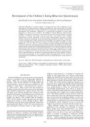
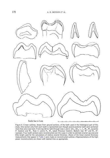
170 A. D. BEYNON ET AL.<br />
Figure 4. Crown outlines, drawn from ground sections, <strong>of</strong> the teeth used in the histological part <strong>of</strong> this<br />
study. One section only from each tooth is represented even though several posterior teeth were sectioned<br />
more than once (see text). From top left to right through rows 1 <strong>and</strong> 2: dm 1 ,dm 2 ,I 1 ,I 2 ,M 1 ,M 2 (juvenile<br />
P. heseloni) <strong>and</strong>M 1 . Row 3: canine P 4 ,M 3 (adult Proconsul heseloni). Bottom row: M 1 <strong>and</strong> M 2 (P.<br />
nyanzae). Four tooth sections were reconstructed over the cusps in order to estimate enamel cap area <strong>and</strong><br />
EDJ length used for the calculations <strong>of</strong> relative enamel thickness. (None <strong>of</strong> the linear enamel thickness<br />
measurements that appear in Table 1 were made on reconstructed outlines.) Representations <strong>of</strong> the high<br />
power reconstructions are shown with dashed lines as appropriate.