Comparative dental development and microstructure of ... - UCL
Comparative dental development and microstructure of ... - UCL
Comparative dental development and microstructure of ... - UCL

You also want an ePaper? Increase the reach of your titles
YUMPU automatically turns print PDFs into web optimized ePapers that Google loves.
188 A. D. BEYNON ET AL.<br />
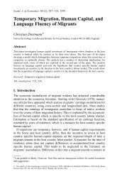
Figure 12. Confocal light image <strong>of</strong> striae <strong>of</strong> Retzius <strong>and</strong> enamel cross striations in the cervical region <strong>of</strong> the<br />
P. nyanzae M 2 . There are six cross striations between adjacent striae. (Fieldwidth 360 μm.)<br />
(S.D.=0·31, range=1·66–2·83). No trend<br />
in values from large to small, or vice versa,<br />
existed through the cuspal dentine <strong>of</strong> these<br />
teeth.<br />
Measurements <strong>of</strong> spacings <strong>of</strong> daily lines in<br />
the more lateral regions <strong>of</strong> the crowns <strong>of</strong><br />
both P. heseloni <strong>and</strong> P. nyanzae were much<br />
smaller than those made in the axis <strong>of</strong> the<br />
cuspal dentine (Table 4). At the EDJ they<br />
were typically 1·5 μm or less. Values around<br />
2 μm were more typical at the EDJ in occlusal<br />
areas between cusps <strong>and</strong> further in<br />
towards the pulp chamber.<br />
The ratio <strong>of</strong> the amount <strong>of</strong> dentine<br />
formed to the amount <strong>of</strong> enamel formed<br />
between the EDJ <strong>and</strong> accentuated lines<br />
common to both tissues appear in Table 4.<br />
This varied between 1:1·6 <strong>and</strong> 1:2·8 in P.<br />
heseloni. The most extreme values occur in<br />
the cervical region <strong>of</strong> the two molars attributed<br />
to P. nyanzae where ratios <strong>of</strong> 1:3 <strong>and</strong><br />
greater can be observed. By way <strong>of</strong> contrast,<br />
in the dm 2 (Individual IV) <strong>and</strong> the M 1<br />
(Individual III) attributed to P. heseloni, that<br />
have clear neonatal lines which mark both<br />
the enamel <strong>and</strong> dentine occlusally, the<br />
ratio <strong>of</strong> dentine to enamel formation is 1:1<br />
either side <strong>of</strong> the dentine horn. This occurs<br />
here partly because the high rate <strong>of</strong> dentine<br />
formation (3·5 μm per day) closely matches<br />
the rate <strong>of</strong> enamel formation (4·0 μm per<br />
day) in this position. However, some decussation<br />
in the enamel prisms here (but in<br />
none <strong>of</strong> the dentine tubules) equalizes the<br />
distance over which each tissue forms in this<br />
time.<br />
Crown completion times<br />
The perikymata counts made from the<br />
replicas <strong>of</strong> all Proconsul tooth surfaces are<br />
presented in detail in Appendix 1 by tooth<br />
type <strong>and</strong> by each aspect <strong>of</strong> each tooth