Comparative dental development and microstructure of ... - UCL
Comparative dental development and microstructure of ... - UCL
Comparative dental development and microstructure of ... - UCL

Create successful ePaper yourself
Turn your PDF publications into a flip-book with our unique Google optimized e-Paper software.
190 A. D. BEYNON ET AL.<br />
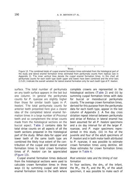
Figure 13. The combined totals <strong>of</strong> cuspal enamel formation times estimated from the histological part <strong>of</strong><br />
the study <strong>and</strong> lateral enamel formation times estimated from perikymata counts from replicas (see in<br />
Appendix 2). The inner vertical lines denote the cuspal enamel formation times. In this chart all<br />
perikymata counts for each tooth type (both upper <strong>and</strong> lower) have been combined <strong>and</strong> the error bars<br />
(1 S.D.) indicate the overall variation for lateral enamel formation only for each tooth type <strong>of</strong> P. heseloni.<br />
surface. The total number <strong>of</strong> perikymata<br />
on any tooth surface appears in the last but<br />
one column. In general the perikymata<br />
counts for P. nyanzae are slightly higher<br />
than those for similar tooth types in P.<br />
heseloni. The total perikymata counts for<br />
anterior teeth presented here give a clearer<br />
idea <strong>of</strong> the completed lateral enamel formation<br />
times in a large number <strong>of</strong> Proconsul<br />
teeth <strong>and</strong> so complement the striae counts<br />
made from the histological sections on the<br />
buccal aspect. Table 2 contains data for<br />
total striae counts on all aspects <strong>of</strong> all the<br />
tooth sections prepared in the histological<br />
part <strong>of</strong> the study. When data for upper <strong>and</strong><br />
lower teeth <strong>of</strong> the same tooth type are<br />
combined the likely true extent <strong>of</strong> the contribution<br />
<strong>of</strong> the cuspal <strong>and</strong> lateral enamel<br />
formation times to total crown formation<br />
times <strong>of</strong> P. heseloni can be appreciated<br />
(Figure 13).<br />
Cuspal enamel formation times deduced<br />
from the histological sections were used to<br />
calculate crown formation times in three<br />
ways (i) by summing cuspal <strong>and</strong> lateral<br />
enamel formation times in the teeth where<br />
complete crowns are represented in the<br />
histological sections (Table 2) <strong>and</strong> (ii) by<br />
summing cuspal formation times with data<br />
for buccal or mesiobuccal perikymata<br />
counts. The average crown formation times,<br />
derived for this purpose from the perikymata<br />
data for each tooth type, appear in the last<br />
column <strong>of</strong> Appendix 2. A five day cross<br />
striation repeat interval between perikymata<br />
<strong>and</strong> striae <strong>of</strong> Retzius in lateral enamel has<br />
been assumed for all P. heseloni specimens<br />
<strong>and</strong> a six day interval for all the larger P.<br />
nyanzae, <strong>and</strong> P. major specimens represented<br />
in this study. (iii) In five <strong>of</strong> the<br />
juvenile <strong>and</strong> four <strong>of</strong> the adult specimens <strong>of</strong><br />
P. heseloni <strong>and</strong> in both molar teeth attributed<br />
to P. nyanzae, it was possible to estimate<br />
crown formation times using dentine. All<br />
these estimates for crown formation times<br />
appear in Table 4.<br />
Root extension rates <strong>and</strong> the timing <strong>of</strong> root<br />
formation<br />
In four sections, the dm 2 <strong>of</strong> the infant,<br />
the M 1 , the P 4 <strong>and</strong> the M 2 <strong>of</strong> the adult<br />
specimen, it was possible to make each <strong>of</strong>