Optical investigation of the electron transfer protein azurinâgold ...
Optical investigation of the electron transfer protein azurinâgold ...
Optical investigation of the electron transfer protein azurinâgold ...

You also want an ePaper? Increase the reach of your titles
YUMPU automatically turns print PDFs into web optimized ePapers that Google loves.
Biophysical Chemistry 139 (2009) 1–7<br />
Contents lists available at ScienceDirect<br />
Biophysical Chemistry<br />
journal homepage: http://www.elsevier.com/locate/biophyschem<br />
<strong>Optical</strong> <strong>investigation</strong> <strong>of</strong> <strong>the</strong> <strong>electron</strong> <strong>transfer</strong> <strong>protein</strong> azurin–gold nanoparticle system<br />
Ines Delfino ⁎, Salvatore Cannistraro<br />
Biophysics and Nanoscience Centre, CNISM, Facoltà di Scienze, Università della Tuscia, Viterbo, Italy<br />
article<br />
info<br />
abstract<br />
Article history:<br />
Received 23 July 2008<br />
Received in revised form 19 September 2008<br />
Accepted 19 September 2008<br />
Available online 30 September 2008<br />
Keywords:<br />
Electron <strong>transfer</strong> <strong>protein</strong><br />
Azurin<br />
Gold nanoparticle<br />
Fluorescence quenching<br />
Energy <strong>transfer</strong><br />
The hybrid system obtained by conjugating <strong>the</strong> <strong>protein</strong> azurin, which is a very stable and well-described<br />
<strong>protein</strong> showing a unique interplay among its <strong>electron</strong> <strong>transfer</strong> and optical properties, with 20-nm sized gold<br />
nanoparticles has been investigated. Binding <strong>of</strong> azurin molecules to gold nanoparticle surface results in <strong>the</strong><br />
red shift <strong>of</strong> <strong>the</strong> nanoparticle resonance plasmon band and in <strong>the</strong> quenching <strong>of</strong> <strong>the</strong> azurin single tryptophan<br />
fluorescence signal. These findings toge<strong>the</strong>r with <strong>the</strong> estimate <strong>of</strong> <strong>the</strong> hydrodynamic radius <strong>of</strong> <strong>the</strong> composite,<br />
obtained by means <strong>of</strong> Dynamic Light Scattering, are consistent with <strong>the</strong> formation <strong>of</strong> a monolayer <strong>of</strong> <strong>protein</strong><br />
molecules, with preserved natural folding, on nanoparticle surface. The fluorescence quenching <strong>of</strong> azurin<br />
bound molecules is explained by an energy <strong>transfer</strong> from <strong>protein</strong> to metal surface and it is discussed in terms<br />
<strong>of</strong> <strong>the</strong> involvement <strong>of</strong> <strong>the</strong> Az <strong>electron</strong> <strong>transfer</strong> route in <strong>the</strong> interaction <strong>of</strong> <strong>the</strong> <strong>protein</strong> with <strong>the</strong> nanoparticle.<br />
© 2008 Elsevier B.V. All rights reserved.<br />
1. Introduction<br />
Integrating biological molecules with nanoparticles into hybrid<br />
devices permits to combine natural biomolecule functions, such as<br />
catalysis, recognition and <strong>electron</strong> <strong>transfer</strong>, with <strong>the</strong> unique <strong>electron</strong>ic,<br />
optical, and catalytic properties <strong>of</strong> nanoparticles, arising from <strong>the</strong>ir<br />
reduced dimension and high surface-to-volume ratio [1–3]. Noble<br />
metal nanoparticles exhibit a strong surface plasmon resonance<br />
absorption, due to <strong>electron</strong> collective oscillations, which is responsible<br />
for <strong>the</strong>ir brilliant colour and is highly dependent on particle size and<br />
surface environment [4,5]. The plasmon resonance can also be<br />
involved in a modulation <strong>of</strong> <strong>the</strong> electric field around <strong>the</strong> nanoparticle,<br />
giving rise to <strong>the</strong> enhancement <strong>of</strong> Raman scattering or fluorescence<br />
signal <strong>of</strong> nearby molecules [6,7]. Moreover, when biomolecules are<br />
conjugated with nanoparticles, <strong>the</strong> mixing <strong>of</strong> <strong>the</strong>ir energy levels has<br />
been shown to favour <strong>the</strong> occurrence <strong>of</strong> ei<strong>the</strong>r energy <strong>transfer</strong> or<br />
<strong>electron</strong> <strong>transfer</strong> phenomena [7,8]. In this way, it is possible to study<br />
fundamental processes characterizing <strong>the</strong> interaction <strong>of</strong> metal<br />
nanoparticles with organic or biological molecules [4,9,10]. Fur<strong>the</strong>rmore,<br />
nanoparticle properties can be exploited for realizing nanohybrid<br />
systems for various applications, also thanks to <strong>the</strong> possibility<br />
to suitably design <strong>the</strong>ir characteristics with a proper selection <strong>of</strong> <strong>the</strong><br />
employed biotic element [3,11–13].<br />
In this framework, <strong>electron</strong> <strong>transfer</strong> (ET) <strong>protein</strong>s are attracting<br />
growing interest. In particular, azurin (Az) is one <strong>of</strong> <strong>the</strong> most promising<br />
ET <strong>protein</strong>s because <strong>of</strong> its very peculiar properties and its capability to<br />
form molecular complexes with natural partners (i.e., cytochrome<br />
c551) [14,15] and o<strong>the</strong>r biomolecules (i.e., p53) [16,17], as recently<br />
⁎ Corresponding author. Tel.: +39 0761 357026; fax: +39 0761 357027.<br />
E-mail addresses: delfino@unitus.it (I. Delfino), cannistr@unitus.it (S. Cannistraro).<br />
outlined. It belongs to <strong>the</strong> class <strong>of</strong> redox blue copper <strong>protein</strong>s acting as<br />
<strong>electron</strong> shuttles in many important biological reactions, for instance<br />
in photosyn<strong>the</strong>sis and in bacterial respiratory redox-chains [18–20].<br />
They are endowed with very efficient ET mechanisms which occur at<br />
<strong>the</strong> level <strong>of</strong> single <strong>electron</strong>, even over long distances, in a very fast,<br />
directional way [21] and can be gated by proper modulation <strong>of</strong> <strong>the</strong><br />
applied potential [22]. Az ET process proceeds via <strong>the</strong> copper (Cu(II))-<br />
containing active site which is characterized by an intense ligand-tometal-charge<br />
<strong>transfer</strong> (LMCT) absorption band located at 628 nm<br />
[23,24]. Interestingly, it has been shown that an <strong>electron</strong> <strong>transfer</strong> well<br />
mimicking <strong>the</strong> <strong>the</strong>rmal one can be induced by light irradiating Az in its<br />
LMCT band [25], similarly to o<strong>the</strong>r blue copper <strong>protein</strong>s [26–28],<br />
pointing out an intriguing interplay between Az optical and electrical<br />
properties. Additionally, Az ET path is hypo<strong>the</strong>sized to involve <strong>the</strong><br />
<strong>protein</strong> single tryptophan residue [29], an intrinsic fluorophore located<br />
at position 48 (Trp48) in a highly hydrophobic environment [30,31],<br />
which is very sensitive to <strong>protein</strong> conformation [30–32].<br />
The integration <strong>of</strong> a biotic element with such extraordinary and<br />
versatile abilities into a nanoparticle-based hybrid system holds potential<br />
for a variety <strong>of</strong> applications ranging from bio-opto<strong>electron</strong>ics (for<br />
designing, as examples, biomemories, optical switches, data storage<br />
systems) [33,34] up to biomedicine, by taking advantage, for example, <strong>of</strong><br />
Az capability to recognize biomolecular analytes as disease markers<br />
[16,17]. However, this potential could be fully exploited only once <strong>the</strong><br />
basic properties <strong>of</strong> Az-based nano-hybrid systems have been described<br />
and <strong>the</strong>ir connection with <strong>the</strong> interaction occurring at <strong>the</strong> interface have<br />
been explored.<br />
In this paper, we report on an optical spectroscopy study <strong>of</strong> <strong>the</strong><br />
system obtained by conjugating <strong>the</strong> <strong>protein</strong> Az to gold nanoparticles<br />
(AuNPs) aiming at a deeper knowledge <strong>of</strong> both <strong>the</strong> interactions <strong>of</strong><br />
<strong>protein</strong> with nanoparticles and <strong>the</strong> resulting photophysical properties <strong>of</strong><br />
0301-4622/$ – see front matter © 2008 Elsevier B.V. All rights reserved.<br />
doi:10.1016/j.bpc.2008.09.016
2 I. Delfino, S. Cannistraro / Biophysical Chemistry 139 (2009) 1–7<br />
<strong>the</strong> hybrid system. Results from fluorescence and absorbance spectroscopy<br />
along with Dynamic Light Scattering (DLS) measurements are<br />
discussed in <strong>the</strong> view <strong>of</strong> <strong>the</strong> formation <strong>of</strong> a <strong>protein</strong> monolayer on<br />
nanoparticle surface and <strong>of</strong> potential involvement <strong>of</strong> <strong>the</strong> <strong>protein</strong> ET site<br />
in <strong>the</strong> interaction between <strong>the</strong> <strong>protein</strong> and <strong>the</strong> metal surface.<br />
2. Experimental methods<br />
Az from Pseudomonas aeruginosa was purchased from Sigma<br />
Aldrich Chemical Co. and used without fur<strong>the</strong>r purification (20-nm<br />
sized). AuNP solution was purchased from Ted Pella Inc., <strong>the</strong><br />
concentration <strong>of</strong> commercial solution being 1.13 nM in AuNPs.<br />
Stock Az solution was prepared by dissolving <strong>the</strong> <strong>protein</strong> in MilliQ<br />
water (Millipore 18 MΩ cm) at a concentration <strong>of</strong> 100 μM. This solution<br />
was fur<strong>the</strong>r diluted in water or in AuNP solution in order to obtain<br />
samples at different Az concentrations with and without AuNPs. In<br />
particular, for absorption and fluorescence spectroscopy measurements,<br />
Az+AuNP solutions at [AuNP]=1.02 nM and Az concentrations<br />
ranging from 0.7 up to 11 μM were investigated. Difference absorption<br />
measurements were carried out on Az+AuNP solutions at [Az]=8 and<br />
14 μM and AuNP concentrations in 0.022–0.20 nM range. DLS<br />
measurements were performed on 1.02-nM AuNP solutions with<br />
([Az]=11 μM) and without Az.<br />
<strong>Optical</strong> absorbance and difference absorbance spectra were recorded<br />
at room temperature by a double beam Jasco V-550 UV/visible<br />
spectrophotometer by using 1-cm path length cuvettes and 2-nm<br />
bandwidth. The investigated spectral range was 250–800 nm. Absorbance<br />
spectra were collected using MilliQ water as reference. For <strong>the</strong><br />
acquisition <strong>of</strong> difference absorbance spectra, <strong>the</strong> cuvette containing <strong>the</strong><br />
Az+AuNP solution was placed on <strong>the</strong> sample beam path, while a cuvette<br />
filled with an AuNP solutionwas on <strong>the</strong> reference path, <strong>the</strong> two solutions<br />
(sample and reference) being at <strong>the</strong> same AuNP concentration.<br />
DLS measurements [35] were taken at room temperature by using a<br />
θ=90° scattering geometry and an Ar laser light source (Ion Laser<br />
Technology Inc.) running at 488 nm (20 mW). The scattered light was<br />
detected by a photomultiplier and <strong>the</strong>n passed to a BI-9000 (Brookhaven<br />
Instruments) digital correlator with a sampling time range <strong>of</strong> 0.1 μs to<br />
1000 ms. The temperature <strong>of</strong> <strong>the</strong> sample was kept at 25.0±0.5 °C by a<br />
<strong>the</strong>rmostatic bath. The scattering intensity data were processed using<br />
<strong>the</strong> BI 9000 s<strong>of</strong>tware in order to obtain <strong>the</strong> hydrodynamic diameter and<br />
<strong>the</strong> size distribution <strong>of</strong> <strong>the</strong> scatterers in each sample. Experiment duration<br />
was in <strong>the</strong> range <strong>of</strong> 5–10 min, and each experiment was repeated<br />
several times on freshly prepared solutions.<br />
Steady-state fluorescence emission spectra, following 280- and<br />
290-nm excitations, were collected at room temperature in <strong>the</strong> range<br />
290–450 nm (or 300–450 nm) by a Spex FluoroMax (Jobin Yvon,<br />
France) spectr<strong>of</strong>luorometer, equipped with 450 W Xenon lamp and<br />
two monochromators. The band-pass width for <strong>the</strong> excitation and<br />
emission monochromators was 5 nm. An integration time <strong>of</strong> 1.0 s, that<br />
relates to 0.5 nm step, was used. The light emitted by 400 μl <strong>of</strong> sample<br />
kept in a 0.2×1.0 cm internal size quartz cuvette was collected at right<br />
angle to <strong>the</strong> excitation radiation. Fluorescence spectra <strong>of</strong> reference<br />
samples were always recorded and subtracted from <strong>the</strong> experimental<br />
data to correct for background. The spectra were also corrected for<br />
variations in lamp intensity by dividing <strong>the</strong> sample signal intensity by<br />
<strong>the</strong> reference (Spex photodiode detector) intensity. A correction for<br />
Inner Filter Effect was introduced, because <strong>of</strong> <strong>the</strong> high absorption due<br />
to AuNP solution in <strong>the</strong> investigated region, according to [31]<br />
Scattering effects on emission spectra were neglected because <strong>of</strong><br />
<strong>the</strong> high ratio between absorption and scattering cross-sections [4,5].<br />
3. Results and discussion<br />
3.1. Absorption<br />
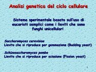
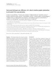
Representative absorption spectra <strong>of</strong> Az and Az+AuNP water<br />
solutions are shown in Fig. 1 along with <strong>the</strong> corresponding spectrum<br />
<strong>of</strong> bare AuNP solution. The absorption spectrum <strong>of</strong> <strong>the</strong> Az solution<br />
(continuous black line) shows two main peaks located at 280 and<br />
628 nm. The former is due to <strong>the</strong> presence <strong>of</strong> <strong>the</strong> aromatic residues,<br />
including <strong>the</strong> lone tryptophan (Trp48) [30,31]. The latter is typical <strong>of</strong><br />
a type-1 blue copper <strong>protein</strong> and is assigned to <strong>the</strong> LMCT transition<br />
(S(Cys-π)→Cu(II) d x<br />
2<br />
− y 2 ), involving <strong>the</strong> copper atom <strong>of</strong> <strong>the</strong> active site<br />
and one <strong>of</strong> <strong>the</strong> five metal ligands (specifically, Cys112) [23,24]. An<br />
intense plasmon resonance band, peaking at 522 nm, is evident in <strong>the</strong><br />
absorption spectrum <strong>of</strong> <strong>the</strong> AuNP solution (continuous gray line).<br />
Plasmon resonance bands have been widely studied and <strong>the</strong>y are now<br />
recognized to arise from <strong>the</strong> superposition <strong>of</strong> all contributing multipole<br />
oscillations peaking at different energies [4,5]. The frequency and<br />
width <strong>of</strong> <strong>the</strong> surface plasmon absorption bands depend on <strong>the</strong> size<br />
and <strong>the</strong> shape <strong>of</strong> metal nanoparticles as well as on <strong>the</strong> dielectric<br />
constant <strong>of</strong> metal itself and surrounding medium. In <strong>the</strong> present case,<br />
<strong>the</strong> features <strong>of</strong> <strong>the</strong> plasmon absorption band shown in Fig. 1 are<br />
consistent with a solution <strong>of</strong> 20-nm sized gold spherical particles, in<br />
agreement with <strong>the</strong> manufacturer specifications.<br />
The spectrum <strong>of</strong> Az+AuNP solution (Fig. 1, dashed line) is dominated<br />
by <strong>the</strong> AuNP plasmon resonance band, being <strong>the</strong> signal due to Az very<br />
small (see absolute absorbance <strong>of</strong> dashed and continuous black-line<br />
spectra <strong>of</strong> Fig. 1,reflecting <strong>the</strong> extinction coefficient values). This band is<br />
slightly shifted at higher wavelengths with respect to AuNP solution<br />
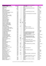
without Az (Az-free). This shift depends on Az concentration as it is clear<br />
from Fig. 2, where <strong>the</strong> normalized absorption spectra <strong>of</strong> Az+AuNP<br />
solutions at different Az concentrations are shown along with a zoom <strong>of</strong><br />
<strong>the</strong> wavelength region <strong>of</strong> plasmon band peaks (inset). A multi-peak<br />
Gaussian fitting procedure points out that red shift and width (FWHM)<br />
<strong>of</strong> <strong>the</strong> peak increase as long as <strong>the</strong> Az concentration increases, until a<br />
maximum value is reached. The trend <strong>of</strong> <strong>the</strong> peak position as a function<br />
<strong>of</strong> Az concentration is shown in Fig. 3. At <strong>the</strong> highest investigated Az<br />
concentration value (11 μM), <strong>the</strong> plasmon band has experienced a small<br />
but consistent red shift (6 nm). The plasmon band is also broadened by<br />
7 nm, which corresponds to a bandwidth increase <strong>of</strong> 230 cm −1 on a<br />
frequency scale.<br />
F corr ¼ F meas d10 ðAexþAem=5Þ=2<br />
ð1Þ<br />
where F corr and F meas are measured and corrected fluorescence<br />
emission spectra, respectively, and A ex and A em are <strong>the</strong> absorbance<br />
<strong>of</strong> <strong>the</strong> AuNP solution at excitation and emission wavelengths,<br />
respectively. A em is divided by a factor 5 for taking into account <strong>the</strong><br />
cuvette geometry.<br />
Fig. 1. Absorption spectra <strong>of</strong> Az solutions ([Az]=2.8 μM) with (dashed line) and without<br />
(continuous black line) 20-nm sized AuNPs ([AuNP]= 1.02 nM). For comparison,<br />
absorption spectrum <strong>of</strong> 1.02-nM bare AuNP solution is shown (continuous gray line).<br />
For dashed black line and continuous gray line <strong>the</strong> vertical scale is on <strong>the</strong> left; for<br />
continuous black line it is on <strong>the</strong> right.
I. Delfino, S. Cannistraro / Biophysical Chemistry 139 (2009) 1–7<br />
3<br />
Fig. 2. Normalized absorption spectra <strong>of</strong> Az+AuNP solutions at [AuNP] =1.02 nM and<br />
various Az concentrations: 0 μM (continuous black line), 0.7 μM (dotted black line),<br />
2.8 μM (dashed black line), 11 μM (continuous gray line). Inset: Zoom <strong>of</strong> <strong>the</strong> wavelength<br />
region <strong>of</strong> plasmon resonance band peaks.<br />
It has been reported that <strong>the</strong> analysis <strong>of</strong> <strong>the</strong> overall characteristics <strong>of</strong><br />
<strong>the</strong> plasmon band can be exploited to elucidate phenomena occurring at<br />
nanoparticle interface, including bioconjugation and molecular recognition<br />
[10,36]. Accordingly, three factors can be invoked as possible causes<br />
<strong>of</strong> <strong>the</strong> plasmon band shift and/or broadening detected in <strong>the</strong> absorption<br />
spectra <strong>of</strong> Az+AuNP solutions: (a) aggregation <strong>of</strong> nanoparticles upon<br />
bioconjugation, (b) <strong>electron</strong>ic communication between <strong>the</strong> Az redox<br />
center and <strong>the</strong> gold nanoparticle, and (c) changes in local dielectric<br />
environments. The hypo<strong>the</strong>sis <strong>of</strong> aggregation caused by Az addition can<br />
be reasonably ruled out by considering <strong>the</strong> absorbance spectra. In fact,<br />
aggregation <strong>of</strong> AuNPs would have lead to <strong>the</strong> formation <strong>of</strong> (at least) 40-<br />
nm sized clusters and, thus, to a red shift <strong>of</strong> <strong>the</strong> plasmon resonance band<br />
larger than that outlined in Az+AuNP solution absorption spectra<br />
(see Fig. 2). Moreover, <strong>the</strong> hypo<strong>the</strong>sis <strong>of</strong> aggregation caused by Az<br />
addition has been definitely ruled out by DLS results showing no<br />
evidence for <strong>the</strong> presence <strong>of</strong> aggregates <strong>of</strong> two or more AuNPs in <strong>the</strong><br />
investigated AZ+AuNP solutions (scatterer size distribution is discussed<br />
in <strong>the</strong> following). The o<strong>the</strong>r two hypo<strong>the</strong>ses are plausible and consistent<br />
with <strong>the</strong> direct interaction between gold surface and Az molecules. In<br />
case (b), interfacial ET from <strong>the</strong> excited Au plasmon to <strong>the</strong> bound Az<br />
molecules could shorten <strong>the</strong> lifetime <strong>of</strong> <strong>the</strong> plasmon excited state,<br />
leading to a corresponding bandwidth increase. This process has<br />
recently emerged as an attractive interpretation for <strong>the</strong> origin <strong>of</strong> <strong>the</strong><br />
observed AuNP plasmon band shift and broadening upon conjugation<br />
with cytochrome c [10]. However, <strong>the</strong> occurrence <strong>of</strong> <strong>the</strong> process has not<br />
been definitely demonstrated. To get deeper insight into <strong>the</strong> role <strong>of</strong> Az<br />
active site (and, hence, into <strong>the</strong> occurrence <strong>of</strong> an interfacial ET process) in<br />
<strong>the</strong> interaction between Az and AuNP, <strong>the</strong> occurrence <strong>of</strong> potential<br />
modifications <strong>of</strong> Az LMCT band upon AuNP addition by means <strong>of</strong><br />
difference absorption measurements has been investigated. Our data<br />
(not shown) indicate that this band remains unperturbed when AuNPs<br />
are added, thus suggesting that no changes take place in copper<br />
oxidation state. However, because <strong>the</strong> extinction coefficient <strong>of</strong> AuNP<br />
plasmon band (10 9 M −1 cm −1 ) greatly exceeds that <strong>of</strong> Az at 628 nm<br />
(5000 M −1 cm −1 ), <strong>the</strong> experiments have been carried out in solution<br />
with an excess <strong>of</strong> Az with respect to AuNP. In fact, in order to obtain a<br />
reliable difference spectrum with well defined LMCT band, it was<br />
necessary to work with high Az concentrations (8 and 14 μM inour<br />
experiments) and low AuNP concentrations (ranging from 0.022 to<br />
0.20 nM) and, consequently, it cannot be excluded that <strong>the</strong> possible<br />
changes induced in LMCT absorption band by <strong>the</strong> interaction <strong>of</strong> Az with<br />
AuNP are hidden by signal from Az molecules not interacting with gold<br />
nanoparticles. In case (c), <strong>the</strong> presence <strong>of</strong> Az near AuNP could cause<br />
significant changes in <strong>the</strong> local dielectric environment <strong>of</strong> <strong>the</strong> nanoparticle<br />
according to <strong>the</strong> well-known single particle plasmon band red shift<br />
with increasing medium dielectric constant [9,37]. Notably, <strong>the</strong><br />
experimental characteristics <strong>of</strong> <strong>the</strong> AuNP plasmon band after Az<br />
addition are consistent with Mie prediction for a 20-nm diameter gold<br />
sphere coated with a uniform Az monolayer having <strong>the</strong> same optical and<br />
geometrical properties <strong>of</strong> an Az monolayer formed on a flat substrate<br />
(properties <strong>of</strong> <strong>the</strong> monolayer have been extracted from Ref. [38]). In fact,<br />
<strong>the</strong> predicted Mie extinction spectrum, in <strong>the</strong> visible range, obtained by<br />
means <strong>of</strong> a dedicated s<strong>of</strong>tware based on a well-known algorithm [39],<br />
shows a plasmon band shifted at 527 nm and broadened up to 70 nm in<br />
fairly good agreement with <strong>the</strong> corresponding values extracted from <strong>the</strong><br />
absorption spectrum <strong>of</strong> <strong>the</strong> solution with <strong>the</strong> highest Az concentration<br />
(band peaked at 528 nm with a FWHM <strong>of</strong> 66 nm). This suggests that <strong>the</strong><br />
changes in plasmon band features upon Az addition are due to local<br />
dielectric environment modification connected to direct interaction <strong>of</strong><br />
<strong>the</strong> Az molecules with gold surface leading to <strong>the</strong> formation <strong>of</strong> a (mono)<br />
layer <strong>of</strong> Az molecules bound to AuNP surface. This hypo<strong>the</strong>sis well<br />
relates with <strong>the</strong> well-known ability <strong>of</strong> Az to link gold surfaces, as also<br />
<strong>the</strong>oretically described by means <strong>of</strong> molecular dynamics simulations<br />
[15,40–42]. Whatever <strong>the</strong> link is, if a monolayer is formed, an estimate <strong>of</strong><br />
<strong>the</strong> <strong>the</strong>oretical number <strong>of</strong> Az <strong>protein</strong>s (N Az ) that each spherical particle<br />
can accomodate on its surface may be derived from steric considerations<br />
using <strong>the</strong> following expression [43]:<br />
N Az ¼ 0:65d<br />
R 3 T −R3 AuNP<br />
R 3 Az<br />
!<br />
where R AuNP is <strong>the</strong> AuNP radius (10 nm) and R T is <strong>the</strong> radius <strong>of</strong> <strong>the</strong><br />
AuNP plus bound Az molecules (R AuNP +2R Az ), with R Az =2 nm [44].<br />
Actually, this description is valid under two assumptions: (i) Az<br />
molecules can be approximated as rigid spheres, close packed around a<br />
central sphere (AuNP); (ii) <strong>the</strong> conformation <strong>of</strong> <strong>the</strong> <strong>protein</strong> interacting<br />
with <strong>the</strong> nanoparticle is not significantly perturbed. In <strong>the</strong> investigated<br />
Az+AuNP system, both <strong>the</strong> assumptions can be thought to be valid<br />
since Az is known to be a quite globular <strong>protein</strong> and it is reasonable to<br />
think that it does not undergo such significant conformation changes<br />
upon <strong>the</strong> interaction with AuNP surface (however, this aspect will be<br />
discussed in <strong>the</strong> following). By introducing <strong>the</strong> expected values for our<br />
Az+AuNP system into Eq. (2), an estimate <strong>of</strong> ≈140 <strong>protein</strong>s per<br />
nanoparticle was obtained. Because <strong>of</strong> <strong>the</strong> occurrence <strong>of</strong> a close and<br />
uniform packing <strong>of</strong> <strong>protein</strong>s around an AuNP seems fairly unlikely, <strong>the</strong><br />
previous calculation has to be viewed as a maximum <strong>the</strong>oretical limit<br />
<strong>of</strong> conjugation, and thus, as a maximum number <strong>of</strong> linking sites per<br />
nanoparticle. This implies <strong>the</strong> presence, in <strong>the</strong> investigated solutions,<br />
<strong>of</strong> a number <strong>of</strong> Az molecules greater than that needed to form a<br />
monolayer on each gold nanoparticle.<br />
Fig. 3. Dependence <strong>of</strong> position <strong>of</strong> plasmon resonance band peak on Az concentration as<br />
obtained by multi-peak Gaussian fitting procedure performed on spectra shown in Fig. 2.<br />
ð2Þ
4 I. Delfino, S. Cannistraro / Biophysical Chemistry 139 (2009) 1–7<br />
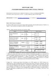
Fig. 4. Size distribution <strong>of</strong> AuNP in solution (at [AuNP] =1.02 nM) before and after Az<br />
addition, as obtained by <strong>the</strong> analysis <strong>of</strong> DLS results; gray bars being referred to bare<br />
AuNP solution and black bars to Az +AuNP solution ([Az]=11 μM). Continuous lines<br />
represent <strong>the</strong> results <strong>of</strong> a Gaussian fit procedure (see text).<br />
3.2. Dynamic Light Scattering<br />
The size distributions <strong>of</strong> AuNPs in solution before and after Az<br />
addition, as obtained by <strong>the</strong> analysis <strong>of</strong> DLS results [35], are shown in<br />
Fig. 4 (gray and black bars, respectively). They point out that diameters<br />
<strong>of</strong> <strong>the</strong> bare AuNP range from 18 to 22 nm and those <strong>of</strong> Az+AuNP<br />
systems range between 22 and 27 nm. A Gaussian fit procedure<br />
performed on both <strong>the</strong> outlined distributions (results are shown as<br />
continuous lines in <strong>the</strong> figure) gives <strong>the</strong> best estimation <strong>of</strong> <strong>the</strong> sizes,<br />
being 19.9±1.6 nm for Az-free solution and 24.1±1.8 nm when an<br />
excess <strong>of</strong> Az is added to AuNP-containing solutions.<br />
The increase in scatter (AuNP) size upon Az addition confirms that<br />
Az molecules have grafted on gold nanoparticles. Comparing <strong>the</strong><br />
diameters <strong>of</strong> Az+AuNP and bare AuNP can determine how far <strong>the</strong> Az<br />
molecule protrudes from <strong>the</strong> AuNP surface. The difference between<br />
<strong>the</strong> two mean diameters is about 4 nm, which can be thought as twice<br />
<strong>the</strong> apparent thickness <strong>of</strong> <strong>the</strong> shell coating <strong>the</strong> AuNP. This estimate is<br />
consistent with <strong>the</strong> thickness <strong>of</strong> an Az monolayer on planar gold [15],<br />
thus confirming <strong>the</strong> formation <strong>of</strong> an Az monolayer on AuNP surface.<br />
Therefore, <strong>the</strong> Az+AuNP system can be thought as a 20-nm sized gold<br />
nanoparticle with a 2-nm shell composed by less than 140 Azurin<br />
molecules around it, as sketched in Scheme 1.<br />
3.3. Fluorescence<br />
Representative fluorescence emission spectra, following a 280-nm<br />
excitation, <strong>of</strong> Az and Az+AuNP solutions are shown in Fig. 5. Az<br />
solution emission spectrum (continuous line) shows <strong>the</strong> well-known<br />
broad peak with a maximum at 308 nm due to <strong>the</strong> single buried<br />
tryptophan (Trp48) [29,30]. Fluorescence spectrum <strong>of</strong> Az+AuNP<br />
Scheme 1. Formation <strong>of</strong> Az monolayer on gold nanoparticles.<br />
Fig. 5. Representative fluorescence emission spectra <strong>of</strong> Az (continuous line) and Az+AuNP<br />
(dashed line) solutions at [Az]=2.8 μM and [AuNP]=1.02 nM.<br />
solution (dashed line) is dominated by signal due to Az, since 20-nm<br />
sized AuNPs are known to have negligible fluorescence signal in <strong>the</strong><br />
investigated region [45], as we have also observed. The addition <strong>of</strong><br />
AuNPs to Az solutions results in (i) a preservation <strong>of</strong> <strong>the</strong> shape <strong>of</strong> <strong>the</strong><br />
spectrum and (ii) a slight decrease <strong>of</strong> <strong>the</strong> Az emission intensity,<br />
suggesting <strong>the</strong> occurrence <strong>of</strong> fluorescence quenching process since <strong>the</strong><br />
spectra have been corrected for trivial intensity effects. In fact, <strong>the</strong><br />
analysis <strong>of</strong> <strong>the</strong> shape <strong>of</strong> Trp emission spectrum can provide useful<br />
information about <strong>the</strong> changes <strong>of</strong> <strong>the</strong> <strong>protein</strong> conformational state,<br />
fluorescence emission spectrum <strong>of</strong> <strong>the</strong> Trp residue in a <strong>protein</strong> being<br />
closely related to its solvent exposure [31,32]. In particular, it was<br />
shown that when unfolding process occurs in Az molecules, 308-nm<br />
emission peak shifts up to 357 nm as a consequence <strong>of</strong> an increased<br />
exposition <strong>of</strong> <strong>the</strong> Trp [46]. Accordingly, <strong>the</strong> outlined preservation <strong>of</strong><br />
<strong>the</strong> shape <strong>of</strong> <strong>the</strong> emission spectrum upon AuNP addition in our<br />
experiments is an indication that Az molecules participating to <strong>the</strong><br />
signal (and, thus, also <strong>the</strong> molecules bound to AuNP surface) remain<br />
presumably in <strong>the</strong>ir native configuration. This is in agreement with<br />
both experimental results [15,41] and molecular dynamics simulation<br />
<strong>of</strong> an Az molecule near a gold surface showing that <strong>the</strong> <strong>protein</strong> undergoes<br />
only small configurational changes even when it is covalently<br />
bound to <strong>the</strong> surface [42].<br />
The examination <strong>of</strong> <strong>the</strong> dependence <strong>of</strong> Az+AuNP solution fluorescence<br />
intensity on Az concentration has to be carried out by taking<br />
into account that we are interested in <strong>the</strong> variation <strong>of</strong> fluorescence<br />
signal occurring as a consequence <strong>of</strong> <strong>the</strong> AuNP addition. Considering<br />
that changes are expected to arise only from bound Az molecules<br />
and that <strong>the</strong> amount <strong>of</strong> free Az molecules in each solution is dependent<br />
on Az concentration, <strong>the</strong> ratio F 0−F<br />
F 0<br />
, where F 0 is <strong>the</strong> integrated<br />
fluorescence emission <strong>of</strong> Az solution and F is <strong>the</strong> same for <strong>the</strong> corresponding<br />
Az+AuNP solution, has been analysed to better evidence<br />
signal variation. Accordingly, in Fig. 6, F 0−F<br />
is shown as a function<br />
F0<br />
<strong>of</strong> <strong>the</strong> ratio <strong>of</strong> <strong>the</strong>oretical maximum number <strong>of</strong> binding sites (from<br />
Eq. (2), N s =140⁎[AuNP]⁎N A , where N A is <strong>the</strong> Avogadro number) to<br />
total number <strong>of</strong> Az molecules available in <strong>the</strong> investigated sample<br />
(N tAz =[Az]⁎N A ). An increase in <strong>the</strong> relative difference <strong>of</strong> fluorescence<br />
intensities as <strong>the</strong> ratio N s /N tAz increases is obtained till a maximum<br />
quenching (≈20%) is reached, for N s /N tAz N≈10%. This behaviour is<br />
consistent with a dependence <strong>of</strong> <strong>the</strong> total quenching on <strong>the</strong> number <strong>of</strong><br />
bound Az, resulting from a direct interaction <strong>of</strong> Az molecule with<br />
nanoparticle gold surface. Under <strong>the</strong> assumption <strong>of</strong> <strong>the</strong> formation <strong>of</strong><br />
Az monolayer on gold nanoparticle, <strong>the</strong> signal <strong>of</strong> Az molecules<br />
belonging to <strong>the</strong> monolayer can be supposed to be <strong>the</strong> only actually<br />
quenched, while free Az molecules preserve <strong>the</strong>ir fluorescence<br />
intensity. The total fluorescence is obviously due to <strong>the</strong> sum <strong>of</strong> signal<br />
<strong>of</strong> quenched and not quenched sources. Accordingly, as <strong>the</strong> relative
I. Delfino, S. Cannistraro / Biophysical Chemistry 139 (2009) 1–7<br />
5<br />
Fig. 6. (F 0 −F)/F 0 vs (# binding sites, N s )/(# total Az molecules, N tAz ) as obtained by<br />
considering integrated emission for <strong>the</strong> all investigated Az solutions (continuous line<br />
being a guide for <strong>the</strong> eye).<br />
number <strong>of</strong> free Az molecules (not quenched) increases, <strong>the</strong> overall<br />
quenching efficiency seems to be reduced. In <strong>the</strong> following discussion,<br />
we reasonably assume that: (i) <strong>the</strong> extinction coefficient in <strong>the</strong> UV<br />
range is <strong>the</strong> same for free and bound Az molecules; (ii) <strong>the</strong> quantum<br />
yield <strong>of</strong> <strong>the</strong> free Az, Q 0 , remains unchanged upon AuNP addition; (iii)<br />
<strong>the</strong> quantum yield <strong>of</strong> Az molecules bound to gold surface, Q, is <strong>the</strong><br />
same for all <strong>the</strong> bound molecules and QbQ 0 since a reduction <strong>of</strong><br />
fluorescence signal has been detected. In this frame, F 0 and F can be<br />
defined as: F 0 =Q 0 N tAz for Az solution without AuNPs (N tAz , <strong>the</strong> total<br />
number <strong>of</strong> Az molecules available in <strong>the</strong> solution, being equal, at<br />
fixed Az concentrations, for solutions containing or not AuNPs), and<br />
F=QN bAz +Q 0 N frAz for Az+AuNP solution, where N bAz is <strong>the</strong> number <strong>of</strong><br />
Az molecules bound to gold surface in Az+AuNP solution, and N frAz is<br />
<strong>the</strong> number <strong>of</strong> Az molecules left not bound (free) in that solution. By<br />
introducing <strong>the</strong> condition N frAz =N tAz −N bAz , <strong>the</strong> following equation is<br />
obtained:<br />
<br />
F 0 −F<br />
¼ Q <br />
0− Q NbAz<br />
ð3Þ<br />
F 0 Q 0 N tAz<br />
which explains <strong>the</strong> relationship between <strong>the</strong> experimental signal and<br />
<strong>the</strong> number <strong>of</strong> bound Az molecules. Obviously, N bAz depends on <strong>the</strong><br />
adsorption processes <strong>of</strong> <strong>the</strong> molecule (Az) onto <strong>the</strong> surface (AuNP's), but<br />
for N tAz ≫N bAz ,thatisathighAzconcentrationsandlowN bAz /N tAz ,we<br />
can roughly consider N bAz /N tAz ∝N s /N tAz . In <strong>the</strong>se conditions, Eq. (3)<br />
qualitatively describes <strong>the</strong> linear dependence <strong>of</strong> F0−F<br />
F 0<br />
on N s /N tAz outlined<br />
in Fig. 6 for N s /N tAz b≈10%, while, behind this threshold value,<br />
remains unchanged upon reducing <strong>the</strong> total number <strong>of</strong> Az molecules.<br />
This saturation effect has to be ascribed to <strong>the</strong> existence <strong>of</strong> a minimum<br />
N bAz /N tAz ratio, which is related to <strong>the</strong> adsorption equilibrium.<br />
These results confirm that Az molecules bound to gold surface<br />
experience a quenching process, i.e. a decrease <strong>of</strong> quantum yield as<br />
compared to that <strong>of</strong> free Az molecules. In order to better understand<br />
such process, it is useful to express <strong>the</strong> quantum yield <strong>of</strong> free (Q 0 ) and<br />
bound Az (Q) molecules as [31]<br />
F 0 −F<br />
F 0<br />
that <strong>the</strong> effect is ascribed to both reduction <strong>of</strong> radiative decay rate<br />
(k r ′bk r ) and increase <strong>of</strong> nonradiative rate (k nr ′Nk nr ), being <strong>the</strong> relative<br />
extent <strong>of</strong> <strong>the</strong> phenomena dependent on both nanoparticle size and<br />
distance between metal surface and fluorophore [47–50]. In <strong>the</strong><br />
Az+AuNP system, <strong>the</strong> decrease <strong>of</strong> <strong>the</strong> fluorophore radiative decay rate<br />
(thus, k r ′bk r ) can be ascribed to <strong>the</strong> so-called phase induced radiative<br />
rate suppression, occurring when <strong>the</strong> molecular dipole and <strong>the</strong> dipole<br />
induced on <strong>the</strong> AuNP radiate out <strong>of</strong> phase if <strong>the</strong> molecules are oriented<br />
tangentially to <strong>the</strong> AuNP surface, leading to a destructive interference<br />
[48]. The lack <strong>of</strong> a preferential orientation <strong>of</strong> Az molecules with respect<br />
to <strong>the</strong> nanoparticle surface suggests that this orientation-dependent<br />
effect cannot fully explain <strong>the</strong> outlined fluorescence quenching <strong>of</strong> bound<br />
Az molecules. The characteristics <strong>of</strong> <strong>the</strong> investigated system suggest that<br />
an important role in fluorescence quenching is played by nonradiative<br />
decay rate increase, which is thought to be induced by <strong>the</strong> opening <strong>of</strong> an<br />
additional nonradiative decay path connected to <strong>electron</strong> and/or energy<br />
<strong>transfer</strong> processes [47,49,51,52] (in such cases, <strong>the</strong> total nonradiative rate<br />
would be k nr ′+k T , where k T is <strong>the</strong> <strong>transfer</strong> decay rate, hence, <strong>the</strong><br />
denominator in Eq. (4b) becoming k r ′+k nr ′+k T ). In fact, on one hand, <strong>the</strong><br />
injection <strong>of</strong> a charge from <strong>the</strong> fluorophore excited state to <strong>the</strong> metal<br />
surface provides <strong>the</strong> additional nonradiative decay route that, in specific<br />
conditions, can compete with <strong>the</strong> naturally available de-excitation<br />
processes [49,52]. Actually, such a process is known to depend on <strong>the</strong><br />
fluorophore-metal energy level interplay, on <strong>the</strong> surface properties and<br />
on <strong>the</strong> distance between <strong>protein</strong> and metal surface, being more probable<br />
for shorter distances. On <strong>the</strong> o<strong>the</strong>r hand, a nonradiative decay rate<br />
increase can be due to <strong>the</strong> occurrence <strong>of</strong> a Resonance Energy Transfer<br />
(RET) process [47,51], a peculiar energy <strong>transfer</strong> occurring whenever <strong>the</strong><br />
emission spectrum <strong>of</strong> a fluorophore, called <strong>the</strong> donor, overlaps with <strong>the</strong><br />
absorption spectrum <strong>of</strong> ano<strong>the</strong>r molecule, called <strong>the</strong> acceptor. The<br />
donor–acceptor distance has to fall within 1–10 nm, values generally<br />
reported in <strong>the</strong> literature for an efficient energy <strong>transfer</strong> process [31].In<br />
<strong>the</strong> Az+AuNP complex, a direct RET between Trp (<strong>of</strong> bound Az<br />
molecules) and gold nanoparticle can be thought to occur, being <strong>the</strong><br />
Trp <strong>the</strong> donor and <strong>the</strong> nanoparticle <strong>the</strong> acceptor. In fact, absorption and<br />
emission spectra <strong>of</strong> AuNPs and Az are overlapping (see Fig. 7)and<strong>the</strong>Trp<br />
is supposed to be inside <strong>the</strong> <strong>protein</strong> monolayer coating <strong>the</strong> AuNP (thus, is<br />
far from <strong>the</strong> metal surface less than 3 nm). Actually, RET efficiency is<br />
dependent on <strong>the</strong> donor–acceptor distance, but it has been shown that<br />
<strong>the</strong> outlined experimental dependence is much weaker than expected<br />
from <strong>the</strong>oretical predictions [48]. Ano<strong>the</strong>r intriguing process involving<br />
energy <strong>transfer</strong> could be invoked to explain <strong>the</strong> outlined quenching<br />
effect. Theoretical work [53] and very recent experimental studies [7,50]<br />
have shown that metal particles could increase <strong>the</strong> efficiency <strong>of</strong> a<br />
naturally occurring RET between a donor and an acceptor when <strong>the</strong>y are<br />
k r<br />
aÞ Q 0 ¼ ;<br />
k r þ k nr<br />
k r V<br />
bÞ Q ¼<br />
k r Vþ k nr V<br />
ð4Þ<br />
where k r and k nr are <strong>the</strong> rate <strong>of</strong> radiative and nonradiative decays for<br />
Az-free molecule, respectively, and k r ′ and k nr ′ are <strong>the</strong> same for Az<br />
bound molecule.<br />
Actually, <strong>the</strong> reduction <strong>of</strong> <strong>the</strong> quantum yield <strong>of</strong> a fluorophore in <strong>the</strong><br />
proximity <strong>of</strong> a metallic nanoparticle has been reported for various<br />
fluorophores. Experimental and <strong>the</strong>oretical studies have demonstrated<br />
Fig. 7. Overlap between Az emission spectrum (continuous line) and absorption spectrum<br />
<strong>of</strong> AuNP solution (dashed line).
6 I. Delfino, S. Cannistraro / Biophysical Chemistry 139 (2009) 1–7<br />
close to a metal particle, leading to <strong>the</strong> so-called RET enhancement. It is<br />
wellknownthatinAz,Trpfluorescence signal is dramatically increased<br />
when Cu is removed, thus Cu itself is thought to operate as fluorescence<br />
quencher. Facile energy <strong>transfer</strong> between <strong>the</strong> Trp and <strong>the</strong> Cu-ligand<br />
complex is thought to be responsible for <strong>the</strong> fluorescence quenching in<br />
Trp. Presumably, this quenching occurs between <strong>the</strong> Trp 1 L ab states and a<br />
Cu-ligand charge <strong>transfer</strong> state [54].Thequenching<strong>of</strong>Trpfluorescence<br />
upon conjugation with AuNPs can be due to an enhancement <strong>of</strong> this<br />
naturally occurring energy <strong>transfer</strong> process. The energy <strong>transfer</strong>-based<br />
hypo<strong>the</strong>sis <strong>of</strong> fluorescence quenching in Az+AuNP system is in agreement<br />
with very recent studies on quenching <strong>of</strong> fluorescence from Bovine<br />
Serum Albumin molecules bound to gold nanoparticles [55]. Interestingly,<br />
in <strong>the</strong> peculiar case <strong>of</strong> Az+AuNP system, <strong>the</strong> occurrence <strong>of</strong> an<br />
energy <strong>transfer</strong> between Trp and gold could be indicative <strong>of</strong> some<br />
changes occurring in <strong>the</strong> <strong>electron</strong> <strong>transfer</strong> path and, thus, in <strong>protein</strong><br />
conductive properties, when Az is in <strong>the</strong> vicinity <strong>of</strong> an AuNP since <strong>the</strong>re<br />
are strong indications that <strong>the</strong> main <strong>electron</strong> <strong>transfer</strong> route in Az is a<br />
path through <strong>the</strong> buried Trp48 [29] and that Trp participates to <strong>electron</strong><br />
<strong>transfer</strong> in biological redox machines [56].<br />
4. Conclusions<br />
The optical spectroscopic study on <strong>the</strong> hybrid system obtained by<br />
conjugating <strong>the</strong> <strong>electron</strong> <strong>transfer</strong> blue copper <strong>protein</strong> Az to 20-nm<br />
sized AuNPs provided interesting information on <strong>the</strong> interactions<br />
occurring at <strong>the</strong> interface between <strong>the</strong> <strong>protein</strong> and <strong>the</strong> metal surface<br />
and on <strong>the</strong> resulting photophysical properties <strong>of</strong> <strong>the</strong> system. The<br />
analysis <strong>of</strong> <strong>the</strong> plasmon resonance band <strong>of</strong> <strong>the</strong> composite along with<br />
DLS results confirmed <strong>the</strong> interaction <strong>of</strong> Az with gold surface, probably<br />
involving a monolayer <strong>of</strong> <strong>protein</strong> molecules grafted on AuNPs. Each<br />
nanoparticle was estimated to bind at maximum 140 Az molecules<br />
which were thought to preserve <strong>the</strong>ir natural folding. The <strong>investigation</strong><br />
<strong>of</strong> fluorescence emission spectrum <strong>of</strong> Az+AuNP system, due to Az<br />
single buried Trp, outlined <strong>the</strong> occurrence <strong>of</strong> a quenching effect<br />
related to <strong>protein</strong>–nanoparticle interactions. This intriguing phenomenon<br />
was explained by <strong>the</strong> increase <strong>of</strong> <strong>the</strong> nonradiative decay rate as a<br />
consequence <strong>of</strong> a direct energy <strong>transfer</strong> between Trp and gold surface<br />
or to <strong>the</strong> enhancement <strong>of</strong> <strong>the</strong> naturally occurring energy <strong>transfer</strong><br />
between Trp and Cu-ligand site, thus suggesting <strong>the</strong> involvement <strong>of</strong><br />
<strong>the</strong> ET path in <strong>the</strong> interactions between Az and gold surface.<br />
These results enable a step forward <strong>the</strong> exploitation <strong>of</strong> <strong>the</strong> potentialities<br />
<strong>of</strong> Az-based nano-hybrid systems which deserve interest for<br />
a variety <strong>of</strong> applications ranging from bio-opto<strong>electron</strong>ics up to<br />
biomedicine.<br />
Acknowledgement<br />
This work has been partially supported by PRIN-MIUR 2006 project<br />
(nr. 2006028219).<br />
References<br />
[1] C.M. Niemeyer, Nanoparticles, <strong>protein</strong>s, and nucleic acids: biotechnology meets<br />
materials science, Angew. Chem., Int. Ed. 40 (2001) 4128–4158.<br />
[2] H. Gu, K. Xu, C. Xu, B. Xu, Bi<strong>of</strong>unctional magnetic nanoparticles for <strong>protein</strong> separation<br />
and pathogen detection, Chem. Commun. (2006) 941–946.<br />
[3] Q. Huo, A perspective on bioconjugated nanoparticles and quantum dots, Colloids<br />
Surf. B: Bionterfaces 59 (2007) 1–10.<br />
[4] S. Link, M.A. El-Sayed, Shape and size dependence <strong>of</strong> radiative, non-radiative and<br />
photo<strong>the</strong>rmal properties <strong>of</strong> gold nanocrystals, Int. Rev. Phys. Chem. 19 (2000)<br />
409–453.<br />
[5] P.V. Kamat, Photophysical, photochemical and photocatalytic aspects <strong>of</strong> metal<br />
nanoparticles, J. Phys. Chem. B 106 (2002) 7729–7744.<br />
[6] K. Kneipp, H. Kneipp, I. Itzkan, R.R. Dasari, M.S. Feld, Ultrasensitive chemical<br />
analysis by Raman spectroscopy, Chem. Rev. 99 (1999) 2957–2975.<br />
[7] J.R. Lakowicz, J. Kusba, Y. Shen, J. Malicka, S. D'Auria, Z. Gryczynski, I. Gryczynski,<br />
Effects <strong>of</strong> metallic silver particles on resonance energy <strong>transfer</strong> between<br />
fluorophores bound to DNA, J. Fluoresc. 13 (2003) 69–77.<br />
[8] I. Willner, R. Baron, B. Willner, Integrated nanoparticle-biomolecule systems for<br />
biosensing and bio<strong>electron</strong>ics, Biosens. Bio<strong>electron</strong>. 22 (2007) 1841–1852.<br />
[9] P.K. Jain, W. Huang, M.A. El-Sayed, On <strong>the</strong> universal scaling behavior <strong>of</strong> <strong>the</strong> distance<br />
decay <strong>of</strong> plasmon coupling in metal nanoparticle pairs: a plasmon ruler equation,<br />
Nano Lett. 7 (2007) 2080–2088.<br />
[10] P.S. Jensen, Q. Chi, F.B. Grumsen, J.M. Abad, A. Horsewell, D.J. Schiffrin, J.J. Ulstrup,<br />
Gold nanoparticle assisted assembly <strong>of</strong> a heme <strong>protein</strong> for enhancement <strong>of</strong> longrange<br />
interfacial <strong>electron</strong> <strong>transfer</strong>, J. Phys. Chem. C. 111 (2007) 6124–6132.<br />
[11] I.H. El-Sayed, X. Huang, M.A. El-Sayed, Surface plasmon resonance scattering and<br />
absorptin <strong>of</strong> anti-EGFR antibody conjugated gold nanoparticles in cancer diagnostics:<br />
applications in oral cancer, Nano Lett. 5 (2005) 829–834.<br />
[12] N.L. Rosi, C.A. Mirkin, Nanostructures in biodiagnostics, Chem. Rev. 105 (2005)<br />
1547–1562.<br />
[13] I. Willner, B. Willner, E. Katz, Biomolecule-nanoparticle hybrid systems for bio<strong>electron</strong>ic<br />
applications, Bioelectrochemistry 70 (2007) 2–11.<br />
[14] M.M. Hervás, J.A. Navarro, A. Díaz, H. Bottin, M.A. De La Rosa, Laser flash kinetic<br />
analysys <strong>of</strong> <strong>the</strong> fast <strong>electron</strong> <strong>transfer</strong> from plastocyanin and cytochrome c6 to<br />
photosystem I. Experimental evidence on <strong>the</strong> evolution <strong>of</strong> <strong>the</strong> reaction mechanism,<br />
Biochemistry 34 (1995) 11321–11326.<br />
[15] B. Bonanni, D. Alliata, L. Andolfi, A.R. Bizzarri, S. Cannistraro, Redox metallo<strong>protein</strong>s<br />
on metal surface as hybrid system for bionanodevices: an extensive<br />
characterization at <strong>the</strong> single molecule level, in: C.P. Norris (Ed.), Surface<br />
Science Research Developments, Nova Science Publishers, Inc., New York, 2005,<br />
pp. 1–73.<br />
[16] Y. Hiroaka, T. Yamada, M. Goto, T.K. Das Gupta, A.M. Charkrabarty, Modulation <strong>of</strong><br />
mammalian cell growth and death by prokaryotic and eukaryotic cytochrome c,<br />
Proc. Natl. Acad. Sci. [USA] 101 (2004) 6427–6432.<br />
[17] V. De Grandis, A.R. Bizzarri, S. Cannistraro, Docking study and free energy<br />
simulation <strong>of</strong> <strong>the</strong> complex between p53 DNA-binding domain and azurin, J. Mol.<br />
Recognit. 20 (2007) 215–226.<br />
[18] A.G. Sykes, Active-site properties <strong>of</strong> <strong>the</strong> blue copper <strong>protein</strong>s, Adv. Inorg. Chem. 36<br />
(1991) 377–408.<br />
[19] E.I. Solomon, M.J. Baldwin, M.D. Lowery, Electronic structures <strong>of</strong> active sites in<br />
copper <strong>protein</strong>s: contributions to reactivity, Chem. Rev. 92 (1992) 521–542.<br />
[20] C. Dennison, Investigating <strong>the</strong> structure and function <strong>of</strong> cupredoxins, Coord. Chem.<br />
Rev. 249 (2005) 3025–3054.<br />
[21] A.R. Bizzarri, S. Cannistraro, Electron <strong>transfer</strong> in metallo<strong>protein</strong>s, in: G. Bassani, G. Liedl,<br />
P. Wyder (Eds.), Encyclopedia <strong>of</strong> Condensed Matter Physics, Elsevier, Amsterdam, The<br />
Ne<strong>the</strong>rlands, 2005, pp. 361–369.<br />
[22] W. Haehnel, T. Jansen, K. Gause, R.B. Klösgen, B. Stahl, D. Michl, B. Huvermann, M.<br />
Karas, R.G. Hermann, Electron <strong>transfer</strong> from plastocyanin to photosystem I, EMBO<br />
J. 13 (1994) 1028–1038.<br />
[23] E.I. Solomon, J.W. Hare, D.M. Dooley, J.H. Dawson, P.J. Stephens, H.B. Gray,<br />
Spectroscopic studies <strong>of</strong> stellacyanin, plastocyanin, and azurin. Electronic<br />
structure <strong>of</strong> <strong>the</strong> blue copper sites, J. Am. Chem. Soc. 102 (1980) 168–178.<br />
[24] E. Fraga, M.A. Webb, G.R. Loppnow, Excited-state charge-<strong>transfer</strong> dynamics <strong>of</strong><br />
azurin, a blue copper <strong>protein</strong>, from resonance Raman intensities, J. Phys. Chem. B<br />
101 (1996) 5062–5069.<br />
[25] T. Cimei, A.R. Bizzarri, S. Cannistraro, G. Cerullo, S. De Silvestri, Vibrational<br />
coherence in azurin with impulsive excitation <strong>of</strong> <strong>the</strong> LMCT absorption band, Chem.<br />
Phys. Lett. 362 (2002) 497–503.<br />
[26] M.D. Edington, W.M. Diffey, W.J. Doria, R.E. Riter, W.F. Beck, Radiationless decay from<br />
<strong>the</strong> ligand-to-metal charge-<strong>transfer</strong> state in <strong>the</strong> blue copper <strong>protein</strong> plastocyanin,<br />
Chem. Phys. Lett. 275 (1997) 119–126.<br />
[27] L.D. Book, D.C. Arnett, H. Hu, N.F. Scherer, Ultrafast pump-probe studies <strong>of</strong> excitedstate<br />
charge-<strong>transfer</strong> dynamics in blue copper <strong>protein</strong>s, J. Phys. Chem. A 102 (1998)<br />
4350–4359.<br />
[28] I. Delfino, C. Manzoni, K. Sato, C. Dennison, G. Cerullo, S. Cannistraro, Ultrafast<br />
pump-probe study <strong>of</strong> excited-state charge-<strong>transfer</strong> dynamics in umecyanin from<br />
horseradish root, J. Phys. Chem. B 110 (2006) 17252–17259.<br />
[29] O. Farver, L.K. Skov, S. Young, N. Bonander, B. Karlsson, T.G. Vanngard, I. Pecht,<br />
Aromatic residues may enhance intramolecular <strong>electron</strong> <strong>transfer</strong> in azurin, J. Am.<br />
Chem. Soc. 119 (1997) 5453–5454.<br />
[30] K.K. Turoverov, I.M. Kuznetsova, V.N. Zaitsev, The environment <strong>of</strong> <strong>the</strong> tryptophan<br />
residue in Pseudomonas aeruginosa azurin and its fluorescence properties, Biophys.<br />
Chemist. 23 (1985) 79–89.<br />
[31] J.R. Lakowicz, Principles <strong>of</strong> Fluorescence Spectroscopy, second ed.Plenum Press,<br />
New York, 1999.<br />
[32] G. Gilardi, G. Mei, N. Rosato, G.W. Canters, A. Finazzi-Agrò, Unique environment <strong>of</strong><br />
Trp48 in Pseudomonas aeruginosa azurin as probed by site-directed mutagenesis<br />
and dynamic fluorescence spectroscopy, Biochemistry 33 (1994) 1425–1432.<br />
[33] D.I. Gittins, D. Be<strong>the</strong>ll, D.J. Schiffrin, R.J. Nichols, A nanometre-scale <strong>electron</strong>ic<br />
switch consisting <strong>of</strong> a metal cluster and redox-addressable groups, Nature 408<br />
(2000) 67–69.<br />
[34] T. Hugel, N.B. Holland, A. Cattani, L. Moroder, M. Seitz, H.E. Gaub, Single-molecule<br />
optomechanical cycle, Science 296 (2002) 1103–1106.<br />
[35] I. Delfino, K. Sato, M.D. Harrison, L. Andolfi, A.R. Bizzarri, C. Dennison, S.<br />
Cannistraro, Dynamic Light Scattering, Wiley-Interscience, New York, 1976;<br />
B.J. Berne, R. Pecora, <strong>Optical</strong> spectroscopic <strong>investigation</strong> <strong>of</strong> <strong>the</strong> alkaline transition<br />
in umecyanin from horseradish root, Biochemistry 44 (2005) 16090–16097.<br />
[36] S. Chan, M.R. Hammond, R.N. Zare, Gold nanoparticles as a colorimetric sensor for<br />
<strong>protein</strong> conformational changes, Chem. biol. 12 (2005) 323–328.<br />
[37] P.K. Jain, M.A. El-Sayed, Surface plasmon resonance sensitivity at metal<br />
nanostructures: physical basis and universal scaling in metal nanoshells, J. Phys.<br />
Chem. C 111 (2007) 17451–17454.<br />
[38] F. Bordi, M. Prato, O. Cavalleri, C. Cametti, M. Canepa, A. Gliozzi, Azurin selfassembled<br />
monolayers characterized by coupling electrical impedance spectroscopy<br />
and spectroscopic ellissometry, J. Phys. Chem. B 108 (2004) 20263–20272.
I. Delfino, S. Cannistraro / Biophysical Chemistry 139 (2009) 1–7<br />
7<br />
[39] C.F. Bohren, D.R. Huffman, Absorption and Scattering <strong>of</strong> Light by Small Particles,<br />
John Wiley & Sons, Inc., New York, 1983.<br />
[40] L. Venkataraman, J.E. Klare, I.W. Tam, C. Nuckolls, M.S. Hybertsen, M.L. Steigerwald,<br />
Single-molecule circuits with well-defined molecular conductance, Nano Lett. 6<br />
(2006) 458–462.<br />
[41] J.J. Davis, H.A.O. Hill, The scanning probe microscopy <strong>of</strong> metallo<strong>protein</strong>s and<br />
metalloenzymes, Chem. Commun. 393 (2002) 200–202;<br />
Q. Chi, J. Zhang, J.U. Nielsen, I. Friis, E.P. Chorkendorff, G.W. Canters, J.E.T. Andersen,<br />
J. Ulstrup, Molecular monolayers and interfacial <strong>electron</strong> <strong>transfer</strong> <strong>of</strong> Pseudomonas<br />
aeruginosa azurin on Au (111), J. Am. Chem. Soc. 122 (2000) 4047–4055.<br />
[42] A.R. Bizzarri, Topological and dynamical properties <strong>of</strong> azurin anchored to a gold<br />
substrate as investigated by molecular dynamics simulation, Biophys. Chemist. 122<br />
(2006) 206–214.<br />
[43] H. Mattoussi, J.M. Mauro, E.R. Goldman, G.P. Anderson, V.C. Sundar, F.V. Mikulec,<br />
M.G. Bawendi, Self-assembly <strong>of</strong> CdSe–ZnS quantum dot bioconjugates using an<br />
engineered recombinant <strong>protein</strong>, J. Am. Chem. Soc. 122 (2000) 12142–12150;<br />
P.P. Pompa, R. Chiuri, L. Manna, T. Pellegrino, L.L. del Mercato, W.J. Parak, F. Calabi,<br />
R. Cingolani, R. Rinaldi, Fluorescence resonance energy <strong>transfer</strong> induced by<br />
conjugation <strong>of</strong> metallo<strong>protein</strong>s to nanoparticles, Chem. Phys. Lett. 417 (2006)<br />
351–357.<br />
[44] H. Nar, A. Messerschmidt, R. Huber, M. Van de Kamp, G.W. Canters, Crystal<br />
structure analysis <strong>of</strong> oxidized Pseudomonas aeruginosa azurin at pH 5.5 and pH 9.0.<br />
A pH-induced conformational transition involves a peptide bond flip, J. Mol. Biol.<br />
221 (1991) 765–772.<br />
[45] J.P. Wilcoxon, J.E. Martin, F. Parsapour, B. Wiedenman, D.F. Kelley, Photoluminescence<br />
from nanosize gold clusters, J. Chem. Phys. 108 (1998) 9137–9143.<br />
[46] P. Guptasarma, Resolving multiple <strong>protein</strong> conformers in equilibrium unfolding<br />
reactions: a time-resolved emission spectroscopic (TRES) study <strong>of</strong> azurin, Biophys.<br />
Chemist. 65 (1997) 221–228.<br />
[47] E. Dulkeith, A.C. Mortcani, T. Niedereichholz, T.A. Klar, J. Feldmann, S.A. Levi, F.C.J.M.<br />
Van Veggel, D.N. Reinhoudt, M. Moller, D.J. Gittins, Fluorescence quenching <strong>of</strong> dye<br />
molecules near gold nanoparticles: radiative and nonradiative effects, Phys. Rev.<br />
Lett. 89 (2002) 2030021–2030024.<br />
[48] E. Dulkeith, M. Ringler, T.A. Klar, J. Feldmann, A. Muñoz, W.J. Parak, Gold nanoparticles<br />
quench fluorescence by phase induced radiative rate suppression, Nano<br />
Lett. 5 (2005) 585–589.<br />
[49] F. Cannone, G. Chirico, A.R. Bizzarri, S. Cannistraro, Quenching and blinking <strong>of</strong><br />
fluorescence <strong>of</strong> a single dye molecule bound to gold nanoparticles, J. Phys. Chem. B<br />
110 (2006) 16491–16498.<br />
[50] J. Zhang, Y. Fu, J.R. Lakowicz, Enhanced Förster Resonance Energy Transfer (FRET)<br />
on a single metal particle, J. Phys. Chem. C 111 (2007) 50–56.<br />
[51] B. Dubertret, M. Calame, A.J. Libchaber, Single-mismatch. detection using goldquenched<br />
fluorescent oligonucleotides, Nat. Biotechnol. 19 (2001) 365–370.<br />
[52] B.I. Ipe, K.G. Thomas, S. Barazzouk, S. Hotchandani, P.V. Kamat, Photoinduced<br />
charge separation in a fluorophore-gold nanoassembly, J. Phys. Chem. B 106 (2002)<br />
18–21;<br />
M.W. Holman, R. Liu, D.M. Adams, Single-molecule spectroscopy <strong>of</strong> interfacial<br />
<strong>electron</strong> <strong>transfer</strong>, J. Am. Chem. Soc. 125 (2003) 12649–12654.<br />
[53] J.I. Gersten, A. Nitzan, Accelerated energy <strong>transfer</strong> between molecules near a solid<br />
particle, Chem. Phys. Lett 104 (1984) 31–37.<br />
[54] J.A. Sweeney, P.A. Harmon, S.A. Asher, C.M. Hutnik, A.G. Szabo, UV resonance<br />
Raman examination <strong>of</strong> <strong>the</strong> azurin tryptophan environment and energy relaxation<br />
pathways, J. Am. Chem. Soc. 113 (1991) 7531–7537.<br />
[55] L. Shang, Y. Wang, J. Jiang, S. Dong, pH-dependent <strong>protein</strong> conformational changes<br />
in albumin:gold nanoparticle bioconjugates: a spectroscopic study, Langmuir 23<br />
(2007) 2714–2721;<br />
N. Wangoo, C.R. Suri, G. Shekhawat, Interaction <strong>of</strong> gold nanoparticles with <strong>protein</strong>:<br />
a spectroscopic study to monitor <strong>protein</strong> conformational changes, Appl. Phys. Lett.<br />
92 (2008) 133104/1-3.<br />
[56] C. Shih, A.K. Museth, M. Abrahamsson, A.M. Blanco-Rodriguez, A.J. Di Bilio, J.<br />
Sudhamsu, B.R. Crane, K.L. Ronayne, M. Towrie, A. Vlcek Jr., J.H. Richards, J.R.<br />
Winkler, H.B. Gray, Tryptophan-accelerated <strong>electron</strong> flow through <strong>protein</strong>s,<br />
Science 320 (2008) 1760–1762.