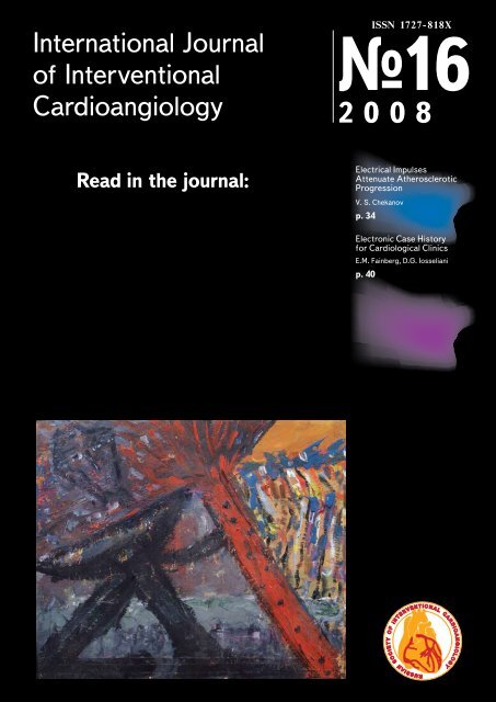
International Journal of Interventional Cardioangiology
International Journal of Interventional Cardioangiology
International Journal of Interventional Cardioangiology
You also want an ePaper? Increase the reach of your titles
YUMPU automatically turns print PDFs into web optimized ePapers that Google loves.
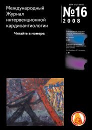
<strong>International</strong> <strong>Journal</strong><br />
<strong>of</strong> <strong>Interventional</strong><br />
<strong>Cardioangiology</strong><br />
ISSN 1727-818X<br />
№16<br />
2008<br />
Read in the journal:<br />
Electrical Impulses<br />
Attenuate Atherosclerotic<br />
Progression<br />
V. S. Chekanov<br />
p. 34<br />
Electronic Case History<br />
for Cardiological Clinics<br />
E.M. Fainberg, D.G. Iosseliani<br />
p. 40
ISSN 1727818X<br />
INTERNATIONAL JOURNAL<br />
OF INTERVENTIONAL CARDIOANGIOLOGY<br />
Quarterly <strong>Journal</strong> <strong>of</strong> the Russian Scientific Society<br />
<strong>of</strong> <strong>Interventional</strong> <strong>Cardioangiology</strong><br />
№ 16, 2008 г.<br />
"<strong>International</strong> <strong>Journal</strong> <strong>of</strong> <strong>Interventional</strong><br />
<strong>Cardioangiology</strong>"<br />
peerreviewed scientific<br />
and practical journal.<br />
Founded in 2002<br />
Address <strong>of</strong> the Editions:<br />
101000, Moscow,<br />
Sverchkov per., 5<br />
Phone: (+ 7 495) 624 96 36<br />
Fax: (+7 495) 624 67 33<br />
Head <strong>of</strong> Editorial Office:<br />
E.D. Bogatyrenko<br />
Editorial Board<br />
Editor-in-Chief D.G. Iosseliani<br />
A.M. Babunashvili (Moscow)<br />
V.V.Chestukhin (Moscow)<br />
V.V. Demin (Orenbourg)<br />
V.A. Ivanov (Krasnogorsk)<br />
Z.A.Kavteladze (Moscow) – Deputy Editor-in-Chief, President <strong>of</strong> Russian<br />
Scientific Society <strong>of</strong> <strong>Interventional</strong> <strong>Cardioangiology</strong><br />
I.V.Pershukov (Voronezh)<br />
A.V.Protopopov (Krasnoyarsk)<br />
A.N. Samko (Moscow)<br />
V.K. Sukhov (St. Petersburg)<br />
B. E. Shakhov (Nijny Novgorod)<br />
B.M.Shukurov (Volgograd) – Deputy Editor-in-Chief<br />
Scientific editors <strong>of</strong> translations:<br />
D.G. Gromov, O.G. Sukhorukov<br />
Editorial Council<br />
Translation:<br />
Medtran<br />
Original layout prepared by:<br />
I. Shishkarev, V. Shelepukhin<br />
Computer typesetting and<br />
makeup:<br />
I. Shishkarev<br />
Corrector:<br />
N. Sheludiakova<br />
Special gratitude to<br />
George Gigineishvili,<br />
doctor and artist, for the <strong>of</strong>fered<br />
opportunity to put the photocopy <strong>of</strong><br />
his painting<br />
"<strong>Interventional</strong> <strong>Cardioangiology</strong>" on<br />
the cover <strong>of</strong> the magazine<br />
S.A. Abugov (Moscow)<br />
Andreas Adam (London)<br />
I.S. Arabadjan (Moscow) A.V.<br />
Arablinsky (Moscow)<br />
T.Batyraliev (Gaziantep)<br />
Yu.V. Belov (Moscow)<br />
S.A. Biriukov (Riazan)<br />
A.S. Bronstein (Moscow)<br />
V.S. Buzaev (Ufa)<br />
Antonio Colombo (Milan)<br />
Carlo Di Mario (London)<br />
Robert Dondelinger (Liege)<br />
D.P.Dundua (Moscow)<br />
Andrejs Erglis (Riga)<br />
A.N.Fedorchenko (Krasnodar)<br />
Francis Fontan (Bordeaux)<br />
V.I. Ganiukov (Novosibirsk)<br />
D.G.Gromov (Moscow)<br />
V.N. Ilyin (Moscow)<br />
Matyas Keltai (Budapest)<br />
Spencer B.King III (Atlanta)<br />
L.S.Kokov (Moscow)<br />
Jan Kovac (Leicester)<br />
V.S. Kuzmenko (Kaliningrad)<br />
V.V.Kucherov (Moscow)<br />
A.N. Maltsev (Ulianovsk)<br />
V.P.Mazaev (Moscow)<br />
Bernhard Meier (Bern)<br />
E.V. Morozova (Penza)<br />
Seung-Jung Park (Seoul)<br />
A.P.Perevalov (Ijevsk)<br />
V.G.Plekhanov (Ivanovo)<br />
A.V.Pokrovsky (Moscow)<br />
Witold Ruzyllo (Warsaw)<br />
Shigeru Saito (Kamakura)<br />
D.B.Sapryguin (Moscow)<br />
S.P. Semitko (Moscow)<br />
Patrick W.Serruys (Rotterdam)<br />
Horst Sievert (Frankfurt)<br />
Rüdiger Simon (Kiel)<br />
A.F.Tsib (Moscow)<br />
Alec Vahanian (Paris)<br />
Jean-Charles Vernhet (Bordeaux)<br />
Yu.D.Volynsky (Moscow)<br />
L. Samuel Wann (Milwaukee)<br />
Petr Widimsky (Prague)<br />
I.P. Zyrianov (Tiumen)<br />
3
Instructions for authors<br />
The <strong>International</strong> <strong>Journal</strong> <strong>of</strong><br />
<strong>Interventional</strong> <strong>Cardioangiology</strong> (IJIC)<br />
publishes peer-reviewed articles on all<br />
aspects <strong>of</strong> cardiovascular disease, as<br />
well as the abstracts <strong>of</strong> communications,<br />
presented at the scientific congresses,<br />
sessions and conferences,<br />
held by the Russian Scientific Society<br />
<strong>of</strong> <strong>Interventional</strong> <strong>Cardioangiology</strong>.<br />
All manuscripts should be<br />
addressed to:<br />
Pr<strong>of</strong>. David G. Iosseliani, Editorin-Chief,<br />
<strong>International</strong> <strong>Journal</strong> <strong>of</strong><br />
<strong>Interventional</strong> <strong>Cardioangiology</strong>,<br />
Sverchkov per., 5, Moscow,<br />
101000, Russia.<br />
Fax: (7 495) 624 67 33<br />
e-mail: davigdi@mail.ru<br />
Manuscripts are considered for review<br />
only under the conditions that they<br />
are not under consideration elsewhere<br />
and that the data presented have not<br />
appeared on the Internet or have not<br />
been previously published. On acceptance,<br />
written transfer <strong>of</strong> copyright to<br />
the IJIC, signed by all authors, will be<br />
required. The IJIC will maintain copyright<br />
records<br />
No part <strong>of</strong> materials published in<br />
IJIC may be reproduced without written<br />
permission <strong>of</strong> the publisher.<br />
Address permission requests to:<br />
Pr<strong>of</strong>. David G. Iosseliani, Editorin-Chief,<br />
<strong>International</strong> <strong>Journal</strong> <strong>of</strong><br />
<strong>Interventional</strong> <strong>Cardioangiology</strong>,<br />
Sverchkov per., 5, Moscow,<br />
101000, Russia. Fax: (7 495) 624<br />
67 33<br />
e-mail: davigdi@mail.ru<br />
The Editors require authors to disclose<br />
any financial associations that might<br />
pose a conflict <strong>of</strong> interest in connection<br />
with the submitted article. If no<br />
conflict <strong>of</strong> interest exists, please state<br />
this in the cover letter.<br />
Along with a cover letter, submit two<br />
complete copies <strong>of</strong> the manuscript, two<br />
sets <strong>of</strong> figures and tables, and two copies<br />
<strong>of</strong> the cover letter. If supplementary<br />
materials such as "in press" references<br />
are included, provide two copies.<br />
The manuscript should be typed<br />
double-spaced throughout, on one<br />
side only, on 22528 cm (8.55II") white<br />
paper with 3-cm margin on all sides<br />
(8-cm at bottom <strong>of</strong> tide page). Please<br />
use a standard 10 cpi font or a laser<br />
printer font no smaller than 12 points.<br />
TITLE PAGE<br />
Include the tittle, authors' names<br />
(including full first name and middle<br />
initial, degrees and, where applicable,<br />
SICA), and a brief title <strong>of</strong> no more than<br />
45 characters. List the departments<br />
and institutions with which the authors<br />
are affiliated, and indicate the specific<br />
affiliations if the work is generated<br />
from more than one institution (use the<br />
footnote symbols). Also provide information<br />
on grants, contracts and other<br />
forms <strong>of</strong> financial support, and list the<br />
cities and states <strong>of</strong> all foundations,<br />
funds and institutions involved in the<br />
work. Under the heading, "Address for<br />
correspondence," give the full name<br />
and complete postal address <strong>of</strong> the<br />
author to whom communications,<br />
printer” s pro<strong>of</strong>s and reprint requests<br />
should be sent. Also provide telephone<br />
and fax numbers and E-mail address.<br />
STRUCTURED ABSTRACT<br />
Provide a structured abstract <strong>of</strong> no<br />
more than 250 words, presenting<br />
essential data in five paragraphs introduced<br />
by separate headings in the following<br />
order: Objectives, Background,<br />
Methods, Results, Conclusions. Use<br />
complete sentences. All data in the<br />
abstract must also appear in the manuscript<br />
text or tables.<br />
CONDENSED ABSTRACT<br />
(for table <strong>of</strong> contents)<br />
Provide a condensed abstract <strong>of</strong> no<br />
more than 100 words, stressing clini-<br />
4<br />
Instructions for authors<br />
(№ 16, 2008)
cal implications, for the expanded<br />
table <strong>of</strong> contents. Include no data that<br />
do not also appear in the manuscript<br />
text or tables.<br />
TEXT<br />
To save space in the <strong>Journal</strong>, up to 10<br />
abbreviations <strong>of</strong> common terms may<br />
be used throughout the manuscript.<br />
On a separate page following the<br />
condensed abstract, list the selected<br />
abbreviations and their definitions.<br />
Editors will determine which lesser<br />
known terms should not be abbreviated.<br />
Use headings and subheadings<br />
in the Methods, Results and, particularly,<br />
Discussion sections. Every reference,<br />
figure and table should be cited<br />
in the text in numerical order according<br />
to order <strong>of</strong> mention.<br />
STATISTICS<br />
All publishable manuscripts will be<br />
reviewed for appropriate accuracy<br />
<strong>of</strong> statistical methods and statistical<br />
interpretation <strong>of</strong> results. Provide in the<br />
Methods a subsection detailing the<br />
statistical methods, including specific<br />
methods used to summarize the<br />
data, method for hypothesis testing<br />
(if any) and the level <strong>of</strong> significance r<br />
hypothesis testing. When using more<br />
sophisticated statistical methods<br />
(beyond t tests, chi-square, simple<br />
linear regression), specify statistical<br />
package used.<br />
REFERENCES<br />
Identity references in the text by<br />
Arabic numerals in parentheses on<br />
the line. The reference list should be<br />
typed double-spaced (separate from<br />
the text; references must be numbered<br />
consececutively in the order in<br />
which they are mentioned in the text.<br />
Do not cite personal communications,<br />
manuscripts in prepation or<br />
other unpublished data in the references;<br />
these may be cited mi in<br />
parentheses.<br />
Use Index Medicus (National<br />
Library <strong>of</strong> Medicine) abbreviations for<br />
journal titles. Use the following style<br />
and punctuation for references:<br />
Periodical<br />
List all authors if six or fewer, otherwise<br />
list the first three and add<br />
the et al.; do not use periods after<br />
the authors' initials. Provide inclusive<br />
page numbers.<br />
Chapter in book<br />
Provide inclusive page numbers,<br />
authors, chapter titles, book title, editor,<br />
publisher and year.<br />
Book (personal author or authors)<br />
Provide a specific (not inclusive) page<br />
number.<br />
FIGURE LEGENDS<br />
Figure legends should be typed double-spaced<br />
on pages separate from<br />
the text; figure numbers must correspond<br />
with the order in which they are<br />
mentioned in the text.<br />
All abbreviations used in the figure<br />
should be identified either after<br />
their first mention in the legend or in<br />
alphabetical order at the end <strong>of</strong> each<br />
legend. All symbols used (arrows, circles,<br />
etc.) must be explained<br />
If previously published figures are<br />
used, written permission from original<br />
publisher and author is required. Cite<br />
the source <strong>of</strong> the figure in the legend.<br />
FIGURES<br />
Submit two sets <strong>of</strong> laser prints or<br />
clean photocopies in two separate<br />
envelopes. Two sets <strong>of</strong> glossy prints<br />
should be provided for all half-tone<br />
or color illustrations. Note: The artwork<br />
<strong>of</strong> published articles will not be<br />
returned to authors.<br />
Figures, particularly graphs,<br />
should be designed to take as little<br />
space as possible. Lettering should<br />
be <strong>of</strong> sufficient size to be legible after<br />
reduction for publication. The optimal<br />
size after reduction is 8 points.<br />
Symbols should be <strong>of</strong> a similar size.<br />
All graphs and line drawings must<br />
be pr<strong>of</strong>essionally prepared or done<br />
on a computer and reproduced as<br />
high quality laser prints. Decimals,<br />
lines and other details must be strong<br />
enough for reproduction. Use only<br />
black and white, not gray, in charts<br />
and graphs.<br />
The first author's last name, the<br />
figure number, and the top location<br />
should be indicated on the back <strong>of</strong><br />
each figure, preferably on an adhesive<br />
label. Figure title and caption<br />
material must appear in the legend,<br />
not on the figure.<br />
TABLES<br />
Tables should be typed doublespaced<br />
on separate sheets, with the<br />
table number and tide centered above<br />
Instructions for authors<br />
5
the table and explanatory notes below<br />
the table. Use Arabic numbers. Table<br />
numbers must correspond with the<br />
order cited in the text.<br />
Abbreviations should be listed in<br />
a footnote under the table in alphabetical<br />
order. Tables should be selfexplanatory,<br />
and the data presented in<br />
them should not be duplicated in the<br />
text or figures. If previously published<br />
tables are used, written permission<br />
from the original publisher and author<br />
is required. Cite the source <strong>of</strong> the<br />
table in the footnote.<br />
OTHER PAPER CATEGORIES<br />
Special materials will be considered<br />
by the Editors. In order to avoid any<br />
conflict <strong>of</strong> interests the authors should<br />
follow the recommendations:<br />
State-<strong>of</strong>-the-Art Papers. The<br />
Editors will consider both invited and<br />
uninvited review articles. Such manuscripts<br />
must adhere to preferred<br />
length guidelines. Authors should<br />
detail in their cover letters how their<br />
submission differs from existing<br />
reviews on the subject.<br />
Editorials and Viewpoints. Succinct<br />
opinion pieces will also be considered.<br />
These papers should have a<br />
brief unstructured abstract.<br />
Editorial Comments. The editors<br />
invite all Editorial Comments published<br />
in the <strong>Journal</strong>.<br />
Letters to the Editor. A limited number<br />
<strong>of</strong> letters will be published. They<br />
should not exceed 500 words and<br />
should focus on a specific article<br />
appearing in IJIC. Type letters doublespaced<br />
and include the cited article as<br />
a reference. Provide a title page that<br />
includes authors' names and institutional<br />
affiliations and a complete<br />
address for correspondence. E-mail<br />
(davigdi@mail.ru) or Mail two copies.<br />
Replies will generally be solicited by<br />
the Editors.<br />
6 Instructions for authors (№ 16, 2008)
Board <strong>of</strong> the Russian Society<br />
<strong>of</strong> <strong>Interventional</strong> <strong>Cardioangiology</strong><br />
President<br />
Kavteladze Z.A. (Moscow)<br />
Vice-Presidents<br />
Arablinsky A.V. (Moscow)<br />
Demin V.V. (Orenburg)<br />
Iosseliani D.G. (Moscow)<br />
Board Members<br />
Abugov S.A. (Moscow)<br />
Babunashvili A.M. (Moscow)<br />
Biriukov S.A. (Riazan)<br />
Bobkov Yu.A. (Moscow)<br />
Buzaev V.S. (Ufa)<br />
Chebotar E.V. (Nijny Novgorod)<br />
Chernyshov S.D. (Yekaterinburg)<br />
Chestukhin V.V. (Moscow)<br />
Dolgushin B.I. (Moscow)<br />
Dundua D.P. (Moscow)<br />
Fedorchenko A.N. (Krasnodar)<br />
Ganiukov V.I. (Novosibirsk)<br />
Gromov D.G. (Moscow)<br />
Ivanov V.A. (Krasnogorsk)<br />
Kapranov S.A. (Moscow)<br />
Karakulov O.A. (Perm)<br />
Khamidullin A.F. (Kazan)<br />
Kokov L.S. (Moscow)<br />
Koledinsky A.G. (Moscow)<br />
Kozlov S.V. (Yekaterinburg)<br />
Krylov A.L. (Tomsk)<br />
Kucherov V.V. (Moscow)<br />
Kuzmenko V.S. (Kaliningrad)<br />
Lopotovsky P.Yu. (Moscow)<br />
Maltzev A.N. (Moscow)<br />
Mazaev V.P. (Moscow)<br />
Melnik A.V. (Irkutsk)<br />
Mironkov B.L. (Moscow)<br />
Mizin A.G. (Khanty-Mansisk)<br />
Morozova E.V. (Penza)<br />
Osiev A.G. (Novosibirsk)<br />
Perevalov A.P. (Ijevsk)<br />
Pershukov I.V. (Voronezh)<br />
Plekhanov V.G. (Ivanovo)<br />
Poliaev Yu.A. (Moscow)<br />
Prokubovsky V.I. (Moscow)<br />
Protopopov A.V. (Krasnoyarsk)<br />
Samko A.N. (Moscow)<br />
Semitko S.P. (Moscow)<br />
Shakhov B.E. (Nijny Novgorod)<br />
Sharabrin E.G. (Nijny Novgorod)<br />
Shebriakov V.V. (Kupavna)<br />
Shipovsky V.N. (Moscow)<br />
Shukurov B.M. (Volgograd)<br />
Sukhorukov O.E. (Moscow)<br />
Sukhov V.K. (St. Petersburg)<br />
Terekhin S.A. (Krasnogorsk)<br />
Volynsky Yu.D. (Moscow)<br />
Yarkov S.A. (Moscow)<br />
Zakharov S.V. (Moscow)<br />
Zyrianov I.P. (Tiumen)<br />
Russia, 101000, Moscow, Sverchkov per., 5.<br />
Moscow City Center <strong>of</strong> <strong>Interventional</strong> <strong>Cardioangiology</strong><br />
(for the Secretary <strong>of</strong> the Society)<br />
Phone.: +7 (495) 6249636, 6244718.<br />
President <strong>of</strong> the Society: +7 (495) 305-34-04.<br />
Fax: +7 (495) 6246733.<br />
email: info@noik.ru<br />
website: www.noik.ru<br />
Russian Society <strong>of</strong> <strong>Interventional</strong> <strong>Cardioangiology</strong> 7
HONORARY MEMBERS<br />
<strong>of</strong> Russian Society <strong>of</strong> <strong>Interventional</strong> <strong>Cardioangiology</strong><br />
COLOMBO Antonio<br />
CONTI, C. Richard<br />
DORROS Gerald<br />
FAJADET Jean<br />
HOLMES David R., Jr.<br />
IOSSELIANI David<br />
KATZEN, Barry T.<br />
KING Spencer B., III<br />
LUDWIG Josef<br />
MEIER Bernhard<br />
PROKUBOVSKY Vladimir<br />
RIENMÜLLER Rainer<br />
SERRUYS Patrick W.<br />
SHAKNOVICH Alexander<br />
SIGWART Ulrich<br />
SIMON Rüdiger<br />
SUKHOV Valentin<br />
VAHANIAN Alec<br />
VOLINSKY Youry<br />
Milan, Italy<br />
Gainesville, USA<br />
Phoenix, Arizona, USA<br />
Toulouse, France<br />
Rochester, Minnesota, USA<br />
Moscow, Russian Federation<br />
Miami, USA<br />
Atlanta, Georgia, USA<br />
Erlangen, Germany<br />
Bern, Switzerland<br />
Moscow, Russian Federation<br />
Graz, Austria<br />
Rotterdam, Netherlands<br />
New York, New York, USA<br />
Geneva, Switzerland<br />
Kiel, Germany<br />
St.Petersburg, Russian Federation<br />
Paris, France<br />
Moscow, Russian Federation<br />
8<br />
Russian Society <strong>of</strong> <strong>Interventional</strong> <strong>Cardioangiology</strong> (№ 16, 2008)
Contents<br />
INTERVENTIONAL CARDIOLOGY<br />
Significance <strong>of</strong> Factors Influencing Optimization <strong>of</strong> Stenting<br />
<strong>of</strong> the Left Main Coronary Artery<br />
V.V. Chestukhin, B.L. Mironkov, A.A. Pokatilov, A.B. Mironkov, I.G. Ryadovoy ................... 10<br />
Types <strong>of</strong> ST Segment Resolution during Thrombolytic Therapy<br />
in Patients with Acute Coronary Syndrome<br />
M.M. Demidova, V.M. Tikhonenko, N.N. Burova ............................................................ 16<br />
Five-Year Results <strong>of</strong> Coronary Stenting in Patients with Different Forms <strong>of</strong> CAD<br />
E.Yu. Khotkevich, D.G. Gromov, S.P. Semitko, Z.A. Aliguisheva, D.G Iosseliani ................. 22<br />
Assessment <strong>of</strong> the Degree <strong>of</strong> Myocardial Revascularization<br />
in Patients with Coronary Heart Disease<br />
B.E. Shakhov, E.B. Shakhova, E.G. Sharabrin, E.B. Shakhov .......................................... 28<br />
EXPERIMENTAL CARDIOLOGY<br />
Electrical Impulses Attenuate Atherosclerotic Progression<br />
V. S. Chekanov .......................................................................................................... 31<br />
MISCELLANEOUS<br />
Electronic Case History for Cardiological Clinics<br />
E.M. Fainberg, D.G. Iosseliani ..................................................................................... 36<br />
Heterophil Anti-Cardiac Antibodies and Cardiovascular Changes<br />
in Children with Viral Infections<br />
M.G. Kantemirova, Е.А. Degtyareva, М.Yu. Tsitsilashvili,<br />
V.А.Аrtamonova, N. Yu. Egorova, O.N. Trosheva ............................................................ 45<br />
JUBILEE<br />
Vladimir Podzolkov...................................................................................................... 51<br />
Contents 9
INTERVENTIONAL CARDIOLOGY<br />
Significance <strong>of</strong> Factors Influencing Optimization<br />
<strong>of</strong> Stenting <strong>of</strong> the Left Main Coronary Artery<br />
V.V. Chestukhin 1 , B.L. Mironkov, A.A. Pokatilov, A.B. Mironkov, I.G. Ryadovoy.<br />
Institute <strong>of</strong> Transplantology and Artificial Organs <strong>of</strong> the Rosmedtechnologies, Moscow, Russia<br />
Key words: CHD, stenting, left main coronary artery<br />
List <strong>of</strong> abbreviations:<br />
CABG - coronary artery bypass grafting<br />
IABP - intra-aortic balloon pump<br />
IVUS - intravascular ultrasound<br />
CHD — coronary heart disease<br />
LV – left ventricle<br />
LCA – left coronary artery<br />
MSCT – multispiral computed tomography<br />
AMI — acute myocardial infarction<br />
CVA – cerebrovascular accident<br />
OTHT – orthotropic heart transplantation<br />
EF – ejection fraction<br />
CRF – chronic renal failure<br />
PTCA – percutaneous transluminal coronary angioplasty<br />
INTRODUCTION<br />
During the initial era <strong>of</strong> implementation <strong>of</strong> angioplasty<br />
procedures into clinical practice, and especially for<br />
the left main coronary artery, this procedure was<br />
performed only if surgical treatment was refused. At<br />
that time it was due to insufficiently high effectiveness<br />
<strong>of</strong> balloon angioplasty: high rate <strong>of</strong> the left main<br />
coronary artery restenosis and considerably high<br />
level <strong>of</strong> procedure-related risk (1). Therefore, only<br />
single observations on the left main coronary artery<br />
PTCA were reported at that time. (2). However, with<br />
the improvement <strong>of</strong> the technique <strong>of</strong> endovascular<br />
treatment (implementation <strong>of</strong> stents and the development<br />
<strong>of</strong> drug-eluting stents), and the accumulation<br />
<strong>of</strong> experience in procedure performance, the<br />
effectiveness and number <strong>of</strong> the left main coronary<br />
artery stenting cases increased, and currently the<br />
left main coronary artery stenting is performed in<br />
many Russian clinics. Thus, competitive ability <strong>of</strong><br />
the endovascular method versus CABG was demonstrated<br />
in the left main coronary artery lesion.<br />
The basic problem <strong>of</strong> the left main coronary artery<br />
stenting seemed to be solved, but there are still some<br />
questions to be solved, and their solution might<br />
allow optimizing the usage <strong>of</strong> this method. These<br />
include: search <strong>of</strong> criteria allowing to determine the<br />
safe time <strong>of</strong> blood flow arrest in LCA, determination<br />
1 Address for correspondence:<br />
Russia, 123182, Moscow, ul. Schukinskaya, 1<br />
for V.V. Chestukhin<br />
Phone:007 499 748-76-58<br />
Fax : 007 499 193-86-09<br />
e-mail: mironkov@rambler.ru, pokatilov@mail.ru<br />
Manuscript received on October 14, 2008<br />
Accepted for publication on November 05, 2008<br />
<strong>of</strong> indications for surgical or endovascular treatment<br />
<strong>of</strong> the left main coronary artery lesion, optimization <strong>of</strong><br />
stenting technique, determination <strong>of</strong> the role <strong>of</strong> IABP<br />
in angioplasty, significance <strong>of</strong> concomitant pathology<br />
for the effectiveness <strong>of</strong> treatment, IVUS and MSCT<br />
role in diagnostics and assessment <strong>of</strong> the treatment<br />
efficacy in the left main coronary artery lesion.<br />
This study was aimed to the search <strong>of</strong> solution for<br />
these problems.<br />
CHARACTERISTICS OF THE EXAMINED GROUP<br />
OF PATIENTS<br />
During the period from 1999 through 2007, PTCA <strong>of</strong><br />
the left main coronary artery was performed in 88<br />
patients. Distribution <strong>of</strong> procedures by year is shown<br />
in Figure 1.<br />
<br />
<br />
<br />
<br />
<br />
<br />
<br />
<br />
<br />
<br />
<br />
<br />
<br />
<br />
<br />
<br />
<br />
<br />
Main clinical characteristics <strong>of</strong> patients are shown<br />
in Table 1.<br />
As evident from the presented data, the majority<br />
<strong>of</strong> patients had risk factors for CHD development.<br />
Many patients were at high risk <strong>of</strong> surgical treatment<br />
(mean Euroscore <strong>of</strong> 5.81).<br />
Clinical manifestations <strong>of</strong> CHD are shown in<br />
Table 2.<br />
It should be noted that approximately one-third <strong>of</strong><br />
patients had symptoms <strong>of</strong> unstable angina and AMI.<br />
In two cases patients in the state <strong>of</strong> cardiogenic<br />
shock were transferred to the cathlab after CABG<br />
performed under extracorporeal circulation. We did<br />
not include these two cases in the examined group<br />
because we did not want to compromise the results<br />
<strong>of</strong> the left main coronary artery stenting, as we<br />
believed that death in these cases was caused by<br />
severe alterations in circulation, metabolism and<br />
myocardial function but not by complications <strong>of</strong> the<br />
left main coronary artery stenting. We paid attention<br />
especially to the fact that IABP use was not effective<br />
<br />
<br />
Figure 1. Number <strong>of</strong> interventions on the left main coronary artery<br />
by year.<br />
10<br />
Significance <strong>of</strong> Factors Influencing Optimization <strong>of</strong> Stenting <strong>of</strong> the Left Main Coronary Artery (№ 16, 2008)
INTERVENTIONAL CARDIOLOGY<br />
Table 1. Baseline clinical characteristics <strong>of</strong> patients.<br />
Age (years) 59.96±9.98<br />
Males, % 78.4<br />
Diabetes mellitus, % 12.5<br />
Arterial hypertension, % 73.9<br />
Smokers, % 67.0<br />
Hypercholesterolemia, (%) 55.7<br />
EF,% 57.2±9.81<br />
Main artery protected, % 9.1<br />
Previous PTCA, % 9.1<br />
Previous MI, % 67.0<br />
Previous CVA, % 5.6<br />
Cardiac failure, % 15.9<br />
Peripheral artery lesion, % 5.7<br />
Euroscore 5.81± 3.27<br />
Table 2. Clinical manifestations <strong>of</strong> CHD.<br />
Stable angina, % 68.76<br />
Unstable angina, % 21.87<br />
AMI, % 9.37<br />
and did not improve hemodynamic parameters in<br />
these cases.<br />
Most patients with stable angina were in functional<br />
class III-IV.<br />
100<br />
90<br />
80<br />
70<br />
60<br />
50<br />
40<br />
30<br />
20<br />
10<br />
0<br />
FC II FC III FC IV<br />
Figure 2. Patients distribution depending on clinical signs <strong>of</strong> stable<br />
angina.<br />
Our study included 8 patients aged over 70<br />
years, 4 patients with severe renal failure, 8 patients<br />
with EF 0.05<br />
Mean stent length, mm 13.7±5.58 18.72±8.56 p0.05<br />
Bifurcation stenting using 2 stents, % 3.12 21.42 p
INTERVENTIONAL CARDIOLOGY<br />
Table 7. Methods <strong>of</strong> bifurcation stenting used in the terminal segment <strong>of</strong><br />
the left main coronary artery.<br />
Ordinary stents<br />
Culotte, n 4<br />
Crush, n 4<br />
Kissing, n 1 1<br />
T-stenting, n 5<br />
area <strong>of</strong> ≥ 6 mm2 was considered sufficient for the left<br />
main coronary artery (Nishioka, 1999). Morphology<br />
<strong>of</strong> lesion, diameter and length <strong>of</strong> required stent were<br />
clarified, and the results <strong>of</strong> performed intervention<br />
were assessed by IVUS as well. IVUS was used in 10<br />
procedures (11.4%). Intravascular manometry was<br />
performed during 2 procedures in order to assess<br />
hemodynamic significance <strong>of</strong> the lesion, particularly<br />
the lesion <strong>of</strong> the left main coronary artery, using Radi<br />
Analyzer device (Radi, Sweden).<br />
Intra-aortic balloon pump (IABP) was used in<br />
28 procedures (31.9%). In 22 (78.7%) out <strong>of</strong> these<br />
cases IABP was installed for preventive purposes<br />
prior to stenting procedure. In 6 cases (21.42%)<br />
IABP was installed during PTCA for indications in<br />
case <strong>of</strong> unstable hemodynamics or prolonged angina<br />
attack, refractory to medication.<br />
Platelet IIb/IIIa receptor blockers were used in<br />
8 procedures. The indications for their usage were<br />
signs <strong>of</strong> acute CA thrombosis development during<br />
the procedure.<br />
IMMEDIATE RESULTS<br />
Success rate <strong>of</strong> procedure was 98.86%. Extravasation<br />
<strong>of</strong> contrast agent requiring urgent CABG occurred in<br />
1 case in the group <strong>of</strong> bare stents.<br />
One patient in the “ordinary” stent group developed<br />
non Q-wave anterior myocardial infarction. One<br />
female patient from the same group and one female<br />
patient from the drug eluting stent group developed<br />
CVA. Vascular complications at the site <strong>of</strong> arterial<br />
access were revealed in 5 patients; subcutaneous<br />
hematoma developed at the puncture site have been<br />
treated conservatively.<br />
LONG-TERM RESULTS<br />
At the initial stage, from 1999 till 2003, patients were<br />
not followed up actively after stenting and appealed<br />
by themselves in case <strong>of</strong> symptoms recurrence.<br />
Later on, 52.2% <strong>of</strong> patients were followed up actively.<br />
Mean observation period was 18.7.1±9.1 months.<br />
Control coronary angiography was performed in<br />
12 patients. In the group <strong>of</strong> bare stents restenosis<br />
<strong>of</strong> the left main coronary artery developed in 4<br />
patients (13.33%). One patient (3.22%) underwent<br />
CABG in 1 year. In the drug-eluting stents group one<br />
patient (1.81%) died <strong>of</strong> non-cardiac complications<br />
at 1 month. One patient developed stent thrombosis<br />
a in 4 months after intervention, b non Q-wave AMI<br />
developed due to spontaneous plavix discontinuation.<br />
PTCA was performed; the postoperative period<br />
DES<br />
was uncomplicated. One female patient developed<br />
ostial restenosis <strong>of</strong> the CxB. PTCA was performed.<br />
One patient underwent CABG 14 months after the<br />
procedure due to appearance <strong>of</strong> new lesions. One<br />
patient developed spontaneous recanalization <strong>of</strong><br />
the occluded artery 6 months after stenting without<br />
recanalization <strong>of</strong> the occluded LAD. PTCA with stenting<br />
was performed. The stent in the left main coronary<br />
artery was patent without signs <strong>of</strong> restenosis.<br />
No hemodynamically significant arterial stenosis was<br />
revealed in this patient. One patient in the “ordinary”<br />
stents group developed CVA 10 months after the left<br />
main coronary artery stenting.<br />
MSCT was performed in 18 patients within 8 – 28<br />
months. No changes in the left main coronary artery<br />
projection were revealed. In one female patient a de<br />
novo LCA branches lesion was revealed, which was<br />
confirmed angiographically; she underwent PTCA.<br />
Thus, stenting <strong>of</strong> the left main coronary artery<br />
proved to be an effective and safe procedure.<br />
Patients showed significant improvement <strong>of</strong> clinical<br />
presentation <strong>of</strong> CHD and increase in physical tolerance.<br />
The use <strong>of</strong> drug-eluting stents allows to reduce<br />
the repeated interventions rate.<br />
DISCUSSION<br />
The analysis <strong>of</strong> 88 stenting procedures performed<br />
in the left main coronary artery allowed us to made<br />
some conclusions and assumptions as well as to<br />
raise some questions about the role <strong>of</strong> endovascular<br />
methods in the treatment <strong>of</strong> the left main coronary<br />
artery lesion and optimization <strong>of</strong> its performance.<br />
Certainly, the fact <strong>of</strong> the efficacy and safety <strong>of</strong> the<br />
left main coronary artery stenting (taking into consideration<br />
that approximately 30% <strong>of</strong> patients were<br />
denied surgical treatment due to severe myocardial<br />
and vascular lesion) is an important conclusion from<br />
the data obtained over 8 years.<br />
The fact <strong>of</strong> discrepancies between our data and<br />
literature data about the influence <strong>of</strong> concomitant<br />
pathology on the effectiveness <strong>of</strong> the left main coronary<br />
artery stenting was important and somewhat<br />
unexpected for us. It was shown that patients with<br />
low ejection fraction, severe mitral regurgitation,<br />
renal failure, with previous CVA had worse immediate<br />
and long-term results <strong>of</strong> stenting compared to<br />
patients without associated pathology (3). In our<br />
opinion, this situation needs to be divided into two<br />
components: performance <strong>of</strong> stenting procedure<br />
itself and the severity <strong>of</strong> concomitant pathology.<br />
In case <strong>of</strong> complications (occlusion or coronary<br />
artery dissection) occurring during the procedure<br />
and leading to a decrease in myocardial blood supply<br />
and ischemia development, a stenting procedure<br />
may be considered as a factor worsening organs’<br />
function and patient’s condition.<br />
In case <strong>of</strong> uncomplicated procedure, revascularization<br />
restores the heart blood supply and improves<br />
the patient’s prognosis. In these situations the cause<br />
<strong>of</strong> fatal complications consists, in particular, in the<br />
severity <strong>of</strong> associated pathology.<br />
12<br />
Significance <strong>of</strong> Factors Influencing Optimization <strong>of</strong> Stenting <strong>of</strong> the Left Main Coronary Artery (№ 16, 2008)
INTERVENTIONAL CARDIOLOGY<br />
The high 1-year mortality rate (up to 18%) in<br />
patients with severe concomitant pathology (4), in<br />
our opinion, should not limit PTCA performance, and<br />
questions <strong>of</strong> treatment and prevention should be<br />
solved together with revascularization.<br />
Our little experience with the left main coronary<br />
artery stenting in 7 patients with ischemic cardiomyopathy<br />
allowed us to understand that it is necessary<br />
to perform PTCA using IVUS in such patients.<br />
The indices <strong>of</strong> EF and LV volume did not change,<br />
but patient’s general condition and working capacity<br />
improved significantly; in our opinion, these was<br />
due to the increase <strong>of</strong> blood supply following PTCA,<br />
the presence <strong>of</strong> functioning but initially ischemic<br />
myocardium, and the increase <strong>of</strong> heart’s functional<br />
reserve (6). 4-year follow-up <strong>of</strong> these patients allows<br />
to consider the left main coronary artery stenting to<br />
be effective in such cases.<br />
In our opinion, one <strong>of</strong> limitations <strong>of</strong> the previous<br />
analyses <strong>of</strong> the results <strong>of</strong> endovascular intervention<br />
in the left main coronary artery consisted in<br />
the fact that the factor <strong>of</strong> initial severity <strong>of</strong> patient’s<br />
condition was not taken into account. However, the<br />
findings <strong>of</strong> our study did not confirm the literature<br />
data. Seven patients aged over 70 years (8%),<br />
4 patients with severe renal failure (4.5%) and 8<br />
patients with EF
INTERVENTIONAL CARDIOLOGY<br />
Figure 3. Change in systolic and pulse pressure during blood flow arrest in the LCA.<br />
14<br />
Significance <strong>of</strong> Factors Influencing Optimization <strong>of</strong> Stenting <strong>of</strong> the Left Main Coronary Artery (№ 16, 2008)
INTERVENTIONAL CARDIOLOGY<br />
We used IABP rather widely not only if needed but<br />
with preventive purposes in patients with marked<br />
clinical manifestations <strong>of</strong> CHD, signs <strong>of</strong> heart failure,<br />
which can be increased or provoked by stenting<br />
procedure. This method allows to increase procedure<br />
safety, to create more comfortable conditions<br />
<strong>of</strong> procedure performance both for patient and<br />
operator, IABP can be used as a certain functional<br />
test for objective choice <strong>of</strong> treatment strategy especially<br />
in marked hemodynamic changes. Therefore,<br />
we perform preventive IABP before stenting in<br />
patients with severe coronary disease, especially<br />
when dealing with the single patent artery. IABP was<br />
used during stenting in 28 patients; in the left main<br />
coronary artery stenting we performed preventive<br />
IABP in 79% <strong>of</strong> cases and afterwards we started<br />
the procedure. In 21% <strong>of</strong> patients with less marked<br />
clinical manifestations we performed puncture <strong>of</strong><br />
two femoral arteries with one arterial introducer left<br />
for urgent IABP placement, prepared IABP device<br />
to start its work rapidly within 5-7 minutes after<br />
appearance <strong>of</strong> indications. There was clear effect<br />
from IABP in all cases.<br />
In our opinion, IABP can be used not only as for<br />
treatment purposes, but also as a functional test<br />
while considering the advisability <strong>of</strong> intervention in<br />
patients with severe myocardial damage. The reason<br />
for this hypothesis were two cases <strong>of</strong> the left main<br />
coronary artery stenting with lethal outcomes in<br />
patients with cardiogenic shock, marked disorders<br />
<strong>of</strong> coronary circulation, metabolism, and mechanical<br />
LV activity, admitted for PTCA following CABG.<br />
It was important that IABP performance practically<br />
did not improve hemodynamic parameters in these<br />
patients, and we qualified this as a total depletion<br />
<strong>of</strong> the heart functional reserve, that made us to put<br />
in question the necessity <strong>of</strong> stenting <strong>of</strong> the left main<br />
coronary artery in such patients. Even short term<br />
interruption in LCA blood flow (within 5-10 minutes)<br />
is worse tolerated by exhausted myocardium than<br />
in stable CHD and can be a decisive factor in such<br />
circumstances.<br />
Observation <strong>of</strong> another patient who was denied<br />
stenting due to high risk (as we believed) <strong>of</strong> its<br />
performance, related with marked CHD manifestations,<br />
unstable hemodynamics and heart rate and<br />
absence <strong>of</strong> effect <strong>of</strong> IABP, in our opinion, proved this<br />
viewpoint. However, in approximately one day on the<br />
background <strong>of</strong> patient condition improvement and<br />
stabilization <strong>of</strong> hemodynamics with IABP use, successful<br />
PTCA with the left main coronary artery stenting<br />
was performed.<br />
We understand that these single observations are<br />
rather supporting the need <strong>of</strong> studying <strong>of</strong> this question<br />
ut not any conclusions, and in our practice we<br />
start performing the left main coronary artery stenting<br />
only if clinical or hemodynamic effect <strong>of</strong> IABP is<br />
obtained, because currently we consider this criteria<br />
to be the only objective one for assessment <strong>of</strong> appropriateness<br />
<strong>of</strong> revascularization performance.<br />
CONCLUSION:<br />
In our opinion, a problem <strong>of</strong> determination <strong>of</strong> indications<br />
for one or another method for treating lesion <strong>of</strong><br />
the left main coronary artery remains inadequately<br />
developed. Such factors as lesion localization, its<br />
morphology, number <strong>of</strong> branches originating from<br />
the left main coronary artery, character <strong>of</strong> coronary<br />
artery lesion, presence <strong>of</strong> concurrent pathology<br />
influences treatment strategy for the left main coronary<br />
artery. A small number <strong>of</strong> observations do not<br />
allow to develop the clear system <strong>of</strong> indications and<br />
contradictions for one or another method <strong>of</strong> treatment.<br />
However, the left main coronary artery stenting<br />
showed itself as the effective and safe method <strong>of</strong><br />
treatment for the left main coronary artery lesion.<br />
References:<br />
1. Gruentzig A.R. Transluminal dilatation <strong>of</strong> coronary artery<br />
stenosis. Lancet, 1978, I,263<br />
2. Ellis S.G., Hill C.M., Lytle B.W. Spectrum <strong>of</strong> surgical<br />
risk for left main coronary stenosis: benchmark for potentially<br />
competing percutaneous therapies. Am. Heart. J., 1998,<br />
135,335-338<br />
3. S. J. Brener, B. W. Lytle, I. P. Casserly et al. Propensity<br />
Analysis <strong>of</strong> Long-Term Survival After Surgical or Percutaneous<br />
Revascularization in Patients With Multivessel Coronary<br />
Artery Disease and High-Risk Features Circulation,<br />
2004,109,2290-2295<br />
4. Silvestri M., Lefèvre T., Labrunie P. et al. on behalf <strong>of</strong> the<br />
FLM registry investigators. The French registry <strong>of</strong> left main<br />
coronary artery treatment: Preliminary results. J. Am. Coll.<br />
Cardiol. (suppl.), 2003,41,45A<br />
5. L.A. Bockeria, B.G. Alekyan, Y.I. Buziashvili et. al Immediate<br />
and long-term results <strong>of</strong> left main coronary artery stenting in<br />
patients with coronary heart disease. Kardiologija, 2006, 46,<br />
№3, 4-12.<br />
6. A.B. Mironkov. Coronary angioplasty in potential recipients<br />
<strong>of</strong> donor heart. Candidate’s thesis. Moscow 2007<br />
7. Brigouri C., Sarais C., Pagnotta P. et al. Elective versus<br />
provisional pumping in high-risk percutaneous transluminal<br />
coronary angioplasty. Am. Heart J., 2003, Apr., 145(4),<br />
700-707<br />
Significance <strong>of</strong> Factors Influencing Optimization <strong>of</strong> Stenting <strong>of</strong> the Left Main Coronary Artery<br />
15
INTERVENTIONAL CARDIOLOGY<br />
Types <strong>of</strong> ST Segment Resolution during Thrombolytic<br />
Therapy in Patients with Acute Coronary Syndrome<br />
M.M. Demidova 1 * V.M. Tikhonenko**, N.N. Burova*<br />
* V.A. Almazov Federal Center <strong>of</strong> Heart, Blood and Endocrinology<br />
**I.M. Mechnikov Saint-Petersburg Medical Academy, St. Petersburg, Russia<br />
Reperfusion therapy is the basic treatment strategy<br />
for patients with acute coronary syndrome (ACS) and<br />
ST segment elevation (1, 2, 3). Choice <strong>of</strong> reperfusion<br />
treatment method is determined by time from onset<br />
<strong>of</strong> pain, patient's prognosis, thrombolytic therapy<br />
risk, availability <strong>of</strong> qualified laboratory for transluminal<br />
balloon angioplasty performance (1, 2, 3). Currently<br />
the thrombolytic therapy is the most widely used<br />
method <strong>of</strong> reperfusion therapy. According to Russian<br />
Scientific Society <strong>of</strong> Cardiology (RSSC) data, the use<br />
<strong>of</strong> thrombolytic therapy allows to save additionally 30<br />
lives per 1000 patients treated during the first 6 hours<br />
after onset <strong>of</strong> disease, and 20 lives per 1000 patients<br />
treated within 7-12 hours after onset (3).<br />
A coronarography with blood flow determination<br />
in the infarct-related artery according to TIMI score is<br />
the gold standard <strong>of</strong> the thrombolytic therapy efficacy<br />
assessment (4). In clinical practice the preference is<br />
given to indirect criteria including disappearance <strong>of</strong><br />
pain, restoration <strong>of</strong> haemodynamical and/or electrical<br />
stability <strong>of</strong> the myocardium, changes in damage biomarker<br />
levels and resolution <strong>of</strong> ST segment on electrocardiogram<br />
(ECG) (2). The evaluation <strong>of</strong> ECG changes<br />
over time is the most available and informative indirect<br />
method <strong>of</strong> thrombolytic therapy efficacy assessment.<br />
The comparison <strong>of</strong> discrete ECG recordings before<br />
and after thrombolytic therapy is a method routinely<br />
used in clinical practice. The decrease <strong>of</strong> ST elevation<br />
by 50% from baseline is the generally accepted criteria<br />
for ECG changes evaluation (2, 3). However, the<br />
methodical aspects <strong>of</strong> the assessment <strong>of</strong> thrombolytic<br />
therapy efficacy by ECG criteria including number <strong>of</strong><br />
leads used for evaluation, timing <strong>of</strong> control ECG registration,<br />
reference degree <strong>of</strong> ST decrease, and benefits<br />
<strong>of</strong> continuous ECG monitoring are discussed actively<br />
in literature (5, 6, 7). This study is dedicated to the<br />
investigation <strong>of</strong> the possibility <strong>of</strong> thrombolytic treatment<br />
efficacy assessment in patients with ACS using<br />
continuous 12-lead ECG monitoring.<br />
MATERIALS AND METHODS<br />
Patients with ACS with ST elevation and without contraindications<br />
to thrombolytic therapy, hospitalized<br />
1 Address for correspondence:<br />
Russia, 194156, St.Petersburg, pr. Parkhomenko, 15<br />
V.A. Almazov Federal Center <strong>of</strong> Heart, Blood and Endocrinology<br />
Marina M. Demidova.<br />
Tel./Fax. 007 (812) 550-49-37<br />
e-mail: marina.demidova@rambler.ru<br />
Manuscript received on September 10, 2008<br />
Accepted for publication on October 1, 2008<br />
within 6 hours after the occurrence <strong>of</strong> symptoms,<br />
were included in the study. Patients with bundle<br />
branch block and baseline scarry changes on ECG<br />
were not enrolled in this study. Investigation group<br />
consisted <strong>of</strong> 36 patients (25 male) aged from 36 to<br />
71 (mean age 55±11 years). Patients were hospitalized<br />
in the intensive care unit in 25 to 360 minutes<br />
(mean 195±72) from pain onset. Forty four percent<br />
<strong>of</strong> examined patients had inferior AMI, 51% - anterior<br />
AMI, circular AMI was revealed in 4% <strong>of</strong> patients.<br />
Ejection fraction estimated by echocardiography<br />
using Simpson’s method was within 33% to 64%<br />
(mean 50±9%). Heart failure (HF) (Killip FC II) was<br />
revealed in 41% <strong>of</strong> examined patients, FC III – in<br />
2.7% <strong>of</strong> patients.<br />
All patients received thrombolytic treatment with<br />
prourokinaza 6 mln U according to standard method<br />
(8). The efficacy <strong>of</strong> thrombolytic therapy was estimated<br />
using indirect criteria, control ECGs were<br />
recorded in 90 and 180 min after initiation <strong>of</strong> therapy.<br />
Continuous 12-lead ECG monitoring was performed<br />
using “Cardioteknika-04" (Inkart, Saint-Petersburg)<br />
cardiomonitor. ECG monitoring was started from the<br />
moment <strong>of</strong> patient’s admission in the intensive care<br />
unit and continued throughout in-hospital period. ST<br />
calculation in recorded leads was performed automatically<br />
along with the mandatory visual medical<br />
control to exclude secondary repolarization changes.<br />
Results were statistically analyzed using standard<br />
Excel and Stats<strong>of</strong>t statistical programs.<br />
RESULTS<br />
Continuous monitoring allowed to reveal some types<br />
<strong>of</strong> ST segment changes over time during systemic<br />
thrombolysis. The ST segment decrease appeared<br />
to be non-monotonous in more than half <strong>of</strong> cases.<br />
Sharp increase in ST elevation appearing as a sharppointed<br />
peak was observed in 54% <strong>of</strong> patients in<br />
37±30 minutes from the start <strong>of</strong> prourokinaza administration.<br />
Elevation increased by 140-500 (mean<br />
203±83) percent from initial values. The increase in<br />
elevation was rapid – within 1-10 minutes (mean 5.1<br />
±3.6), then ST decreased immediately, reaching the<br />
baseline values within 1-15 (mean 10.1±4.5) minutes<br />
and continued to decrease further.<br />
Typical character <strong>of</strong> ST changes over time with<br />
peak <strong>of</strong> increase in elevation is shown in figure 1.<br />
Initially, ST elevation in leads V3-V6 takes place;<br />
degree is maximal in lead V3. Sharp peak <strong>of</strong> increase<br />
in ST elevation is recorded in 15 minutes after initia-<br />
16<br />
Types <strong>of</strong> STSegment Resolution during Thrombolytic Therapy<br />
in Patients with Acute Coronary Syndrome<br />
(№ 16, 2008)
INTERVENTIONAL CARDIOLOGY<br />
tion <strong>of</strong> thrombolytic therapy –ST elevation rises up<br />
to 160% from baseline within 9 minutes. Further<br />
ST-segment starts to decrease reaching the prepeak<br />
values within 9 minutes, and in 95 minutes ST<br />
segment decreased staying at the same level within<br />
the next day. Development <strong>of</strong> the sharp peak <strong>of</strong><br />
increase in ST-segment elevation in 15 minutes after<br />
thrombolytic administration was accompanied by the<br />
episodes <strong>of</strong> idioventricular rhythm with heart rate 58<br />
to 78 beats per minute lasting up to 1 minute, which<br />
were considered to be the reperfusion arrhythmias.<br />
The example <strong>of</strong> similar changes in ST-segment in<br />
inferior AMI is shown on figures 2, 3. A 59 years old<br />
patient diagnosed with the peracute phase <strong>of</strong> inferior<br />
AMI was hospitalized within 5 hours 20 minutes from<br />
the onset <strong>of</strong> first in life angina attack. Maximal degree<br />
<strong>of</strong> ST elevation was 180 uV and was observed in lead<br />
III. Thrombolytic therapy was started. In 20 minutes<br />
dramatic peak <strong>of</strong> increase in ST elevation to 280 uV was<br />
recorded, which corresponds to 155% <strong>of</strong> the baseline<br />
value. ST elevation raised to maximal values within 3<br />
minutes, and then ST-segment decreased rapidly to<br />
baseline level within 7 minutes; and within 36 minutes<br />
ST was stabilized at 50 uV level remaining the same<br />
within the next day. STIII level was 50 uV on the control<br />
ECG recorded in 90 minutes after the drug administration,<br />
ST-segment decreased by 72% from baseline suggesting<br />
the effective thrombolytic therapy. Reperfusion<br />
arrhythmias were recorded in time period near the ST<br />
peak, in this patient the arrhythmias were manifested<br />
as episodes <strong>of</strong> severe sinus bradycardia up to 41 beats<br />
per minutes, frequent ventricular extrasystoles, and<br />
episodes <strong>of</strong> unstable ventricular tachycardia.<br />
It is worth noting that there were no recurrent<br />
angina attacks at the moment <strong>of</strong> sharp-pointed ST<br />
peak recording in any patient.<br />
Dramatic intermittent character <strong>of</strong> increase in<br />
ST elevation suggested an immediate relationship<br />
between the peak and reperfusion moment. The<br />
data from the experimental studies provided basis<br />
<strong>of</strong> this hypothesis. Thus, studies using intramyocardial<br />
electrodes described rapid hyperpolarization <strong>of</strong><br />
cells during reperfusion, greater decrease in action<br />
potential duration compared to ischemia period<br />
that was accompanied by superficial ECG changes<br />
expressed as a positive shift <strong>of</strong> TQ, ST and T-wave<br />
peak (9).<br />
If the observed sharp-pointed peak is caused by<br />
blood flow restoration in the infarct-related artery,<br />
then rapid decrease in ST level was to be expected.<br />
Indeed, when sharp reperfusion ST peak was recorded<br />
during thrombolytic therapy, ST level was normalized<br />
more rapidly in 79% <strong>of</strong> patients as compared to<br />
the group without specific peak (table 1).<br />
In the group with peak, the ST-segment decreased<br />
fully and stabilized at the level close to the isoelectric<br />
line – within 100±51 minutes from the initiation <strong>of</strong><br />
thrombolysis. On the contrary, in the group without<br />
“reperfusion” peak, the time <strong>of</strong> the ST-segment<br />
decrease was 220±149 minutes, but in 5 patients ST<br />
decrease was not revealed during 36 hours. In the<br />
group without typical sharp pointed peak, the time<br />
<strong>of</strong> ST decrease to isoelectric line was more than 140<br />
minutes in 76% <strong>of</strong> patients, whereas in the group<br />
with peak – in 26% only (differences between groups<br />
p=0.00095 by Fisher method).<br />
<br />
<br />
<br />
<br />
<br />
<br />
<br />
<br />
<br />
<br />
<br />
<br />
<br />
<br />
<br />
<br />
<br />
<br />
<br />
<br />
<br />
<br />
<br />
<br />
<br />
<br />
<br />
<br />
<br />
<br />
<br />
<br />
<br />
<br />
<br />
Figure 1. Typical character <strong>of</strong> ST changes over time with peak <strong>of</strong><br />
increase in ST elevation during thrombolytic therapy. The<br />
arrow indicates the moment <strong>of</strong> prourokinasa administration.<br />
Typical ST pattern characteristics are shown on the magnified<br />
fragment <strong>of</strong> the diagram.<br />
Figure 2. Example <strong>of</strong> ST changes over time with specific peak <strong>of</strong><br />
increase in ST elevation in inferior AMI. The arrow indicates<br />
the moment <strong>of</strong> prourokinaza administration. See explanations<br />
in the text.<br />
Types <strong>of</strong> ST Segment Resolution during Thrombolytic Therapy<br />
in Patients with Acute Coronary Syndrome<br />
17
INTERVENTIONAL CARDIOLOGY<br />
<br />
<br />
<br />
Figure 3. ECG examples during effective thrombolytic therapy in inferior AMI. Patient A., 59 years old. ECG: baseline, at the moment <strong>of</strong> maximal<br />
elevation, in 90 and 180 min. The arrows indicate the ST shift degree.<br />
The example <strong>of</strong> absence <strong>of</strong> ST segment resolution<br />
during ineffective thrombolytic therapy is shown<br />
in figures 4, 5. Sixty years old patient C. was hospitalized<br />
in the intensive care unite within 4 hours 40 minutes<br />
after the onset <strong>of</strong> prolonged angina attack. Fig.<br />
4 shows ST shift diagram from the moment <strong>of</strong> hospitalization,<br />
isoelectric line in leads V 1<br />
-V 3<br />
is shown by<br />
dashed line. ST elevation is well seen in leads V 1<br />
-V 3<br />
,<br />
maximal – in lead V 2<br />
– 600 uV (see ECG in fig.5). No<br />
peak <strong>of</strong> increase in ST elevation or decrease <strong>of</strong> ST<br />
elevation level is observed during thrombolytic therapy.<br />
In 90 and 180 minutes after initiation <strong>of</strong> therapy,<br />
ST segment remains at the same level: lead V 2<br />
– 600<br />
uV. The patient refused from coronary angiography;<br />
ST elevation was kept at the same level during the<br />
first day <strong>of</strong> observation, then ECG changes slowed,<br />
but later postinfarction aneurism developed.<br />
On the stage <strong>of</strong> thrombolytic therapy the multiple<br />
dynamic painless episodes <strong>of</strong> ST shift were recorded<br />
in 13% <strong>of</strong> cases (Fig. 6). Probably, these series <strong>of</strong><br />
episodes resembling ST shift <strong>of</strong> angiospastic origin<br />
can be explained by fluctuations in vessel tonus <strong>of</strong><br />
the open infarct-related artery, or can be consequence<br />
<strong>of</strong> angiospastic reaction due to distal vessels<br />
embolization during thrombus fragmentation in<br />
thrombolytic therapy.<br />
In 10% <strong>of</strong> patents recurrent ST elevation episodes<br />
were observed already after thrombolytic therapy<br />
completion. These episodes differed from reperfusion<br />
peak <strong>of</strong> increase in ST elevation as follows:<br />
absence <strong>of</strong> temporal association <strong>of</strong> episodes <strong>of</strong><br />
recurrent elevation to the moment <strong>of</strong> thrombolysis<br />
start, there was no rapid reverse decrease in ST,<br />
segment; such episodes were <strong>of</strong>ten associated with<br />
recurrent angina attack in these patients.<br />
DISCUSSION<br />
The analysis <strong>of</strong> the degree <strong>of</strong> ST depression is a commonly<br />
accepted way <strong>of</strong> indirect assessment <strong>of</strong> the<br />
thrombolytic therapy efficacy. Comparison <strong>of</strong> two<br />
discrete electrocardiogram recordings is used most<br />
<strong>of</strong>ten. It is considered that ST decline by 50% and more<br />
in the lead with the maximal degree <strong>of</strong> initial elevation<br />
within 180 minutes after therapy initiation indicates<br />
the successful reperfusion (3). The approaches are<br />
also suggested that use assessment <strong>of</strong> changes over<br />
time not by one lead, but by calculation <strong>of</strong> the overall<br />
ST shift in all leads with initial elevation, or the overall<br />
ST shift in all leads where elevation is recorded and<br />
where the reciprocal ST depression is revealed (5).<br />
In literature, the reference degree <strong>of</strong> ST decrease<br />
during thrombolytic therapy is discussed actively.<br />
Table 1. Baseline characteristics, ST changes over time and reperfusion arrhythmias in patients with ACS depending on presence <strong>of</strong> "reperfusion”<br />
peak <strong>of</strong> increase in ST elevation.<br />
Age<br />
(years)<br />
Localisation<br />
(% inferior AMI)<br />
size <strong>of</strong> Baseline<br />
ST shift (uV)<br />
Interval from pain onset to<br />
STL (min)<br />
Reperfusion<br />
arrhythmias<br />
Time to ST decrease (min)<br />
Group 1 (“reperfusion peak”) 54±8 63 343±222 220±81 68*** 100±52**<br />
Group 2 (no peak) 56±10 47 327±200 226±69 12 220±149<br />
Note: **-p
INTERVENTIONAL CARDIOLOGY<br />
<br />
<br />
<br />
<br />
<br />
<br />
<br />
<br />
<br />
<br />
<br />
<br />
<br />
<br />
Figure 4. Absence <strong>of</strong> ST changes over time during ineffective<br />
thrombolytic therapy. Patient C., 60 years old. Diagram <strong>of</strong><br />
ST segment shift during thrombolytic therapy. The dashed<br />
line indicates the isoelectric line in leads V 1<br />
, V 2<br />
, V 3<br />
.<br />
Thus, there is an opinion that ST decline by 50%<br />
is sufficient in anterior infarctions, and decrease<br />
by 70% is optimal in inferior infarctions (5). G.<br />
Schroder suggested grading <strong>of</strong> ST resolution degree<br />
as total (≥70%), partial (30% to 70%) and absence<br />
<strong>of</strong> decrease (ST changes over time less than by 30%)<br />
(7). It is well known that degree <strong>of</strong> ST decrease during<br />
reperfusion therapy is closely related to patients<br />
prognosis (6, 10).<br />
Another relevant issue is the time <strong>of</strong> ECG recording<br />
in order to assess the changes over time. In HIT-4<br />
study it was shown that in the group where streptokinase<br />
was administered as fibrinolytic, thrombolytic<br />
therapy efficacy criteria assessed in 90 minutes<br />
were achieved only in 25% <strong>of</strong> patents, whereas in<br />
the group <strong>of</strong> tissue plasminogen activators – in 35%<br />
<strong>of</strong> patients. Proportion <strong>of</strong> patients with successful<br />
thrombolysis assessed in 180 minutes was 50% in<br />
both groups (7). Thus, the time <strong>of</strong> ST decrease and<br />
the time interval for assessment <strong>of</strong> changes over<br />
time depends among others on the type <strong>of</strong> fibrinolytic<br />
drug used. Thrombolytic therapy efficacy assessment<br />
performed in 180 minutes provides more reliable<br />
results than that performed in 90 minutes and<br />
much less in 60 minutes.<br />
However, if thrombolytic therapy fails, repeated<br />
administration <strong>of</strong> thrombolytic drug is not very effective,<br />
and such patients demand transluminal balloon<br />
angioplasty (3, 11). Since the volume <strong>of</strong> salvaged<br />
myocardium depends closely on the time from angina<br />
attack onset to coronary blood flow restoration,<br />
decision about “saving” PCI performance should be<br />
taken in shorter time. In the current guidelines this<br />
<br />
Figure 5. Examples <strong>of</strong> ECG recordings in Patient C performed before initiation <strong>of</strong> thrombolytic therapy and in 90 and 180 min. The arrows indicate<br />
the ST shift degree in leads with maximal elevation, there are no changes over time.<br />
Types <strong>of</strong> ST Segment Resolution during Thrombolytic Therapy<br />
in Patients with Acute Coronary Syndrome<br />
19
INTERVENTIONAL CARDIOLOGY<br />
Figure 6. Dynamic episodes <strong>of</strong> ST shift on the stage <strong>of</strong> thrombolytic therapy completion. The decrease <strong>of</strong> ST elevation is clearly seen in leads II, III,<br />
the decrease <strong>of</strong> ST reciprocal depression in leads V 2<br />
, V 3<br />
, then series <strong>of</strong> painless episodes <strong>of</strong> ST shift are recorded (magnified fragment<br />
<strong>of</strong> the diagram and ECG examples during and outside the shift episode are shown).<br />
time interval is defined as 60 minutes from initiation<br />
<strong>of</strong> thrombolytic therapy (3). There are no reliable<br />
electrocardiographic criteria allowing evaluation <strong>of</strong><br />
the thrombolytic therapy efficacy in this time interval.<br />
Our study showed that continuous ECG monitoring<br />
allows to reduce substantially the time <strong>of</strong> assessment<br />
<strong>of</strong> the thrombolytic therapy efficacy. In the<br />
examined group, the assessment <strong>of</strong> thrombolytic<br />
therapy efficacy defined as ST decrease by 50%<br />
and more on the basis <strong>of</strong> discrete ECG recordings,<br />
the therapy was considered to be successful in 33%<br />
<strong>of</strong> patients in 90 minutes, and in 63% <strong>of</strong> patients in<br />
180 minutes. During continuous ECG monitoring it<br />
is possible to predict the efficacy <strong>of</strong> thrombolytic<br />
therapy after sharp-pointed peak recording followed<br />
by ST decrease. Using the analysis <strong>of</strong> ST changes<br />
pattern in the same patients, the conclusion about<br />
the efficacy <strong>of</strong> thrombolytic therapy could be made<br />
within up 90 minutes, and in 43% within up to 60<br />
minutes.<br />
Continuous 12-lead ECG monitoring with online<br />
ST shift assessment during thrombolytic therapy is<br />
used rarely in routine practice. However, there are<br />
descriptions <strong>of</strong> ST elevation increase during reperfusion<br />
therapy in literature (12, 13). In the cited works<br />
the vectorcardiography method was used for assessment<br />
<strong>of</strong> ST segment resolution, overall ST shift was<br />
calculated from orthogonal leads X, Y, Z. The episodes<br />
<strong>of</strong> ST elevation increase were considered<br />
to be a manifestation <strong>of</strong> the reperfusion syndrome<br />
contributing to formation <strong>of</strong> greater final focal area<br />
<strong>of</strong> necrosis (14). According to our data, patients with<br />
“reperfusion peak” <strong>of</strong> increase in ST elevation had<br />
shorter ST normalization time which was proved to<br />
be associated with better myocardial perfusion, better<br />
contractility preservation, lower mortality values<br />
and recurrent infarctions rate (15, 16). We believe<br />
that the specific peak is revealed more <strong>of</strong>ten in initially<br />
greater myocardial mass involvement in ischemia/reperfusion<br />
process, which can determine the<br />
resulting infarction area, but this hypothesis requires<br />
further investigations.<br />
Aside from thrombolytic therapy efficacy assessment,<br />
continuous ECG monitoring allows to reveal<br />
recurrent ST shift episodes. Data about frequency<br />
and prognostic value <strong>of</strong> transient ischemic attacks<br />
in ACS without ST elevation are well known (17, 18,<br />
19). The role <strong>of</strong> dynamic ST shift episodes, especially<br />
painless, in ACS is less studied. Further investigations<br />
<strong>of</strong> pathogenesis and prognostic value <strong>of</strong> series<br />
<strong>of</strong> dynamic ST shift episodes are needed on the stage<br />
20<br />
Types <strong>of</strong> STSegment Resolution during Thrombolytic Therapy<br />
in Patients with Acute Coronary Syndrome<br />
(№ 16, 2008)
INTERVENTIONAL CARDIOLOGY<br />
<strong>of</strong> systemic thrombolysis completion. Recording <strong>of</strong><br />
the ST elevation recurrences during ECG monitoring<br />
after reperfusion therapy completion influences the<br />
choice <strong>of</strong> the further treatment strategy.<br />
CONCLUSIONS<br />
1. Detection <strong>of</strong> sharp-pointed peak <strong>of</strong> increase in ST<br />
elevation with rapid further ST depression is highly<br />
suggestive <strong>of</strong> the efficacy <strong>of</strong> thrombolytic therapy<br />
performed. Typical pattern is characterized by<br />
the following parameters: ST segment amplitude<br />
increase to 140% and more from baseline over<br />
time not exceeding 10 minutes and restoration to<br />
initial level in not more than 15 minutes.<br />
2. Typical ST pattern recording during continuous<br />
ECG monitoring allows to reduce substantially<br />
the time <strong>of</strong> assessment <strong>of</strong> the reperfusion therapy<br />
efficacy – less than 90 minutes in all patients, less<br />
than 1 hour almost in half <strong>of</strong> patients.<br />
References:<br />
1. Management <strong>of</strong> acute myocardial infarction in patients<br />
presenting with ST-segment elevation/ ESC guidelines. Eur.<br />
Heart J., 2003, 24, 28-66.<br />
2. ACC/AHA guidelines fot the Management <strong>of</strong> Patients with<br />
ST-Elevation Myocardial Infarction. Circulation, 2004, 110,<br />
e82-e293.<br />
3. Diagnosis and treatment <strong>of</strong> patients with AMI with ST segment<br />
<strong>of</strong> ECG elevation Russian recommendations <strong>of</strong> RSCS.<br />
Moscow, 2007, p.152.<br />
4. Chesebro J.H., Katterud G., Roberts R., Borer J., Cohen<br />
L.S., Dalen J. et. al. Thrombolysis in myocardial infarction<br />
(TIMI) trial. Phase I: a comparison between intravenous tissue<br />
plasminogen activator and intravenous streptokinase. Clinical<br />
findings through hospital discharge. Circulation, 1987, 76,<br />
142-154.<br />
5. de Lemos J.A., Antman E.M., McCabe C.H.,et.al.<br />
ST-segment resolution and infarct related artery patency<br />
and flow after thrombolytic therapy. Am.J.Cardiol., 2000, 85,<br />
299-304.<br />
6. de Lemos J.A., Braunwald E. ST segment rsolution as a<br />
tool for assessing the efficacy <strong>of</strong> reperfusion therapy. J.Am.<br />
Coll.Cardiol., 2001, 38, 5, 1283-1294.<br />
7. Schroder R., Zeymer U., Wegscheider K., Neuhaus K.L.<br />
Comparison <strong>of</strong> the predictive value <strong>of</strong> ST segment elevation<br />
resolution at 90 and 180 min after start <strong>of</strong> streptokinase in<br />
acute myocardial infarction: a substudy <strong>of</strong> the Hirudin for<br />
improvement <strong>of</strong> Thrombolysis (HIT)-4 study. Eur. Heart J.,<br />
1999, 20, 1563-1571.<br />
8. I.I. Staroverov, K.L. Kotkin. Purolasa – Russian thrombolytic<br />
drug <strong>of</strong> third generation. Treatment in acute myocardial<br />
infarction. Russkij Meditsinkij Zhurnal (Kardiologia), 2004, 12,<br />
9, 3-7.<br />
9. Carmeliet E. Cardiac ionic currents and acute ischemia:<br />
from channels to arrhythmias Physiol. Rev. 1999, 79 (3),<br />
917-1017.<br />
10. Wilcox R.G. ST-segment elevation resolution – a surrogate<br />
for infarct vessel patency or myocardial perfusion, but a call<br />
for rescue? Eur.Heart J., 2001, 22, 722-724.<br />
11. ESC Guidelines for percutaneous coronary interventions<br />
Eur. Heart J., 2005, 26, 804-847.<br />
12. Johanson P., Fu Y., Goodman S.G., Dellborg M. et al. A<br />
dynamic model forecasting myocardial infarct size before,<br />
during, and after reperfusion therapy: an ASSENT-2 ECG/VCG<br />
substudy. Eur.Heart.J., 2005, 26, 1726-1733.<br />
13. Naslund U., Haggmark S., Johansson G., Reiz S.<br />
Quantification model <strong>of</strong> myocardium at risk and detection<br />
<strong>of</strong> reperfusion by dynamic vectorcardiographic ST segment<br />
monitoring in a pig occlusion-reperfusion. Cardiovasc.Res.,<br />
1993, 27, 12, 2170-2178.<br />
14. Feldman L.J., Himbert D., Juliard J.M. et. al. Reperfusion<br />
syndrome: relationship <strong>of</strong> coronary blood flow reserve to left<br />
ventricular function and infarct size. J.Am.Coll. Cardiol., 2000,<br />
35, 1162-1169.<br />
15. Andrews A., Straznicky I.T., French J.K., Green C.L., Maas<br />
A.C.P., Lund M., Kruc<strong>of</strong>f M.W., White H.D. ST-segment recovery<br />
adds to the assessment <strong>of</strong> TIMI 2 and 3 flow in predicting<br />
infarct wall motion after thrombolytic therapy. Circulation,<br />
2000, 101, 2138-2143.<br />
16. Dong J., Ndrepepa G, Schmitt C. et. Al. Early resolution<br />
<strong>of</strong> ST-segment elevation correlates with myocardial salvage<br />
assessed by Tc-99m sestamibi scintigraphy in patients with<br />
acute myocardial infarction after mechanical or thrombolytic<br />
reperfusion therapy. Circulation, 2002, 105, 2946-2949.<br />
17. Aguiar C., Ferreira J., Seabra-Gomes R. Prognostic<br />
value <strong>of</strong> continuous ST-segment monitoring in patients with<br />
non-ST-segment elevation acute coronary syndromes Ann.<br />
Noninvasive Electrocardiol., 2002, 7,1:29-39.<br />
18. Akkerhuis K.M., Maas A.C., Klootwijk P.A., Kruc<strong>of</strong>f M.W.,<br />
Meij S., Califf R.M., Simoons M.L. Recurrent ischemia during<br />
continuous 12-lead ECG-ischemia monitoring in patients with<br />
acute coronary syndromes treated with eptifibatide: relation<br />
with death and myocardial infarction. PURSUIT ECG-Ischemia<br />
Monitoring Substudy Investigators. Platelet glycoprotein IIb/<br />
IIIa in Unstable angina: Receptor Suppression Using Integrilin<br />
Therapy. J.Electrocardiol., 2000, 33, 2, 127-36.<br />
19. Jernberg T., Lindahl B., Wallentin L. ST-segment monitoring<br />
with continuous 12-lead ECG improves early risk stratification<br />
in patients with chest pain and ECG nondiagnostic <strong>of</strong><br />
acute myocardial infarction. J.Am.Coll.Cardiol., 1999, 34, 5,<br />
1413-1419.<br />
Types <strong>of</strong> ST Segment Resolution during Thrombolytic Therapy<br />
in Patients with Acute Coronary Syndrome<br />
21
INTERVENTIONAL CARDIOLOGY<br />
Five-Year Results <strong>of</strong> Coronary Stenting in Patients with<br />
Different Forms <strong>of</strong> CAD.<br />
E.Yu. Khotkevich, D.G. Gromov, S.P. Semitko, Z.A. Aliguisheva, D.G. Iosseliani 1<br />
Moscow City Center <strong>of</strong> <strong>Interventional</strong> Cardiolangiology, Russia<br />
The introduction <strong>of</strong> stents with antiproliferative drugeluting<br />
coating into the clinical practice certainly<br />
reduced the rate <strong>of</strong> coronary restenoses (1-3). However<br />
with the accumulation <strong>of</strong> experience and the generalization<br />
<strong>of</strong> results several limitations to the use <strong>of</strong> these<br />
stents became evident. Firstly, some patients show<br />
hypersensitivity to their components. Secondly, in<br />
view <strong>of</strong> high risk for bleeding complications, prolonged<br />
use <strong>of</strong> double disaggregant therapy creates problems<br />
related to non-planned surgical interventions. Thirdly,<br />
several authors have shown that one year or more after<br />
the procedure the risk <strong>of</strong> thromboses development<br />
in stents with antiproliferative drug-eluting coating is<br />
higher than after the use <strong>of</strong> bare metal stents (4-8).<br />
Let us remind, that the endothelization <strong>of</strong> bare metal<br />
stents is achieved, as a rule, by six months, while<br />
the same process in the stents with antiproliferative<br />
drug-eluting coating is more time-consuming (9-10).<br />
Besides, according to some authors, in some cases<br />
the rate <strong>of</strong> restenosis development after the use <strong>of</strong><br />
bare metal and drug-eluting stents is not significantly<br />
different (for example, in cases with non-complicated<br />
coronary lesions <strong>of</strong> types А or В1 according to АНА/<br />
АСС classification). For these reasons bare metal<br />
stents are still widely used in routine clinical practice.<br />
Thus, in the USA the share <strong>of</strong> stents without drugeluting<br />
coating is about 50%. However only a careful<br />
analysis <strong>of</strong> long-term results <strong>of</strong> stenting procedure<br />
can give the final answer concerning the place <strong>of</strong> any<br />
given type <strong>of</strong> the stents in the treatment <strong>of</strong> coronary<br />
artery disease. Unfortunately, at present few works<br />
on this subject are available, and most <strong>of</strong> them deal<br />
only with clinical results, while angiographic results are<br />
poorly covered [11-15]. Meanwhile just the analysis <strong>of</strong><br />
angiographic data can provide a real idea <strong>of</strong> the state<br />
<strong>of</strong> stents at different time spread after the treatment.<br />
Taking into account, on the one hand, the relevance<br />
<strong>of</strong> the above-mentioned problems, and,<br />
on the other hand, a many years’ experience with<br />
therapeutic procedures in Moscow City Center <strong>of</strong><br />
<strong>Interventional</strong> <strong>Cardioangiology</strong>, we decided to conduct<br />
a retrospective study <strong>of</strong> the results <strong>of</strong> coronary<br />
arteries stenting with bare metal stents in patients<br />
with different forms <strong>of</strong> CAD at least five years after<br />
the implantation.<br />
1 Address for correspondence:<br />
Russia, 101000, Moscow, Sverchkov per., 5<br />
Moscow City Center <strong>of</strong> <strong>Interventional</strong> <strong>Cardioangiology</strong><br />
Tel. +7 (495) 624-96-36<br />
Fax +7 (495) 624-67-33<br />
e-mail: davidgi-@mail.ru<br />
Manuscript received on August 15, 2008.<br />
Accepted for publication on September 21, 2008<br />
DESIGN OF STUDY<br />
We have studied long-term results <strong>of</strong> examination and<br />
treatment in 283 patients who underwent 353 procedures<br />
<strong>of</strong> coronary stenting in our Center from 1997<br />
through 2002. The main conditions for patients’ inclusion<br />
into the study were optimal immediate results and<br />
known six months’ results <strong>of</strong> endovascular intervention.<br />
208 patients out <strong>of</strong> 283 agreed to undergo control<br />
selective coronary angiography in five or more years’<br />
time after stenting; 39 refused in-hospital examination<br />
in view <strong>of</strong> absence <strong>of</strong> complaints and good<br />
physical tolerance (these patients hade been interviewed<br />
and their data were taken into account for the<br />
evaluation <strong>of</strong> their clinical state), and 36 patients died<br />
at different time spread after the procedure.<br />
CHARACTERISTICS OF STUDIED PATIENTS AND<br />
METHODS OF INVESTIGATIONSTUDY<br />
Some historical and clinical data <strong>of</strong> our patients are<br />
summarized in Table 1.<br />
Table 1. Clinical and historical data <strong>of</strong> the studied patients.<br />
Number <strong>of</strong> patients 283<br />
Age, years 55.4±5.6<br />
Males, % 77<br />
History <strong>of</strong> MI, % 53.7<br />
Duration <strong>of</strong> CAD, months 30.4±6.3<br />
Before stenting 128 (45,2%) patients had exertional<br />
angina <strong>of</strong> functional class II-IV, another 37<br />
(13,1%) had unstable angina <strong>of</strong> clinical form II, and<br />
118 (41,7%) patients were within the first 24 hours<br />
after the onset <strong>of</strong> AMI (fig. 1).<br />
<br />
<br />
<br />
<br />
<br />
<br />
<br />
<br />
EA – exertional angina<br />
UA – unstable angina<br />
<br />
197 (68,6%) patients had the history <strong>of</strong> arterial<br />
hypertension, 172 (60,8%) had hypercholester-<br />
<br />
<br />
Figure 1. Patients’ distribution depending on their initial diagnosis, %.<br />
<br />
<br />
<br />
22<br />
Five-Year Results <strong>of</strong> Coronary Stenting in Patients with Different Forms <strong>of</strong> CAD (№ 16, 2008)
INTERVENTIONAL CARDIOLOGY<br />
olemia, 19 (6,7%) had diabetes mellitus. More than<br />
one <strong>of</strong> these risk factors for CAD were present in 54<br />
(19,1%) patients.<br />
24-hours ECG monitoring revealed the changes<br />
in the terminal portion <strong>of</strong> QRS complex (ST segment<br />
depression or elevation by over 1 mm, T wave inversion)<br />
in over 50% <strong>of</strong> patients.<br />
Positive results <strong>of</strong> bicycle ergometry (development<br />
<strong>of</strong> typical anginal attack and/or ischemic changes<br />
on ECG) were obtained in 141 (85,5%) out <strong>of</strong> 165<br />
patients, negative – in 13 (7,9%). The remaining 11<br />
(6,7%) patients had non-informative stress test.<br />
Left ventriculography and selective coronary<br />
angiography revealed left ventricular ejection fraction<br />
(LV EF) <strong>of</strong> 59,8 ±11,7%. The lesions affecting ><br />
70% <strong>of</strong> one coronary artery lumen were seen in 79<br />
(27,9%) patients, <strong>of</strong> two arteries – in 160 (56,6%),<br />
<strong>of</strong> three arteries – in 44 (15,5%). LAD lesion was<br />
revealed in 192 (67,8%) patients, CxB or OMB lesion<br />
– in 102 (36%) and RCA lesion – in 123 (43,5%)<br />
patients. No cases <strong>of</strong> left main coronary artery lesion<br />
were encountered (Table 2).<br />
Table 2. Data <strong>of</strong> selective coronary angiography in the studied group<br />
before PCI performance.<br />
Number <strong>of</strong> patients 283<br />
LAD lesion 192 (67.8%)<br />
CxB or OMB lesion 102 (36%)<br />
RCA lesion 123 (43.5%)<br />
Lesion <strong>of</strong> the proximal LAD 153 (54.1%)<br />
True bifurcation lesion 28 (9.9%)<br />
Major artery occlusion 62 (21.9%)<br />
Before the PCI patients received, besides aspirin<br />
(125 mg daily), the following drugs: nitrates– 241<br />
(85,2%), β-adrenoblockers – 232 (82%), Са antagonists<br />
– 226 (79,9%) patients. Complex antianginal<br />
therapy was conducted in 178 (62,9%) patients.<br />
Before the procedures all patients underwent<br />
routine complex examination, adopted in our Center,<br />
which included ECG, 24-hours ECG monitoring,<br />
EchoCG (stress-EchoCG in cases <strong>of</strong> necessity),<br />
bicycle ergometry – in the absence <strong>of</strong> contraindications.<br />
All PCIs were performed in conformity with the<br />
generally adopted technique. In total, 283 patients<br />
received 353 bare metal stents: 187 (53%) in the<br />
LAD, 59 (16,7%) in the CxB or the OMB and 107<br />
(30,3%) in the RCA. In the majority <strong>of</strong> cases we<br />
used MultiLink, CrossFlex, BiodYvisio, Bx Velocity, Bx<br />
Sonic and AngioStent stents.<br />
Immediate angiographic results <strong>of</strong> the procedure<br />
were optimal in all patients. No cases <strong>of</strong> stent<br />
thrombosis were revealed during in-hospital stay.<br />
Complete myocardial revascularization (blood flow<br />
restoration in all major arteries and dominating side<br />
branches with ≥ 70% lumen injury) was achieved<br />
in 229 (80,9%) patients. In most cases incomplete<br />
myocardial revascularization was due to pronounced<br />
diffuse lesion and/or chronic occlusion <strong>of</strong> one coronary<br />
artery.<br />
Depending on the diagnosis patients were discharged<br />
at day 2-10 after the procedure (patients<br />
with AMI were followed in hospital for 10 days). The<br />
continuation <strong>of</strong> daily intake <strong>of</strong> aspirin (permanently)<br />
and ticlopidine (for 1 month at least) was an indispensable<br />
condition <strong>of</strong> treatment in all patients without<br />
exclusion.<br />
According the protocol adopted in our Center,<br />
the first control examination was recommended in 6<br />
months after the discharge. In cases <strong>of</strong> detection <strong>of</strong><br />
coronary arterial restenosis or reocclusion, as well<br />
as <strong>of</strong> a de-novo lesion, repeated PCI have been performed,<br />
or surgical treatment was recommended.<br />
Statistical analysis was performed using SPSS for<br />
Windows 10.0.5 s<strong>of</strong>tware. P< 0,05 was considered<br />
significant. All data related to mean values are given<br />
as M ± m, where M is the arithmetic mean <strong>of</strong> the set<br />
sample, m – standard error <strong>of</strong> the mean.<br />
RESULTS OF STUDY<br />
283 patients were examined in 64,3 months on<br />
the average, that is, in over 5 years after stenting.<br />
By this time the survival was 87,3% (247 patients).<br />
Survival without MACE (AMI and ACVA) was 83,7%<br />
(237 patients).<br />
During this period 36 (12,7%) patients died,<br />
among them 16 (5,7%) – from AMI, 8 (2,8%) –<br />
from ACVA, and 6 (2,1%) patients – from progressive<br />
cardiovascular failure. Mortality from other reasons<br />
(cancer, accident, renal failure) was 2,1% (6<br />
patients).<br />
Two (0,7%) <strong>of</strong> 16 patients dead from cardiovascular<br />
causes (Q-wave MI) died within the first 30 days<br />
after PCI from subacute occlusive in-stent thrombosis<br />
<strong>of</strong> the LAD, and 14 (4,9%) died later from occlusion<br />
<strong>of</strong> a non-stented coronary artery (in the majority<br />
<strong>of</strong> cases – the LAD or the RCA).<br />
Non-fatal (in all cases Q-wave) myocardial infarction<br />
occurred in 10 (3,5%) patients. Among them 3<br />
(1,1%) had MI in the territory <strong>of</strong> the stented artery<br />
(resulting from in-stent occlusive thrombosis in the<br />
LAD and the RCA), and 7 (2,9%) – in the territory <strong>of</strong><br />
a native coronary artery (as a result <strong>of</strong> progressive<br />
lesion) (fig. 2).<br />
<br />
<br />
<br />
<br />
<br />
<br />
<br />
<br />
<br />
<br />
Figure 2. Mortality and the rate <strong>of</strong> MI on the average 64,3 months after PCI.<br />
<br />
<br />
Five-Year Results <strong>of</strong> Coronary Stenting in Patients with Different Forms <strong>of</strong> CAD<br />
23
INTERVENTIONAL CARDIOLOGY<br />
The cause <strong>of</strong> myocardial infarction was established<br />
in all cases on the base <strong>of</strong> clinical and laboratory<br />
studies, including control selective coronary<br />
angiography, or at autopsy. Herewith 4 out <strong>of</strong> 5<br />
patients had stent occlusion within the first 30 days<br />
after the intervention, and 1 – after this period. Thus,<br />
the estimated rate <strong>of</strong> subacute in-stent thrombosis<br />
was 1,1%, <strong>of</strong> the late thrombosis– 0,3%.<br />
By the moment <strong>of</strong> repeated control examination<br />
in the long-term follow up 113 patients (45,7%) out<br />
<strong>of</strong> 247 surviving patients (i.e., almost a half <strong>of</strong> the<br />
studied patients) were angina-free. Forty-nine among<br />
them (43,3%) previously (on the average, in 6 months<br />
after the first procedure) underwent repeated (in most<br />
cases endovascular) interventions for in-stent stenosis<br />
and/or progressive lesion <strong>of</strong> the native coronary<br />
arteries. Hence, angina was seen in 134 (54,2%)<br />
out <strong>of</strong> 247 patients, among them unstable angina <strong>of</strong><br />
clinical form II – in 4 (1,6%), exertional angina (in most<br />
cases <strong>of</strong> functional class II) – in 130 (52,6%) (fig. 3).<br />
<br />
<br />
<br />
<br />
<br />
<br />
<br />
EA – exertional angina<br />
UA – unstable angina<br />
<br />
<br />
<br />
Figure 3. Distribution <strong>of</strong> patients depending on the diagnosis by the<br />
<br />
moment <strong>of</strong> control examination (64,3 months after stenting).<br />
Probable causes <strong>of</strong> angina (as judged by the<br />
data <strong>of</strong> clinical and angiographic examination) in 122<br />
(91%) out <strong>of</strong> 134 patients were progressive lesion<br />
and/or initially incomplete myocardial revascularization<br />
with the preservation <strong>of</strong> good results <strong>of</strong> previous<br />
endovascular interventions, while restenosis <strong>of</strong><br />
the coronary artery was revealed only in 12 (8,9%)<br />
patients: for the first time – in 8 and repeatedly (in the<br />
site <strong>of</strong> PTCA performed in 6 months after the first PCI<br />
for in-stent stenosis) – in 4 patients.<br />
The absence <strong>of</strong> angina or its equivalents, the<br />
decrease <strong>of</strong> the rate or <strong>of</strong> the intensity <strong>of</strong> anginal<br />
attacks, that is, the improvement <strong>of</strong> the quality <strong>of</strong><br />
life in comparison with the baseline (before stenting)<br />
were noted in 201 (81,4%) out <strong>of</strong> 247 surviving<br />
patients, i.e., in the vast majority <strong>of</strong> the studied<br />
population. The state without significant dynamics in<br />
comparison with the baseline was seen in 32 (12,9%)<br />
patients, while a deterioration, mainly in the form <strong>of</strong><br />
increased attacks frequency <strong>of</strong> decrease <strong>of</strong> physical<br />
tolerance – in 14 (5,7%) patients.<br />
In the long-term follow up we noted a significant<br />
decrease <strong>of</strong> the amount <strong>of</strong> antianginal medications<br />
used (nitrates and Ca antagonists) in comparison<br />
with the baseline (Table 3).<br />
<br />
<br />
<br />
<br />
Table 3. Changes <strong>of</strong> antianginal therapy.<br />
Period Before PCI (n=283) At 5 years (n=247) р<br />
Nitrates 241 (85.2%) 115 (46.5%) < 0.05<br />
Ca antagonists 226 (79.9%) 68 (27.5%) < 0.05<br />
β-adrenoblockers 232 (82%) 176 (71.2%) < 0.1<br />
As one can see from this table, the change <strong>of</strong> the<br />
number <strong>of</strong> patients taking β-adrenoblockers was<br />
less significant, which can be explained by the fact,<br />
that these drugs had been prescribed not only for<br />
antianginal, but also for hypotensive purposes (most<br />
patients had history <strong>of</strong> arterial hypertension).<br />
During bicycle ergometry performed in the longterm<br />
follow-up the criterion for the termination <strong>of</strong><br />
study in 134 (64,4%) out <strong>of</strong> 208 patients was the<br />
achievement <strong>of</strong> sub-maximal HR without the development<br />
<strong>of</strong> anginal attacks and/or ischemic ECG<br />
changes. Positive results <strong>of</strong> bicycle ergometry were<br />
obtained in 74 (35,6%) patients. One has no note<br />
that before stenting positive results <strong>of</strong> stress testing<br />
were seen in 141 (85,5%) out <strong>of</strong> 165 patients (р <<br />
0,05).<br />
Comparative analysis <strong>of</strong> baseline and control bicycle<br />
ergometry performed in the long-term follow-up<br />
revealed that the increase <strong>of</strong> physical tolerance by at<br />
least one step occurred in over 50% <strong>of</strong> cases. On the<br />
average its value increased from 60,6±26 at baseline<br />
to 83,4 ±27 Wt (р < 0,05) (fig. 4).<br />
90<br />
75<br />
60<br />
Вт 45<br />
30<br />
15<br />
0<br />
60.6<br />
83.4<br />
before PCI 5 years later<br />
Figure 4. Dynamics <strong>of</strong> physical tolerance in studied patients.<br />
Thus, control examination revealed the increase<br />
<strong>of</strong> physical tolerance by at least one step in comparison<br />
with the baseline in the majority <strong>of</strong> patients.<br />
Physical tolerance increased on the average by over<br />
20 Wt.<br />
The results <strong>of</strong> control left ventriculography performed<br />
in the long-term follow-up revealed general<br />
decrease <strong>of</strong> the ejection fraction in comparison with<br />
the baseline from 62,8 ±12,8 to 54,4 ±11,7% (р ><br />
0,05), which theoretically can be explained by several<br />
causes. Firstly, before PCI the majority <strong>of</strong> patients<br />
with AMI had compensatory hyperkynesis <strong>of</strong> the<br />
intact portions <strong>of</strong> the left ventricle, which was neutralized<br />
after some time (with scar development and<br />
redistribution <strong>of</strong> myocardial functions). Secondly,<br />
some patients had myocardial infarction after stenting,<br />
which also could contribute to the decrease <strong>of</strong><br />
ejection fraction indices in the whole group. Besides,<br />
24<br />
Five-Year Results <strong>of</strong> Coronary Stenting in Patients with Different Forms <strong>of</strong> CAD (№ 16, 2008)
INTERVENTIONAL CARDIOLOGY<br />
one cannot exclude the natural process <strong>of</strong> progressing<br />
<strong>of</strong> coronary arterial lesions in some patients; this<br />
process could affect left ventricular function as a<br />
result <strong>of</strong> so called atherosclerotic cardiosclerosis.<br />
The increase <strong>of</strong> LV EF on the average from 56,5%<br />
to 62,4% (p < 0,05) was seen only in patients with<br />
good results <strong>of</strong> previous endovascular procedures<br />
and without signs <strong>of</strong> progressive stenotic-occlusive<br />
lesions <strong>of</strong> the coronary arteries by the moment <strong>of</strong><br />
control study (fig. 5).<br />
<br />
<br />
<br />
<br />
<br />
<br />
<br />
<br />
<br />
<br />
<br />
Figure 5. Dynamics <strong>of</strong> total LV EF in patients with good results <strong>of</strong><br />
previously performed PCIs and without signs <strong>of</strong> progressive<br />
coronary arterial lesions by the moment <strong>of</strong> control study.<br />
DATA OF SELECTIVE CORONARY<br />
ANGIOGRAPHY IN THE LONG-TERM PERIOD OF<br />
FOLLOW-UP<br />
In the long-term follow-up control coronary<br />
angiography was performed in 208 patients. As a<br />
result, the state <strong>of</strong> 258 stents was evaluated. The<br />
preservation <strong>of</strong> the optimal state <strong>of</strong> the stented segment<br />
for the whole follow-up duration was noted in<br />
176 (68,2%) cases, unsatisfactory results <strong>of</strong> the procedure<br />
were seen in 82 (31,8%) cases: restenosis ≥<br />
50% – in 72 (27,9%) and occlusion within the stent or<br />
in the stented segment <strong>of</strong> the coronary artery – in 10<br />
(3,9%) cases (fig. 6).<br />
<br />
<br />
<br />
<br />
<br />
<br />
<br />
<br />
<br />
<br />
<br />
<br />
<br />
On the average in 6 months after the procedure,<br />
that is, during the first control coronary angiographic<br />
examination, good results <strong>of</strong> stenting were seen in 184<br />
(71,3%) out <strong>of</strong> 258 cases. In 176 (95,7%) out <strong>of</strong> these<br />
cases angiographic results were preserved in the longterm,<br />
while in the remaining 8 (4,3%) cases restenosis<br />
≥ 50% was revealed. No cases <strong>of</strong> in-stent occlusion<br />
<br />
<br />
Figure 6. Angiographic results <strong>of</strong> coronary stenting in 5 years (on the<br />
<br />
average 64,3 months) after the procedure.<br />
(provided good six months’ results <strong>of</strong> stenting) were<br />
seen in the long-term follow-up.<br />
Unsatisfactory angiographic results <strong>of</strong> stenting at<br />
6 months were seen in 74 (28,7%) out <strong>of</strong> 258 cases:<br />
restenosis ≥ 50% - in 64 (24,8%) and in-stent occlusion<br />
– in 10 (3,9%) cases. Balloon angioplasty within<br />
the stent was performed in 64 cases with optimal<br />
immediate angiographic results, in the remaining 10<br />
cases in view <strong>of</strong> concomitant multiple coronary lesions<br />
and/or high risk <strong>of</strong> endovascular treatment surgical<br />
myocardial revascularization was recommended. In<br />
the long-term follow-up good angiographic results <strong>of</strong><br />
in-stent balloon angioplasty were seen in 60 (93,8%)<br />
cases, unsatisfactory results – in 4 (6,2%) cases: restenosis<br />
≥ 50% - in 2 (3,1%) and reocclusion in 2 (3,1%)<br />
cases.<br />
Thus, the preservation <strong>of</strong> good results <strong>of</strong> coronary<br />
stenting for 5 years was seen in the great majority <strong>of</strong><br />
patients (almost in 70% <strong>of</strong> cases). The rate <strong>of</strong> so called<br />
late restenosis (6 months and more after stenting) was<br />
less than 4,5%, the rate <strong>of</strong> repeated restenosis (in the<br />
site <strong>of</strong> in-stent balloon angioplasty) – 6,2% (including<br />
reocclusion rate – about 3% <strong>of</strong> cases).<br />
Hemodynamically significant de novo coronary<br />
lesions were seen in 83 (39,9%) patients. Herewith<br />
within the first 6 months progressive atherosclerotic<br />
lesions were revealed only in 2 (0,96%) patients, while<br />
in later periods – in 81 (38,9%). In 10 (4,8%) cases a<br />
newly formed > 50% stenosis <strong>of</strong> the left main coronary<br />
artery was found at control examination. It is worth<br />
noting that the patients in the group with progressive<br />
atherosclerotic lesions received hypolipidemic agents<br />
(statins), significantly less frequently that in the group<br />
without signs <strong>of</strong> progressive lesions – in 36% and<br />
81% <strong>of</strong> cases, respectively (р < 0,01). Meanwhile the<br />
number <strong>of</strong> patients with baseline dyslipidemia in these<br />
groups was similar<br />
Surgical myocardial revascularization was recommended<br />
to 24 (28,9%) out <strong>of</strong> 83 patients with newly<br />
formed hemodynamically significant coronary lesions,<br />
repeated endovascular interventions – to 46 (55,4%),<br />
and 13 (15,7%) patients (with distal coronary lesions,<br />
absence <strong>of</strong> clinical signs <strong>of</strong> angina or exertional angina<br />
<strong>of</strong> func. class I) received the recommendations for<br />
conservative therapy with mandatory prescription <strong>of</strong><br />
hypolipidemic agents.<br />
Repeated myocardialum revascularisation.<br />
During the follow-up period (on the average - 64,3<br />
months) repeated interventions had been recommended<br />
<br />
to 146 (51,6%) out <strong>of</strong> 283 patients: surgical<br />
myocardial revascularization – to 32 (11,3%) (in<br />
most cases for progressive lesions) and endovascular<br />
interventions – to 114 (40,3%) (in 68 cases<br />
for in-stent restenosis or reocclusion and in 46<br />
cases – for the lesions <strong>of</strong> native coronary arteries)<br />
(fig. 7).<br />
Within the first six months after stenting the main<br />
indication for repeated interventions was restenosis,<br />
after that – progressive atherosclerotic lesions <strong>of</strong><br />
native coronary arteries (fig. 8).<br />
Five-Year Results <strong>of</strong> Coronary Stenting in Patients with Different Forms <strong>of</strong> CAD<br />
25
INTERVENTIONAL CARDIOLOGY<br />
<br />
<br />
<br />
<br />
<br />
<br />
<br />
<br />
<br />
<br />
<br />
<br />
<br />
<br />
<br />
<br />
<br />
<br />
<br />
Figure 7. Rate <strong>of</strong> repeated interventions on the average in 64,3 months<br />
after coronary stenting.<br />
CONCLUSION<br />
Thus, stenting <strong>of</strong> the coronary arteries is a clinically<br />
effective and safe method for the treatment <strong>of</strong> CAD,<br />
providing high 5-year survival and low rate <strong>of</strong> cardiovascular<br />
complications.<br />
During 5 years good angiographic results <strong>of</strong> stenting<br />
are preserved in almost 70% <strong>of</strong> cases, restenosis<br />
is revealed in about 30% (among them in-stent<br />
occlusion – in less than 4%). Herewith in the vast<br />
majority <strong>of</strong> patients restenosis develops within the<br />
first six months after the procedure, while the probability<br />
<strong>of</strong> restenosis development in later follow-up<br />
periods (provided good results <strong>of</strong> stenting at six<br />
months) is under 4,5%.<br />
In the vast majority <strong>of</strong> patients it is possible to<br />
eliminate restenosis by in-stent balloon angioplasty,<br />
without recurring to surgical methods. In doing so,<br />
in over 90% <strong>of</strong> cases the results are preserved for<br />
many years.<br />
In the whole, within 5 years about 50% <strong>of</strong> patients<br />
need repeated myocardial revascularization.<br />
Endovascular procedures can be performed in the<br />
vast majority <strong>of</strong> them with good clinical effect. Within<br />
the first six months after stenting the main indication<br />
for repeated myocardial revascularization is restenosis,<br />
while in later follow-up periods – progressive<br />
atherosclerotic lesions <strong>of</strong> native coronary arteries.<br />
<br />
<br />
<br />
<br />
<br />
<br />
Figure 8. Relation <strong>of</strong> the rate <strong>of</strong> repeated interventions to the rate <strong>of</strong><br />
restenosis and progressive lesions at different follow-up<br />
periods.<br />
References:<br />
1. Cordis, Johnson & Johnson. The Cypher Stent Evidence<br />
Base. Summary <strong>of</strong> Pivotal Clinical Trials. May, 2005.<br />
2. Fajadet J., Morice M., Bode C. et al. Maintenance <strong>of</strong> longterm<br />
clinical benefit with sirolimus-eluting coronary stents:<br />
three-year results <strong>of</strong> the RAVEL trial. Circulation, 2005, 111<br />
(8), 1040-4.<br />
3. Alfredo E. et al. Revascularization strategies <strong>of</strong> coronary<br />
multiple vessel disease in the Drug Eluting Stent Era: one-year<br />
follow-up results <strong>of</strong> the ERACI-III Trial. Euro Intervention, 2006,<br />
2, 1, 53-61.<br />
4. Camenzind E., Steg P., Wijns W. Stent thrombosis late after<br />
implantation <strong>of</strong> first-generation drug-eluting stents: a cause<br />
for concern. Circulation, 2007, 115, 1440-55.<br />
5. Holmes D. jr, Moses J., Sch<strong>of</strong>er J. et al. Cause <strong>of</strong> death<br />
with bare metal and sirolimus-eluting stents. Eur. Heart J.,<br />
2006, 27, 2815-22.<br />
6. Bavry A., Kumbhani D., Helton T. et al. Late thrombosis <strong>of</strong><br />
drug-eluting stents: a meta-analysis <strong>of</strong> randomized clinical trials.<br />
Am. J. Med., 2006, 119, 1056-61.<br />
7. Ellis S., Colombo A., Grube E. et al. Incidence, timing and<br />
correlates <strong>of</strong> stent thrombosis with the polymeric paclitaxel<br />
drug-eluting stent: a TAXUS II, IV, V and VI meta-analysis <strong>of</strong><br />
3.445 patients followed for up to 3 years. J. Am. Coll. Cardiol.,<br />
2007, 49, 1043-51.<br />
8. Kastrati A., Mehilli J. et al. Analysis <strong>of</strong> 14 trials comparing<br />
sirolimus-eluting stents with bare-metal stents. N. Engl. J.<br />
Med., 2007, 356, 1030-9.<br />
9. Morice M., Serruys P., Sousa J. et al. A randomized comparison<br />
<strong>of</strong> a sirolimus-eluting stent with a standard stent for coronary<br />
revascularization. N. Engl. J. Med., 2002, 346, 1773-80.<br />
10. Serruys P., Degertekin M., Tanabe K. et al. Intravascular<br />
ultrasound findings in the multicenter randomized, doubleblind<br />
RAVEL trial. Circulation, 2002, 106, 798-803.<br />
11. Kornowski R., Mehran R., Hong M., Satler L. et al.<br />
Procedural results and late clinical outcomes after placement<br />
<strong>of</strong> three or more stents in single coronary lesions. Circulation,<br />
1998, 97, 1355-1361.<br />
12. Abizaid A., Costa M.A., Centemero M. et al. for the Arterial<br />
Revascularization Therapy Study Group. Coronary arteries<br />
bypass surgery versus percutaneous coronary intervention<br />
with stent implantation in patients with multivessel coronary<br />
artery disease (the Stent or Surgery trial): a randomised controlled<br />
trial. Lancet, 2002, 360, 965-70.<br />
26<br />
Five-Year Results <strong>of</strong> Coronary Stenting in Patients with Different Forms <strong>of</strong> CAD (№ 16, 2008)
INTERVENTIONAL CARDIOLOGY<br />
13. Sedlis S. et al. The trial Angina With Extremely Serious<br />
Operative Mortality Evaluation (AWESOME). Am. J. Cardiol.,<br />
2004, 94, 118-20.<br />
14. Serruys P.W., Ong A.T.L., van Herwerden L.A. et al. Five-<br />
Year Outcomes after Coronary Stenting Versus Bypass Surgery<br />
for the Treatment <strong>of</strong> Multivessel Disease: The Final Analysis<br />
<strong>of</strong> the Arterial Revascularization Therapies Study (ARTS)<br />
Randomized Trial. J. Am. Coll. Cardiol., 2005, Aug. 16, 46,<br />
575-81.<br />
15. Rodriguez A.E., Baldi J., Pereira C.F. Five-Year Follow-Up<br />
<strong>of</strong> the Argentine Randomized Trial <strong>of</strong> Coronary Angioplasty<br />
With Stenting Versus Coronary Bypass Surgery in Patients<br />
With Multiple Vessel Disease (ERACI II). J. Am. Coll. Cardiol.,<br />
2005, Aug. 16, 46, 582–8.<br />
Five-Year Results <strong>of</strong> Coronary Stenting in Patients with Different Forms <strong>of</strong> CAD<br />
27
INTERVENTIONAL CARDIOLOGY<br />
Assessment <strong>of</strong> the Degree <strong>of</strong> Myocardial Revascularization<br />
in Patients with Coronary Heart Disease<br />
B.E. Shakhov, E.B. Shakhova 1 , E.G. Sharabrin, E.B. Shakhov<br />
Nizhniy Novgorod State Medical Academy, Nizhniy Novgorod, Russia<br />
Kew words: Coronary heart disease, cumulative<br />
parameter <strong>of</strong> coronary lesion, myocardial revascularization,<br />
degree <strong>of</strong> revascularization, revascularization<br />
index.<br />
Abbreviations<br />
CHD – coronary heart disease,<br />
MI – myocardial infarction,<br />
CABG – coronary artery bypass grafting,<br />
MCBG – mammary coronary bypass grafting,<br />
PTCA – percutaneous transluminal coronary angioplasty<br />
LV – left ventricle,<br />
EchoCG — echocardiography,<br />
SCG – selective coronarography,<br />
CA – coronary arteries,<br />
ADA – anterior descending artery,<br />
CA – circumflex artery,<br />
RCA – right coronary artery,<br />
LCA – left coronary artery,<br />
EF – ejection fraction,<br />
DD – diastolic dysfunction,<br />
AV – aortic valve,<br />
MV – mitral valve,<br />
LCII – local contractility impairment index<br />
1 Address for correspondence:<br />
Ekaterina Shakhova,<br />
10/1, ul. Minina, Nizhniy Novgorod, 603005, Russia;<br />
Tel. 007 (831) 439-02-95,<br />
e-mail: skkb@yandex.ru<br />
Manuscript received on October 10, 2008<br />
Accepted for publication on November 12, 2008<br />
Currently, surgical restoration <strong>of</strong> coronary blood<br />
flow is the basic therapeutic modality in CHD patients<br />
(1). Surgical revascularization is aimed mainly at the<br />
restoration <strong>of</strong> cardiac muscle function, since in the<br />
absence <strong>of</strong> coronary blood flow improvement the<br />
remaining areas <strong>of</strong> viable myocardium will be inevitably<br />
necrotised (2). Surgical techniques <strong>of</strong> treatment<br />
in CHD patients are being improved continually; the<br />
new approaches to the myocardial reconstruction<br />
and revascularization are being developed (3). The<br />
most important methods <strong>of</strong> MI and CHD treatment<br />
are CABG, MCBG, PTCA and stenting. Endovascular<br />
intervention on coronary vessels was proven to be<br />
non-inferior to CABG (4, 5). In these cases the efficacy<br />
criteria for coronary stenting and bypass grafting<br />
are the completeness <strong>of</strong> blood flow restoration<br />
in involved arteries and improvement <strong>of</strong> the left ventricle<br />
functional parameters leading to the improvement<br />
<strong>of</strong> clinical manifestation <strong>of</strong> the disease.<br />
In many scientific works attention is paid to complete<br />
and incomplete myocardial revascularization<br />
(6, 7, 8), as well as to its influence on LV function<br />
changes, however, there is no objective criterion<br />
reflecting the degree <strong>of</strong> revascularization in available<br />
literature.<br />
Thus, the aim <strong>of</strong> this study is to develop the<br />
objective criterion for the assessment <strong>of</strong> myocardial<br />
revascularization after endovascular intervention on<br />
coronary arteries and to demonstrate the importance<br />
<strong>of</strong> this parameter for the assessment <strong>of</strong> changes <strong>of</strong><br />
LV myocardial function over time.<br />
MATERIAL AND METHODS<br />
We analyzed the results <strong>of</strong> treatment <strong>of</strong> 94 CHD<br />
patients hospitalized in Nizhniy Novgorod Regional<br />
Specialized Cardiosurgery Clinical Hospital, including<br />
14 (15%) women and 80 (85%) men aged from<br />
35 to 73 (mean age 54.1±0.9 years).<br />
Ain addition to physical examination, all patients<br />
underwent electrocardiography, Doppler and<br />
echocardiography as well as stress echocardiography<br />
and selective coronary angiography.<br />
Doppler and echocardiography were performed<br />
using SonoAce 8000 EX system (Korea), with transducers<br />
3.5 MHz and 7 MHz. The echocardiographic<br />
end-volumes were examined by standard methods,<br />
LV ejection fraction, LV local contractility impairment<br />
index, vE/vA parameter describing the severity <strong>of</strong> LV<br />
diastolic dysfunction were determined as well.<br />
Selective coronary angiography as well as therapeutic<br />
endovascular procedures were performed in<br />
cathlabs equipped with Angioscop – 3D angiography<br />
systems (Siemens, Germany).<br />
All patients underwent myocardial revascularization<br />
using instruments for coronary angioplasty<br />
(Cordis, Medtronic, Occam, Terumo).<br />
For a quantitative assessment <strong>of</strong> coronary arteries<br />
the cumulative parameter <strong>of</strong> coronary lesion was<br />
calculated (9, 11).<br />
We were first to implement the conception <strong>of</strong><br />
"revascularization degree" for objective assessment<br />
<strong>of</strong> coronary vessels after blood flow correction<br />
(Patent No. 2322188 dated April 20, 2008).<br />
The known way <strong>of</strong> determination <strong>of</strong> cumulative<br />
parameter <strong>of</strong> coronary lesion was chosen as a prototype<br />
<strong>of</strong> prospective method for the assessment <strong>of</strong><br />
28<br />
Assessment <strong>of</strong> the Degree <strong>of</strong> Myocardial Revascularization<br />
in Patients with Coronary Heart Disease<br />
(№ 16, 2008)
INTERVENTIONAL CARDIOLOGY<br />
coronary arteries after endovascular correction (10).<br />
The scores for affected arteries were determined<br />
using a scale specially developed at the A.N. Bakulev<br />
Scientific Center for Cardiovascular Surgery under<br />
the direction <strong>of</strong> pr<strong>of</strong>essor Yu.S. Petrosyan. Further,<br />
after the intervention the scores were summated so<br />
that only treated arteries were included in this sum.<br />
The ratio <strong>of</strong> the sum <strong>of</strong> operated arteries scores<br />
to absolute maximal sum <strong>of</strong> all arteries scores (240)<br />
represents the parameter we proposed to call "revascularization<br />
index”.<br />
Revascularization<br />
index<br />
The ratio <strong>of</strong> revascularization index to preoperative<br />
total score <strong>of</strong> coronary lesion is expressed<br />
as percentage and represents the indicator <strong>of</strong> the<br />
degree <strong>of</strong> revascularization.<br />
Degree <strong>of</strong><br />
revascularization<br />
=<br />
=<br />
Sum <strong>of</strong> operated arteries<br />
scores<br />
240<br />
Revascularization index<br />
Total score <strong>of</strong> coronarylesion<br />
) 100<br />
Thus, the method we have suggested allowed<br />
to assess the efficacy <strong>of</strong> endovascular treatment in<br />
CHD, and to determine a plan <strong>of</strong> further treatment in<br />
cases <strong>of</strong> incomplete revascularization.<br />
STUDY RESULTS AND DISCUSSION<br />
The following results were obtained during performed<br />
study.<br />
One hundred seventy nine (83%) out <strong>of</strong> 216 coronary<br />
lesions were corrected. Stenting <strong>of</strong> 154 stenoses<br />
was performed, among these 114 (74%) stenoses<br />
were eliminated by direct stenting, 40 (26%) – by<br />
indirect stenting. Eighteen (12%) antiproliferative<br />
drug-eluting stents (Cypher, Axion) were implanted.<br />
Twenty five (60%) out <strong>of</strong> 42 occlusion lesions<br />
were eliminated by stenting with predelatation.<br />
Correction <strong>of</strong> 20 (9%) stenoses and 17 (40%)<br />
occlusions failed due to different reasons.<br />
Analysis <strong>of</strong> the relation between the LCII, EF,<br />
DD and the degree <strong>of</strong> revascularization in all operated<br />
patients at 24 months after stenting showed<br />
that the best results for myocardial contractility<br />
restoration and normalization <strong>of</strong> diastolic function<br />
and ejection fraction were obtained with complete<br />
revascularization (67%, 66%, and 82% <strong>of</strong> patients,<br />
respectively). Similar results were obtained with the<br />
revascularization degree <strong>of</strong> more than 80% (64%,<br />
67%, 75% <strong>of</strong> patients, respectively). With the revascularization<br />
degree <strong>of</strong> 50-80% LCII improved in 36%<br />
<strong>of</strong> cases, EF improved in 33%, and diastolic function<br />
improved in 57% <strong>of</strong> patients. With incomplete revascularization<br />
<strong>of</strong> less than 50% the positive changes in<br />
LCII over time were revealed only in 11% <strong>of</strong> patients,<br />
in EF – in 25%, and in diastolic function – in 44% <strong>of</strong><br />
patients (Fig.1).<br />
Thus, in incomplete restoration <strong>of</strong> coronary<br />
blood flow, the more pronounced positive changes<br />
over time in the main functional parameters were<br />
achieved with the degree <strong>of</strong> revascularization <strong>of</strong><br />
more than 80%.<br />
<br />
LCII, EF, DD normalization depending on the degree <strong>of</strong><br />
revascularization in Q-wave and non Q-wave MI patients<br />
in long-term follow-up after stenting<br />
<br />
<br />
<br />
<br />
<br />
<br />
<br />
<br />
<br />
<br />
<br />
<br />
<br />
<br />
<br />
<br />
Figure 1. LCII, EF, DD improvement depending on the degree <strong>of</strong><br />
CONCLUSIONS<br />
Conception <strong>of</strong> the degree <strong>of</strong> revascularization,<br />
proposed for the first time in this work, and its quantitative<br />
characteristics allow to assess the efficacy <strong>of</strong><br />
endovascular treatment in CHD patients.<br />
Calculation <strong>of</strong> this parameter after surgical intervention<br />
allows for the determination <strong>of</strong> the strategy <strong>of</strong><br />
further patient treatment.<br />
With the degree <strong>of</strong> revascularization <strong>of</strong> 80%<br />
and more, the changes in LV myocardial functional<br />
parameters over time are comparable with those <strong>of</strong><br />
100% coronary blood flow restoration.<br />
When complete correction <strong>of</strong> coronary atherosclerotic<br />
lesion is impossible, achievement <strong>of</strong> partial<br />
luminal restoration in coronary arteries (with the<br />
degree <strong>of</strong> revascularization <strong>of</strong> at least 80%) should<br />
be reasonably considered as suboptimal result <strong>of</strong><br />
surgical intervention.<br />
References:<br />
1. Moustapha A., Anderson H.V. Revascularization interventions<br />
for ischemic heart disease. Curr. Opin. Cardiol., 2000,<br />
15, 463 – 471.<br />
2. F.E. Ageev, A.A. Skvortsov, V.Yu Mareev, Yu.N. Belenkov.<br />
Heart failure in coronary heart disease: Some issues <strong>of</strong> epidemiology,<br />
pathogenesis and treatment. Russkij Medicinskij<br />
Zhurnal, 2000, 15 -16, 622 – 626.<br />
3. G.M. Solovjev, O.Yu. Shaenko. Actual issues <strong>of</strong> surgical<br />
treatment in coronary heart disease. Kardiologija, 1997, 4,<br />
76 – 79.<br />
4. N.A. Mazur. Effective and safe treatment methods<br />
in patients with chronic coronary heart disease. Russkij<br />
Meditsinkij Zhurnal, 1998, 6, 14, 908 – 913.<br />
Assessment <strong>of</strong> the Degree <strong>of</strong> Myocardial Revascularization<br />
in Patients with Coronary Heart Disease<br />
29
INTERVENTIONAL CARDIOLOGY<br />
5. A.N. Samko. Use <strong>of</strong> intracoronary stents in treatment<br />
<strong>of</strong> patients with coronary heart disease. Russkij Meditsinkij<br />
Zhurnal, 1998, 6, 14, 923 – 927.<br />
6. L.A. Bokeria, V.S. Rabotnikov, Yu.I. Buziashvili, S.K.<br />
Chinaliev, E.U. Asymbekova, S.T. Matskeplishvili, Coronary<br />
heart disease in patients with low left ventricle myocardial contractility<br />
(diagnosis, treatment strategy). Moscow, Publishing<br />
house <strong>of</strong> A.N. Bakoulev SCCVS, 2001.<br />
7. D.G. Iosseliani, S.V. Rogan, S.P. Semitko, A.V. Arablinsky,<br />
M.V.Yanitskaya, O.P. Soloviev. Comparative results <strong>of</strong> early (up<br />
to 24 hours) and delayed (up to 21 days) stenting in patients<br />
with acute myocardial infarction. <strong>International</strong> <strong>Journal</strong> <strong>of</strong><br />
<strong>Interventional</strong> <strong>Cardioangiology</strong>, 2005, 8, 23-27.<br />
8. Yu.M. Saakyan, R.S. Polyakov, S.A. Abugov. Influence <strong>of</strong><br />
nonviable myocardium revascularization on coronary stenting<br />
results in patients with coronary heart disease and ejection<br />
fraction less than 35%. Patologiya krovoobrasheniya i kardiokhirurgiya,<br />
2004, 2, 23 – 28.<br />
9. Yu.S. Petrosyan, D.G. Iosseliani. On cumulative assessment<br />
<strong>of</strong> coronary circulation in patients with coronary heart<br />
disease, Kardiologija, 1976, 16, 12, 41 – 46.<br />
10. Yu.S. Petrosyan, B.E. Shakhov. Coronary circulation in<br />
patients with post-infarction aneurism <strong>of</strong> heart left ventricle.<br />
Gorkij, 1983, p.18 – 20.<br />
30<br />
Assessment <strong>of</strong> the Degree <strong>of</strong> Myocardial Revascularization<br />
in Patients with Coronary Heart Disease<br />
(№ 16, 2008)
EXPERIMENTAL CARDIOLOGY<br />
Electrical Impulses Attenuate Atherosclerotic Progression<br />
V. S. Chekanov 1<br />
Milwaukee Heart Institute, USA<br />
INTRODUCTION<br />
A large body <strong>of</strong> research has been aimed at the finding<br />
<strong>of</strong> the most effective way to manage atherosclerosis,<br />
and yet, as a cause <strong>of</strong> stroke and myocardial infarction,<br />
it remains one <strong>of</strong> the leading causes <strong>of</strong> mortality<br />
and morbidity in the western world (1). Current<br />
recommendations include medical management and/<br />
or an interventional procedure. Medical management<br />
relies on drugs that inhibit intima thickening to reduce<br />
the luminal narrowing <strong>of</strong> the vessel (2) and on dietary<br />
restrictions and/or drugs to reduce plasma low-density<br />
lipoprotein (LDL) concentration. Patients requiring<br />
mechanical intervention undergo percutaneous<br />
balloon angioplasty with stent placement, coronary<br />
atherectomy, or bypass grafting <strong>of</strong> arteries. Our previous<br />
investigation showed that the application <strong>of</strong> low<br />
frequency electrical impulses (EI) and the subsequent<br />
creation <strong>of</strong> an electrical field, in rabbits with the early<br />
stages <strong>of</strong> atherosclerosis, may inhibit intima thickening,<br />
affect newly-formed atherosclerotic plaque in the<br />
intima <strong>of</strong> the vessels, and decrease the extent <strong>of</strong> previous<br />
pathologic damage in these structures (3, 4, and<br />
5). We used different voltage (2, 3, 4 V) and different<br />
rate <strong>of</strong> impulses (30, 60, 120 impulses per minute).<br />
Best results we obtained when 3V and 30 impulses per<br />
minute were used. The present study evaluates the<br />
effect <strong>of</strong> EI on different stage <strong>of</strong> atherosclerosis.<br />
METHODS<br />
Animal studies in this investigation were approved by<br />
our institution’s Animal Care Committee, which is in<br />
compliance with the “Principles <strong>of</strong> Laboratory Animal<br />
Care” formulated by National Society for Medical<br />
Research “AHA position”, and to all federal laws. All<br />
animal studies were conducted in a full AAALACaccredited<br />
research facility. We used New Zealand<br />
White Rabbits divided into seven different series.<br />
Study Population<br />
Series 1 (6 rabbits): 8 weeks <strong>of</strong> high cholesterol diet<br />
(HCD) without electrical impulses (EI).<br />
Series 2 (6 rabbits): 11 weeks <strong>of</strong> HCD without EI<br />
Series 3 (6 rabbits): 8 weeks <strong>of</strong> HCD, followed by 8<br />
weeks <strong>of</strong> normal diet (ND)<br />
Series 4 (6 rabbits): 8 weeks <strong>of</strong> HCD, followed by 8<br />
weeks <strong>of</strong> ND with EI<br />
1 Address for correspondence:<br />
Pr<strong>of</strong>. Valery S. Chekanov,<br />
7693 Mission Woods Court, Franklin, Wisconsin 53132, USA<br />
FIMEX, Foundation for <strong>International</strong> Medical Exchange, Vice President<br />
E-mail: valerichekanov@yahoo.com<br />
Phone 414-427-0056<br />
Manuscript received on October 21, 2008<br />
Accepted for publication on December 07, 2008<br />
Series 5 (6 rabbits): 11 weeks <strong>of</strong> HCD with EI<br />
Series 6 (6 rabbits): 11 weeks <strong>of</strong> HCD, followed by 8<br />
weeks <strong>of</strong> ND<br />
Series 7 (6 rabbits): 11 weeks <strong>of</strong> HCD, followed by 8<br />
weeks <strong>of</strong> ND with EI<br />
High Cholesterol Diet (HCD) and blood sampling.<br />
All rabbits were put on a 2% cholesterol diet and<br />
were fed the same quantity <strong>of</strong> food at the same time.<br />
Blood samples (series 2, 6, and 7) were taken prior<br />
to implant and prior to painless sacrifice to assess<br />
blood cholesterol, triglyceride and iron levels. Blood<br />
was taken from the peripheral artery <strong>of</strong> the ear.<br />
OPERATIVE TECHNIQUE<br />
Prior to surgery, rabbits were anesthetized with a<br />
mixture <strong>of</strong> Ketamine (25 mg/kg IM), Acepromazine<br />
(1 mg/kg IM), and Glycopyrrolate (0.02 mg/kg IM).<br />
Once sedated, rabbits were placed on a semi-open,<br />
non-rebreathing ventilation system with Halothane<br />
gas (0.75–2%) mixed with 2–3 L O2 via mask. Then<br />
they were given Buprenorphine (0.05 mg/kg IM) for<br />
pain (one-thirds <strong>of</strong> the dose after induction <strong>of</strong> anesthesia,<br />
and two-thirds after surgery). Additional pain<br />
medication was given as needed (Buprenorphine<br />
0.02–0.05 mg/kg BID). Chloramphenicol Succinate<br />
(30 mg/kg IM SID for 10 days) was given for prophylactic<br />
postoperative treatment <strong>of</strong> infection. Incision<br />
sites were checked at least once daily for any abscess<br />
formation. Appropriate action, including drainage,<br />
additional sutures, and antibiotic treatment were taken<br />
as necessary under the direction <strong>of</strong> a veterinarian. No<br />
infections or erosion <strong>of</strong> pacers or leads occurred<br />
Stimulator and lead implantation<br />
Surgery (series 4, 5, and 7) was performed using<br />
strict sterile technique. After induction <strong>of</strong> anesthesia,<br />
a Lateral incision was made and the stimulating lead<br />
implanted into the left Psoas Major muscle close<br />
to the upper part <strong>of</strong> abdominal aorta. The lead was<br />
tunneled behind the vertebral column on the right<br />
side and connected to the pacemaker (Thera 8966,<br />
Medtronic), which was implanted between the right<br />
Psoas Major muscle and the Abdominal Oblique<br />
muscle, close to the upper part <strong>of</strong> the abdominal<br />
aorta. The pacemaker was programmed to 30 contractions<br />
per minute, 0.5 Hz and 3 V for 24 hours/day.<br />
The incision was closed in layers.<br />
PATHOLOGICAL STUDIES<br />
Iron and cholesterol investigations<br />
The spectrophotometry method was used to investigate<br />
the iron in blood. Cholesterol and triglyceride<br />
Electrical Impulses Attenuate Atherosclerotic Progression<br />
31
EXPERIMENTAL CARDIOLOGY<br />
in the blood were measured using the enzymatic<br />
rate method; these investigations were performed at<br />
Marshfield Laboratories, Marshfield, Wisconsin.<br />
Evaluation <strong>of</strong> surface area affected by athero<br />
sclerosis<br />
Aortas were opened longitudinally, stretched onto a<br />
piece <strong>of</strong> mounting board and fixed using 10% formalin.<br />
Following fixation, samples were immersed<br />
in 70% ethanol for 24 hours. Samples were stained<br />
using 70% ethanol and 2 gm Sudan IV for 24 hours<br />
and rinsed using 70% ethanol. The Rabbit’s aorta<br />
was photographed with a digital camera. ADOBE<br />
PhotoShop was used to calculate the atherosclerotic<br />
area and its percentage <strong>of</strong> involvement in the aorta.<br />
The aorta was divided into two segments: thoracic<br />
and abdomen. Total pixel numbers for each area<br />
were calculated using the rectangular marquee tool.<br />
From “SELECT” tool bar (“COLOR RANGE”), the eyedropper<br />
tool was used to choose the intermediate<br />
color in the segment that we selected. We adjusted<br />
the fuzziness to 200 in order to calculate pixels for the<br />
entire segment. To calculate pixel numbers we used<br />
the “IMAGE” tool bar and selected “HISTOGRAM”,<br />
using channel “LUMINOSITY”. Atherosclerotic area<br />
was calculated for each segment by using the pixel<br />
number <strong>of</strong> the atheroma and pixel number <strong>of</strong> whole<br />
segment. We calculated the percentage surface area<br />
<strong>of</strong> the atherosclerosis by dividing the atheroma’s<br />
pixel number by the whole segment’s pixels. These<br />
steps were repeated for all segments <strong>of</strong> all rabbits in<br />
the study.<br />
STATISTICAL ANALYSIS<br />
The results are expressed as the mean ± standard<br />
deviation <strong>of</strong> the mean. All analyses were performed<br />
with appropriate s<strong>of</strong>tware (SAS Institute, Inc, Version<br />
8.0, Windows application). Resultant fraction was<br />
found to be significant if p0.05)<br />
Weight <strong>of</strong> Heart, Liver and Spleen<br />
Weights <strong>of</strong> heart, liver, and spleen (series 2, 6, and<br />
7) were measured after sacrifice <strong>of</strong> the animals.<br />
Weight <strong>of</strong> the heart after 11 weeks <strong>of</strong> HCD (series<br />
2) was 7.0±0.4 g, after 11 weeks <strong>of</strong> HCD, followed<br />
by 8 weeks <strong>of</strong> ND (series 6) – 6.9±0.3 g (p>0.05)<br />
and after 11 weeks <strong>of</strong> HCD followed by 8 weeks<br />
ND with EI (series 7) – 6.3±0.2 g (p0.05);<br />
and after 11 weeks <strong>of</strong> HCD followed by ND with EI<br />
(series 7) – 136.7±5.1g (p0.05) and after 11 weeks<br />
<strong>of</strong> HCD followed by ND with EI (series 7) – 2.9±0.5<br />
g (p>0.05).<br />
Level <strong>of</strong> blood cholesterol<br />
Level <strong>of</strong> blood cholesterol was investigated in series<br />
2, 6 and 7. Serum cholesterol at baseline was<br />
71.8±32.4 mg/dL for all rabbits. This increased to<br />
1449.6±419.1 mg/dL (p0.05). However, in series 7<br />
rabbits with 8 weeks <strong>of</strong> normal diet and EI, the iron<br />
level in the blood decreased to 116.0±16.4 ug/dL<br />
(p
EXPERIMENTAL CARDIOLOGY<br />
Atherosclerotic area<br />
Thoracic aorta<br />
After 8 weeks <strong>of</strong> HCD (series 1) area covered by<br />
atherosclerotic plaque was 46.9±7.5%. After 11<br />
weeks <strong>of</strong> HCD (series 2) this percentage increased to<br />
57.5±5.0% (p>0.05). When electrical stimulation was<br />
applied simultaneously with HCD (series 5), after 11<br />
weeks there was 6 times less atherosclerotic plaques<br />
in thoracic aorta (7.6±7.2% vs. 57.5±5.0%, p<br />
< 0.0001). When normal diet was applied for 8<br />
weeks after preliminary 8 or 11 weeks <strong>of</strong> HCD, there<br />
was no statistically significant decrease in atherosclerotic<br />
area. In series 3 this area decreased to<br />
40.6±6.1% (vs. 46.9±7.5%., p>0.05), and in series 6<br />
to 49.1±7.9% (vs. 57.5±5.0%, p>0.05).<br />
The results completely changed when electrical<br />
impulses were applied. In series 4 (8 weeks <strong>of</strong> HCD<br />
after 8 weeks <strong>of</strong> ND plus EI) the surface covered by<br />
atherosclerotic plaque decreased to 18.4±6.3%<br />
(p
EXPERIMENTAL CARDIOLOGY<br />
Morphological investigations <strong>of</strong> surface area covered<br />
by atherosclerotic plague were more informative<br />
and confirmed some very well known knowledge<br />
about atherosclerosis in rabbit, and also supported our<br />
hypothesis that electrical impulses may prevent the<br />
development <strong>of</strong> atherosclerosis or decrease the size<br />
<strong>of</strong> already existing moderate or advanced plaques.<br />
In all series (paced and non-paced animals) surface<br />
area covered by atherosclerosis every time was large<br />
in thoracic aorta in comparison with abdominal aorta.<br />
Surface area covered by plaques was the largest after<br />
11 weeks <strong>of</strong> HCD in comparison with 8 weeks <strong>of</strong> HCD.<br />
Surface area decreased in all cases when HCD was<br />
changed to normal diet. However, the speed <strong>of</strong> elimination<br />
or decreasing in size <strong>of</strong> atherosclerotic plaques<br />
in abdominal aorta was greater than in thoracic aorta.<br />
Here are data from thoracic aorta. Surface area was<br />
46.9±7.5% after 8 weeks <strong>of</strong> HCD and decreased to<br />
40.6±6.1% (minus 6.3%) after 8 weeks <strong>of</strong> normal<br />
diet. Surface area was 57.5±5.0% after 11 weeks <strong>of</strong><br />
HCD and decreased to 9.1±7.9% (minus 6.4%) after<br />
8 weeks <strong>of</strong> normal diet. In both series here was the<br />
same speed <strong>of</strong> elimination <strong>of</strong> plaques.<br />
Here are the data from abdominal aorta. Surface<br />
area covered by atherosclerosis was 24.7±3.7% after<br />
8 weeks <strong>of</strong> HCD and decreased to 13.3±2.3% (minus<br />
11.4%) after 8 weeks <strong>of</strong> normal diet. After 11 weeks<br />
<strong>of</strong> HCD, surface area covered by atherosclerosis<br />
was 32.5±4.2% and decreased to 19.6±4.8% (minus<br />
12.9%) after 8 weeks <strong>of</strong> normal diet. In both series, the<br />
speed <strong>of</strong> elimination <strong>of</strong> atherosclerosis was mostly the<br />
same and near two times higher than in thoracic aorta.<br />
These data support the opinion, that atherosclerotic<br />
plaques in rabbit after HCD are more stable and more<br />
difficult eliminated under influence <strong>of</strong> normal diet in<br />
thoracic aorta than in abdominal aorta.<br />
Electrical impulses have been shown to markedly<br />
influence the decrease and even the elimination<br />
<strong>of</strong> atherosclerotic plaques. When electrical<br />
impulses were applied in the same day with HCD,<br />
after 11 weeks, only 7.6±7.2% <strong>of</strong> surface <strong>of</strong> thoracic<br />
aorta and 3.0±2.9% <strong>of</strong> surface <strong>of</strong> abdominal aorta<br />
were covered by atherosclerotic plaques (versus<br />
57.2±5.0% and 32.5±4.2% without EI, respectively).<br />
These data let us to consider that electrical impulses<br />
prevent the development <strong>of</strong> atherosclerosis.<br />
When electrical impulses were applied after 8<br />
weeks <strong>of</strong> HCD (moderate atherosclerosis ) for another<br />
8 weeks, the surface area covered by atherosclerotic<br />
plaque was 18.4±6.3% in thoracic aorta and<br />
3.0±2.9% in abdominal aorta (versus 46.9±7.5% and<br />
24.7±3.7% after HCD only, respectively). These data<br />
led us to conclude, that in case <strong>of</strong> moderate, non stable<br />
atherosclerotic process electrical impulses may<br />
almost completely eliminate atherosclerotic plaques<br />
in abdominal aorta and considerably decrease their<br />
number and size in thoracic aorta. As we mentioned<br />
previously, the plaques are more stable in thoracic<br />
aorta, but we must also consider, that in our experiments<br />
the site <strong>of</strong> electrical impulses applications was<br />
more close to abdominal than to thoracic aorta.<br />
When electrical impulses were applied after 11<br />
weeks <strong>of</strong> HCD (advanced atherosclerosis) for another<br />
8 weeks, the surface area covered by atherosclerotic<br />
plaque was 31.0±5.8% in thoracic aorta and 10.1±2.9%<br />
in abdominal aorta (versus 57.5±5.0% and 32.5±4.2%<br />
without EI, respectively). The surface area continue to<br />
be larger than after 8 weeks <strong>of</strong> HCD followed by 8 weeks<br />
EI, but atherosclerosis was more advanced. Maybe, in<br />
this case 8 weeks <strong>of</strong> EI are not enough for complete<br />
elimination <strong>of</strong> atherosclerosis in rabbits. We believe, that<br />
continuation <strong>of</strong> EI for additional several weeks will contribute<br />
to more effective cleaning <strong>of</strong> the aortic surface<br />
from atherosclerotic plaque. New experimental investigations<br />
are needed to confirm this suggestion.<br />
LIMITATIONS<br />
A limitation <strong>of</strong> this study is that we have yet to clarify<br />
the mechanism by which EI prevents atherosclerosis<br />
plaque formation and fat deposition. The assumption,<br />
that EI changes iron-ion concentration in the<br />
aortic wall requires histological and biochemical<br />
investigations <strong>of</strong> the aortic wall. We also realize that it<br />
is necessary to investigate cholesterol deposition in<br />
the aortic wall in areas within the electrical field.<br />
Although this is not strictly a limitation, but an indication<br />
for future works, some questions remained to be<br />
solved: does EI affect macrophage and leukocyte deposition<br />
in the aortic intima , does EI prevent accumulation<br />
<strong>of</strong> lipid material, cholesterol clefts, cellular debris, lipidladen<br />
foam cells, fibrin, and thrombi, does EI increase<br />
the permeability <strong>of</strong> the endothelium to plasma lipids,<br />
thus preventing the adherence <strong>of</strong> blood monocytes and<br />
platelets to the endothelial surface, does EI prevent<br />
smooth muscle cells from migrating from the media to<br />
the intima, does EI depress LDL receptors so that they<br />
no longer can recognize and find LDL?<br />
We also realize that our approaches (even with<br />
intravenous catheter application) is still far from<br />
being completely suitable for clinical application.<br />
Furthermore, it needs to be determined whether EI<br />
can just as effectively be applied transcutaneously.<br />
Another limitation is that it is difficult to apply this<br />
method in patients who suffer from coronary atherosclerosis.<br />
We have started to study the effect <strong>of</strong> EI on coronary<br />
atherosclerosis, but have only preliminary results.<br />
References:<br />
1. Reynolds, G.A. Rational therapy <strong>of</strong> familial hypercholesterolemia.<br />
Circulation, 1989, 79: 1146-1148.<br />
2. Rekhter M.D. Collagen synthesis in atherosclerosis: too<br />
much and not enough. Cardiovasc. Res., 1999, 41, 376-384.<br />
3. Chekanov V., Tchekanov G., Mortada M.E. et al. Use <strong>of</strong><br />
electrical stimulation to prevent atherosclerosis in the abdominal<br />
aorta: experimental study [abstract]. J. Am. Coll. Cardiol.,<br />
2000, 35 (suppl A),1022.<br />
4. Chekanov V.S., Mortada M.E., Tchekanov G.V. et al.<br />
Pathologic and histologic results <strong>of</strong> electrical impulses in a<br />
rabbit model <strong>of</strong> atherosclerosis: 24-hour versus 8–hour regimen.<br />
J. Vasc. Surg., 2002, 35, 554-562.<br />
34<br />
Electrical Impulses Attenuate Atherosclerotic Progression (№ 16, 2008)
EXPERIMENTAL CARDIOLOGY<br />
5. Chekanov V., Mortada M., Maternowski M. et al. Slowed<br />
progression or elimination <strong>of</strong> atherosclerosis by low-frequency<br />
electrical impulses. J. Card. Surg., 2003, 18, 47-58.<br />
6. Martins e Silva J., Saldanha C. Diet, atherosclerosis and<br />
atherothrombotic events.<br />
Rev. Port. Cardiol., 2007, 26 (3), 277-94.<br />
7. Saleh S.A., El-Kemery T.A., Farrag K.A. et al. Ramadan<br />
fasting: relation to atherogenic risk among obese Muslims. J.<br />
Egypt. Public Health Assoc., 2004, 79(5-6), 461-83<br />
8. Roberts C.K., Chen A.K., Barnard R.J. Effect <strong>of</strong> a shortterm<br />
diet and exercise intervention in youth on atherosclerotic<br />
risk factors. Atherosclerosis, 2007, 191 (1),98-106.<br />
9. Thijssen M.A., Mensink R.P. Fatty acids and atherosclerotic<br />
risk. Handb. Exp. Pharmacol., 2005, (170),165-94.<br />
10. Akahane K., Furuhama K., Onodera T. Simultaneous<br />
occurrence <strong>of</strong> hypercholesterolemia andhemolytic anemia in<br />
rats fed cholesterol diet. Life science, 1985, 39, 499-505<br />
11. Pessina GP, Paulesu L, Bocci V. Red cell modifications in<br />
cholesterol-fed rabbits. Int. J. Biochem., 1986,13,805-810<br />
12. Diaz M, Frei B, Vita J, et al. Antioxidants and atherosclerotic<br />
heart disease. N. Engl. J. Med., 1997, 337, 408-16.<br />
13. Smith C., Mitchinson M., Arnoma O. et al. Stimulation <strong>of</strong><br />
lipid peroxidation and hyroxyl-radical generation by the contents<br />
<strong>of</strong> human atherosclerotic lesions. Biochem. J., 1992,<br />
286,901-905.<br />
14. Cooper E. Nitric oxide and iron proteins. Biochem. Biophys.<br />
Aeta., 1999, 1411, 290-309.<br />
15. Duffy S., Biegelsen E., Holbrook M. et al. Iron chelation<br />
improves endothelial function in patients with coronary artery<br />
disease. Circulation, 2001, 103, 2799-804.<br />
16. Biemond P., Swaak A., Beindorff C., et al. Superoxidedependent<br />
and independent mechanisms <strong>of</strong> iron mobilization<br />
from ferritin by xanthine oxidase. Biochem. J., 1986, 239,<br />
169-73.<br />
17. Ross R. Atherosclerosis: an inflammatory disease. N.<br />
Engl. J. Med., 1999, 340,115-26.<br />
18. Stany H.C. Natural history <strong>of</strong> calcium deposits in atherosclerosis<br />
progression and regression. Z. Kardiol., 2000,<br />
89(suppl 2), 28-35.<br />
19. Farb A., Burke A.P., Tang A.L. et al. Coronary plaque erosion<br />
without rupture into a lipid core: a frequent cause <strong>of</strong> coronary<br />
thrombosis in sudden coronary death. Circulation, 1996,<br />
93, 1354-1363<br />
20. Burke A.P., Taylor A., Farb A., Malcom G.T., Virmani R.<br />
Coronary calcification: insights from sudden coronary death<br />
victims. Z. Kardiol., 2000, 89(suppl 2), 49-53<br />
21. Schmermund A., Schwartz R.S., Adamzik M. et al. Coronary<br />
atherosclerosis in unheralded sudden coronary death under<br />
age 50: histopathologic comparison with “healthy’ subjects<br />
dying out <strong>of</strong> hospital. Atherosclerosis, 2001,155, 499-608<br />
8 weeks HCD: Surface Involved in Atherosclerosis<br />
Abdominal Aorta ( A-control. B-electrical stimulation)<br />
11 weeks HCD: Surface Involved in Atherosclerosis<br />
Abdominal Aorta ( A-control. B-electrical stimulation)<br />
Electrical Impulses Attenuate Atherosclerotic Progression<br />
35
MUSCELLANONEOUS<br />
Electronic Case History for Cardiological Clinics<br />
E.M. Fainberg* 1 , D.G. Iosseliani**<br />
*Russian Scientific Center “Kurchatov Institute” **, Moscow City Center <strong>of</strong> <strong>Interventional</strong><br />
<strong>Cardioangiology</strong>, Moscow, Russia<br />
INTRODUCTION<br />
Modern cardiological clinic is a high-technology<br />
medical institution comprising multiple departments<br />
and services aimed at enhancement <strong>of</strong> diagnostic<br />
and therapeutic care using the newest methods.<br />
It comprises outpatient and rehabilitation departments.<br />
The purposes <strong>of</strong> medical information system<br />
(MIS) in such clinic are to ensure concerted<br />
functioning <strong>of</strong> all these departments, exclusion <strong>of</strong><br />
information doubling and minimization <strong>of</strong> medical<br />
staff routine job, providing access to the results <strong>of</strong><br />
examinations for physicians whatever department<br />
they are located at, using <strong>of</strong> uniform regulatory system.<br />
Benefit and necessity <strong>of</strong> such systems development<br />
seems nowadays to be obvious. Convenience<br />
and efficacy <strong>of</strong> working with electronic case histories<br />
(ECH), which are created using MIS, are already<br />
acknowledged by physicians from many countries<br />
worldwide. Hundreds <strong>of</strong> enterprises abroad and in<br />
our country work on developing <strong>of</strong> complexes and<br />
facilities for keeping <strong>of</strong> electronic CHs. If there was<br />
a necessity to convince physicians <strong>of</strong> the expediency<br />
<strong>of</strong> adoption <strong>of</strong> electronic CHs as early as 10-15<br />
years ago, then National Standard “Electronic Case<br />
History. General Provisions” (GOST R 52636 – 2006)<br />
has come into operation since January 01, 2008.<br />
It binds medical institutions over mandatory use <strong>of</strong><br />
such systems.<br />
Main advantages <strong>of</strong> ECHs were mentioned above.<br />
However, ECH has some fundamental peculiarities<br />
for cardiological clinics:<br />
• Medical information system, which provides keeping<br />
<strong>of</strong> electronic CH in cardiological clinic, should<br />
be designed for large flow <strong>of</strong> patients requiring<br />
planned and urgent hospitalization. Door-totreatment<br />
time should be minimized in acute cardiovascular<br />
diseases. Sometimes every minute<br />
counts for successful treatment, and a patient<br />
should be registered, physician should examine<br />
him/her and make decision concerning therapeutic<br />
approach and policy <strong>of</strong> treatment during<br />
this time. It is virtually impossible to achieve this<br />
without powerful informational support, including<br />
physician insight into patient’s history and results<br />
<strong>of</strong> previous examinations.<br />
1 Address for correspondence:<br />
Evgueny M. Fainberg,<br />
Russia,123182 г. Moscow, Kurchatov square, 1<br />
Phone: 007 499 196 98 69<br />
Fax: 007 499 196 81 11<br />
e-mail: fainberg.evg@ivtem.kiae.ru<br />
Manuscript received on October 15, 2008<br />
Accepted for publication on November 20, 2008<br />
• Angiography is one <strong>of</strong> the important examination<br />
methods in patients suffering from cardiovascular<br />
diseases. The last 20 years were marked with wide<br />
spreading <strong>of</strong> surgical and endovascular methods <strong>of</strong><br />
cardiovascular diseases treatment. Thus far, angioplasty<br />
and stenting as well as coronary artery bypass<br />
grafting became leading methods for coronary heart<br />
disease management. More than 2 million <strong>of</strong> angiographic<br />
procedures and interventions are performed<br />
annually worldwide, their use became popular by<br />
its nature, thus determining the expediency and in<br />
many instances even the necessity <strong>of</strong> automation <strong>of</strong><br />
the whole diagnostic and treatment process, which<br />
is characterized by the necessity <strong>of</strong> processing, analyzing<br />
and storing <strong>of</strong> large volumes <strong>of</strong> video data.<br />
• Along with the above-stated, the necessity to perform<br />
diagnostic procedures in large quantities,<br />
including the performance and the analysis <strong>of</strong> the<br />
results <strong>of</strong> different clinical examinations, X-ray, CT<br />
scan etc. is an important aspect <strong>of</strong> treatment <strong>of</strong><br />
cardiovascular and other diseases<br />
• At any time <strong>of</strong> the day cardiologist and/or cardiac<br />
surgeon should be able to look through all the<br />
set <strong>of</strong> the angiographic scenes obtained during<br />
examination at any clinic and in any time period.<br />
This imposes additional requirements to the construction<br />
<strong>of</strong> medical information system archive<br />
and to the creation <strong>of</strong> specialized subsystems <strong>of</strong><br />
procurement and conversion <strong>of</strong> angiographic data<br />
during the performance <strong>of</strong> both a study itself and<br />
an examination <strong>of</strong> a patient referred from another<br />
clinic. Similar requirements are imposed on ability<br />
<strong>of</strong> reviewing and analysis <strong>of</strong> X-ray and tomographic<br />
images, results <strong>of</strong> clinical analyses and other data.<br />
• Many examinations have to be repeated again<br />
and again when a patient is transferred from one<br />
clinic to another. Treatment cost increases significantly.<br />
Moreover, many examinations are not<br />
harmless for the organism. Therefore, a possibility<br />
to repeatedly refer to primary results <strong>of</strong> diagnostics,<br />
irrespective <strong>of</strong> clinic and time they were<br />
obtained, seems to be important.<br />
• Cardiological patients need long-time follow-up<br />
at the outpatient or rehabilitation department<br />
post-treatment. During this follow-up physicians<br />
should have a possibility to not only control their<br />
current condition using their diagnostic equipment<br />
and other facilities, but also to compare the<br />
results <strong>of</strong> previous examinations.<br />
All these peculiarities are taken into consideration<br />
by medical information system, DIMOL-IK, which is<br />
installed in a number <strong>of</strong> largest cardiological clinics<br />
36<br />
Electronic Case History for Cardiological Clinics (№ 16, 2008)
MUSCELLANONEOUS<br />
<strong>of</strong> Moscow. Below we will focus on the principles <strong>of</strong><br />
building <strong>of</strong> DIMOL-IK complexes, which are intended<br />
for diagnostics and management <strong>of</strong> cardiovascular<br />
diseases and provide keeping <strong>of</strong> electronic case history<br />
and electronic outpatient's cards in hospitals and<br />
outpatient departments. It will be demonstrated how<br />
the activities <strong>of</strong> several cardiological clinics including<br />
those geographically remote from each other were<br />
successfully combined into a common informational<br />
space on the basis <strong>of</strong> DIMOL-IK complexes using<br />
information and telecommunication technologies.<br />
DIMOL-IK complex, which is suggested in this<br />
paper, provides complex automation and computation<br />
<strong>of</strong> medical technologies in the cardiological<br />
and cardiosurgical clinics, provides keeping <strong>of</strong> full<br />
electronic case history in hospitals, and outpatient's<br />
card in outpatient departments, permits exchanging<br />
digital data with different domestic and foreign<br />
specialists. A patient is given a disk, which can be<br />
read on any computer, containing case records and<br />
results <strong>of</strong> main examinations.<br />
Structure and principles <strong>of</strong> functioning <strong>of</strong> complex<br />
DIMOL-IK STATSIONAR<br />
Complex DIMOL-IK STATSIONAR provides complex<br />
automation <strong>of</strong> all diagnostic and treatment processes<br />
including:<br />
• registration <strong>of</strong> patients;<br />
• creation and keeping <strong>of</strong> electronic case histories;<br />
• availability <strong>of</strong> uniform regulatory system and automated<br />
system <strong>of</strong> classification and coding <strong>of</strong><br />
medical information;<br />
• Obtaining and processing <strong>of</strong> diagnostic data<br />
(angiography, X-ray, CT scan, EchoCG, ECG,<br />
ECG-Holter etc.)<br />
• generation <strong>of</strong> output documents;<br />
• performing <strong>of</strong> statistical analysis;<br />
• input, transfer, storing, processing and displaying<br />
video information (angioscenes, X-ray, tomographic<br />
images etc.)<br />
Structure <strong>of</strong> complex and principles <strong>of</strong> its functioning<br />
are demonstrated at diagrams presented on<br />
Fig. 1 and Fig. 2.<br />
The complex consists <strong>of</strong> workstations (PC) installed<br />
in different rooms <strong>of</strong> the clinic and integrated through<br />
network means into an unified system <strong>of</strong> hardware<br />
and s<strong>of</strong>tware resources. The server provides work <strong>of</strong><br />
the complex database and processing <strong>of</strong> requests<br />
for provision <strong>of</strong> information to network abonents.<br />
Hierarchical archive system is used for storing and<br />
operative issue <strong>of</strong> data, dynamic and static images,<br />
obtained during performing <strong>of</strong> diagnostic procedures<br />
and surgical interventions. Functional workstations<br />
are intended for automation <strong>of</strong> functioning <strong>of</strong> physicians<br />
<strong>of</strong> corresponding specialties. These PCs permit<br />
work with full case history, angiographic movies, X-ray<br />
and tomographic images. Possibility <strong>of</strong> analysis and<br />
processing <strong>of</strong> dynamic images, generating <strong>of</strong> reports<br />
according to the results <strong>of</strong> angiographic and other<br />
examinations and procedures is provided.<br />
PC “Director” is installed at the workplace <strong>of</strong><br />
director or head physician <strong>of</strong> a clinic. PC “Director”<br />
possesses the same features as any other functional<br />
PC and has access to the administrative subsystems<br />
<strong>of</strong> the complex. The most important feature <strong>of</strong> PC lies<br />
in providing to the director <strong>of</strong> a clinic the possibility<br />
to follow the course <strong>of</strong> angiographic procedure “online”<br />
not leaving his/her <strong>of</strong>fice and to interact with<br />
operating crew <strong>of</strong> in the mode <strong>of</strong> video conference.<br />
<br />
<br />
<br />
<br />
<br />
<br />
<br />
<br />
<br />
<br />
<br />
<br />
<br />
<br />
<br />
<br />
<br />
<br />
<br />
<br />
<br />
<br />
<br />
Figure 1. Structural diagram <strong>of</strong> the “DIMOL-IK STATSIONAR” complex.<br />
<br />
<br />
<br />
<br />
Electronic Case History for Cardiological Clinics<br />
37
MUSCELLANONEOUS<br />
<br />
<br />
<br />
<br />
<br />
<br />
<br />
<br />
<br />
<br />
<br />
<br />
<br />
<br />
<br />
<br />
<br />
<br />
<br />
<br />
<br />
<br />
<br />
<br />
<br />
<br />
<br />
<br />
<br />
<br />
<br />
<br />
<br />
<br />
Figure 2. Functional diagram <strong>of</strong> the complex “DIMOL-IK STATSIONAR”.<br />
PC “Technologist” is the workplace <strong>of</strong> the complex<br />
administrator. The complex includes printers for<br />
printout <strong>of</strong> patient case histories, including the most<br />
informative diagnostic images.<br />
Possibility <strong>of</strong> communication and information<br />
exchange using specifically organized protected<br />
channels with the complexes DIMOL-IK POLIKLINIKA,<br />
DIMOL-IK REABILITATSIA and other similar complexes<br />
at any medical institutions is provided by the<br />
“DIMOL-IK STATSIONAR” complex.<br />
All the complex workstations are built on the basis<br />
<strong>of</strong> IBM-compatible personal computers combined<br />
in a local area network with a capacity 100 Mbit per<br />
second. Central control complex is built on the basis<br />
<strong>of</strong> two high-end servers. Two Raid-controllers in<br />
each server are connected with corresponding Raidmassive<br />
rack intended for archiving <strong>of</strong> all medical<br />
and administrative information. Recordable CD-RW,<br />
DVD-RW and additional Hard-driver are used for<br />
permanent archive. Three “Angiography” PC, which<br />
can work with devices having both analogous and<br />
digital output, workstations “X-Ray” and “Computed<br />
tomography” are included in the complex. General<br />
s<strong>of</strong>tware includes Windows 2000 at servers and<br />
Windows XP at workstations. Applied mathematical<br />
s<strong>of</strong>tware is designed using products <strong>of</strong> Sybase company<br />
and programming language C++ as well.<br />
WORKSTATION “ANGIOGRAPHY”<br />
PC “Angiography” works in two main modes: «online»<br />
and «<strong>of</strong>f-line». In “on-line” mode “live” image,<br />
which comes to its input from X-ray operative room,<br />
can be viewed on PC display. PC adjustment, compression<br />
and recording <strong>of</strong> angioscenes to hard disk<br />
and viewing <strong>of</strong> movies during monitoring and recording<br />
can be performed in this mode. Possibility <strong>of</strong> data<br />
entry to system from VCR or compact disc is provided<br />
by PC for viewing and analyzing <strong>of</strong> previously<br />
recorded angiographic movies.<br />
“Off-line” mode permits following:<br />
• Viewing <strong>of</strong> video films using video blaster both at<br />
the point <strong>of</strong> input and at the point <strong>of</strong> output – viewing<br />
<strong>of</strong> digitized video films recorded on PC hard<br />
disc.<br />
38<br />
Electronic Case History for Cardiological Clinics (№ 16, 2008)
MUSCELLANONEOUS<br />
• Cyclic viewing with adjustable speed including<br />
manual step-by-step mode.<br />
• Viewing <strong>of</strong> a sequence <strong>of</strong> film frames in the mode<br />
<strong>of</strong> multiimage.<br />
• Simultaneous viewing on the display <strong>of</strong> 2 and<br />
more frames from different angioscenes.<br />
• Hardware and s<strong>of</strong>tware decompression <strong>of</strong> movies<br />
(scenes).<br />
• Mathematical processing <strong>of</strong> angioscenes.<br />
Following main algorithms are realized in the PC<br />
“Angiography”:<br />
• Quantitative measurements within separate<br />
frames, changing <strong>of</strong> their contrast and edge<br />
enhancement.<br />
• Subtraction (different variants) <strong>of</strong> the angiographic<br />
movie.<br />
• Calculations needed for determination <strong>of</strong> the<br />
extent <strong>of</strong> the coronary vessels narrowing.<br />
• Ventriculographic examinations, needed for evaluation<br />
<strong>of</strong> the left ventricle function, calculation <strong>of</strong><br />
total and segmental ejection fraction.<br />
Methods and algorithms <strong>of</strong> processing are divided<br />
into general-purpose and special. The generalpurpose<br />
algorithms are as follows:<br />
• Calculation <strong>of</strong> area within closed contour on the<br />
image.<br />
• Calculation <strong>of</strong> the given area focal point coordinates<br />
on the image.<br />
• Generating <strong>of</strong> the given area contrast histogram.<br />
• Enhancement <strong>of</strong> image contrast and smoothing<br />
<strong>of</strong> image brightness.<br />
• Edge enhancement and sharpening.<br />
• Smoothing contours and curvature calculation.<br />
Algorithms <strong>of</strong> special-purpose, realized in the PC<br />
“Angiography” are as follows:<br />
• Subtraction.<br />
• Contouring <strong>of</strong> coronary vessels, accentuation and<br />
counting <strong>of</strong> stenotic segments <strong>of</strong> vessels.<br />
• Determination <strong>of</strong> the left ventricle volumes in different<br />
phases <strong>of</strong> cardiac cycle and ejection fraction,<br />
both total and segmental.<br />
• Calculation <strong>of</strong> area enclosed within closed contour<br />
on the image.<br />
• Calculation <strong>of</strong> the given area focal point coordinates<br />
on the image.<br />
• Generating <strong>of</strong> the given area blackening histogram.<br />
• Enhancement <strong>of</strong> image contrast etc.<br />
Prepared report based on the results <strong>of</strong> angiographic<br />
examination is automatically entered in electronic<br />
case history and corresponding section <strong>of</strong><br />
summary. Examples <strong>of</strong> angiographic frames and<br />
results <strong>of</strong> their subsequent mathematical processing<br />
obtained using complex “DIMOL-IK” during angiographic<br />
procedures in real patients <strong>of</strong> a clinic are<br />
presented in Fig. 3-5. Machine form for generating<br />
<strong>of</strong> report for one <strong>of</strong> angiographic examinations is<br />
shown in Fig. 6. Results <strong>of</strong> ventriculographic analysis<br />
and calculation <strong>of</strong> ejection fraction are presented in<br />
Fig. 7. Possibilities <strong>of</strong> image subtraction algorithm<br />
realized in the DIMOL-IK complex are demonstrated<br />
in Fig. 8.<br />
ARCHIVE SYSTEM<br />
Archive system <strong>of</strong> the complex “DIMOL-IK” is built<br />
according to hierarchical approach. There is a local<br />
data archive on each workstation which is necessary<br />
for routine work <strong>of</strong> physician. Current archive<br />
includes all registration and medical data (including<br />
angiographic movies, X-ray and tomographic images<br />
over the last 10 years). Current archive allows to<br />
obtain necessary information on a patient, which is<br />
now being treated or has whenever been treated in<br />
the clinic, at any moment and nearly instantaneously.<br />
Permanent archive provides reliability <strong>of</strong> the whole<br />
archive system by doubling <strong>of</strong> current archive data<br />
on optical media. Data deleted from current archive<br />
are stored in permanent archive as well.<br />
Archive system plays the most important role in<br />
information support <strong>of</strong> a clinic. Facing the patient<br />
admitted with an acute complication <strong>of</strong> cardiovascular<br />
disease, the physician has only a few minutes<br />
to find a unique appropriate solution concerning<br />
the approach and policy <strong>of</strong> treatment. If this patient<br />
has been already treated in a hospital equipped with<br />
electronic archive system, then his/her data are<br />
stored in global archive. Request and receiving <strong>of</strong><br />
а<br />
Figure 3. The circumflex artery before (a) and after (b) PTCA.<br />
а<br />
Figure 4. The left subclavian artery before (a) and after (b) PTCA.<br />
а<br />
Figure 5. The renal artery before (a) and after (b) PTCA.<br />
b<br />
b<br />
b<br />
Electronic Case History for Cardiological Clinics<br />
39
MUSCELLANONEOUS<br />
Figure 6. Generating <strong>of</strong> reports according to the results <strong>of</strong> ventriculography.<br />
Figure 7. Calculation <strong>of</strong> ejection fraction and segmental characteristics.<br />
40<br />
Electronic Case History for Cardiological Clinics (№ 16, 2008)
MUSCELLANONEOUS<br />
Figure 8. Subtraction <strong>of</strong> images obtained in diastole and systole.<br />
The structure <strong>of</strong> the workstation “ANGIOGRAPHY”<br />
and chart <strong>of</strong> its interaction with different subsystems<br />
<strong>of</strong> the complex is presented in Fig. 9.<br />
<br />
<br />
<br />
<br />
<br />
<br />
<br />
<br />
<br />
<br />
<br />
<br />
<br />
<br />
<br />
<br />
<br />
Figure 9. Chart <strong>of</strong> interaction <strong>of</strong> the PC “ANGIOGRPHY” with subsystems <strong>of</strong> the complex.<br />
Electronic Case History for Cardiological Clinics<br />
41
MUSCELLANONEOUS<br />
them require only several minutes or even seconds.<br />
In such a case, physician obtains systematized and<br />
generalized information, sees data <strong>of</strong> all previous<br />
functional examinations, can judge on objective condition<br />
<strong>of</strong> the cardiovascular system at the time <strong>of</strong><br />
patient’s previous stay at the hospital. The possibility<br />
to view angiographic films demonstrating heart performance<br />
and vessels’ condition in details is <strong>of</strong> special<br />
significance. The physician can select an optimal<br />
schedule for further examinations after analysis <strong>of</strong><br />
the results stored in archive only. In case <strong>of</strong> need,<br />
there is a possibility to return to analysis <strong>of</strong> previously<br />
obtained angiographic data.<br />
The advantages <strong>of</strong> the electronic archive system<br />
consist in the fact that it allows to perform the<br />
researches on the basis <strong>of</strong> rich factual material. If<br />
research assistants <strong>of</strong> a clinic have formerly spent<br />
many months for search and selection <strong>of</strong> necessary<br />
data, now it takes minutes or tens <strong>of</strong> minutes.<br />
Technology <strong>of</strong> digital archiving realized in the<br />
“DIMOL-IK” complex combined with workstations<br />
located in different rooms <strong>of</strong> a clinic allows to perform<br />
an operative and repetitive detailed analysis<br />
and processing <strong>of</strong> obtained angiographic data.<br />
Patient capacity <strong>of</strong> a clinic and the effectiveness <strong>of</strong><br />
the use <strong>of</strong> expensive angiographic equipment are<br />
significantly increased due to parallel work <strong>of</strong> operative<br />
team and physicians performing the analysis<br />
<strong>of</strong> the results <strong>of</strong> examinations, while the time spent<br />
for searching and analysis <strong>of</strong> required information<br />
decreases sharply.<br />
SYSTEM OF CLASSIFICATION AND CODING OF<br />
MEDICAL INFORMATION<br />
Automated system <strong>of</strong> classification and coding <strong>of</strong><br />
medical information is an important element <strong>of</strong> the<br />
“DIMOL-IK” complex. The system <strong>of</strong> classification<br />
and coding allows creation <strong>of</strong> digital classifiers for<br />
different examinations based on common international<br />
and domestic practice. Collection <strong>of</strong> classifiers<br />
and coding tables represents a medical regulatory<br />
system which includes main types <strong>of</strong> diagnostics and<br />
treatment as follows:<br />
1. Examination (catheterization).<br />
2. Procedures (catheterization).<br />
3. Interventions (catheterization).<br />
4. Complications (catheterization).<br />
5. Diagnoses (referring, underlying, surgical).<br />
6. Medicinal remedies (surgery and anesthesiology).<br />
7. Procedures and surgical interventions (surgery<br />
and anesthesiology).<br />
8. Duplex scanning <strong>of</strong> vessels.<br />
9. Clinical and laboratory examinations.<br />
10. Drug treatment.<br />
11. Referral for clinical and laboratory examinations.<br />
12. Complications.<br />
13. ECG-Holter monitoring.<br />
14. 12-lead electrocardiography.<br />
15. Veloergometry.<br />
16. Ergonovine test.<br />
17. Echocardiography.<br />
Due to introduction <strong>of</strong> the system <strong>of</strong> classification<br />
and coding, report generation is performed by physician<br />
via selection <strong>of</strong> variants from a tree <strong>of</strong> conjugate<br />
alternatives. Such work technology provides uniformity<br />
<strong>of</strong> regulatory system used by physicians and<br />
accuracy <strong>of</strong> information entry. Information retrieval<br />
system integrated in the “DIMOL-IK” complex allows<br />
to find any classified data concerning examinations<br />
performed in any time period in a matter <strong>of</strong> seconds.<br />
Thus, physicians got the possibility to carry unique<br />
researches on the basis <strong>of</strong> materials actually collected<br />
in digital archive.<br />
STRUCTURE AND PRINCIPLES OF<br />
FUNCTIONING OF THE DIMOL-IK POLIKLINIKA<br />
COMPLEX<br />
The structure and principles <strong>of</strong> functioning <strong>of</strong> the<br />
DIMOL-IK STATSIONAR and DIMOL-IK POLIKLINIKA<br />
complexes are similar. Therefore, we will dwell on<br />
some peculiarities <strong>of</strong> complex work in outpatient<br />
department.<br />
Functional diagram <strong>of</strong> the complex is presented<br />
in Fig. 10.<br />
Outpatient services equipped with workstations<br />
<strong>of</strong> the “DIMOL-IK POLIKLINIKA” complex are demonstrated<br />
in Fig. 10.<br />
Main form for completing electronic outpatient’s<br />
card is shown in Fig. 11.<br />
The “DIMOL-IK POLIKLINIKA” complex can function<br />
both stand-alone and together with the “DIMOL-<br />
IK STATSIONAR” complex. Complex automation<br />
<strong>of</strong> diagnostic and treatment process in outpatient<br />
department is provided under stand-alone mode.<br />
Work <strong>of</strong> the record department is significantly simplified.<br />
Cardiologists are permitted to work with electronic<br />
outpatient’s card; a process <strong>of</strong> statistical card<br />
keeping and other functions are automated.<br />
However, the main advantages <strong>of</strong> the complex are<br />
evident during its work together with the “DIMOL-IK<br />
STATSIONAR” complex. In this case, the technology<br />
<strong>of</strong> outpatient department work appears to be as follows.<br />
Patient’s electronic outpatient card is initiated<br />
during his/her initial visit. If hospitalization is required<br />
according to the results <strong>of</strong> examination doctor puts a<br />
patient on a waiting list and if free place is available<br />
he/she presses function key “Hospitalization”. In<br />
this case, all registration and medical data including<br />
examination results are automatically transferred<br />
to hospital. On patient’s admission to the hospital,<br />
case history with the results <strong>of</strong> his/her examination<br />
in outpatient department is automatically generated.<br />
Hospitalization process for such patients takes several<br />
minutes. There is also a mode <strong>of</strong> urgent hospitalization<br />
from outpatient department, which is similar<br />
to that described above.<br />
Taking into consideration that cardiological<br />
patients need outpatient follow-up after being<br />
discharged from the hospital there is a mode <strong>of</strong><br />
automated generating <strong>of</strong> outpatient card for such<br />
patients. At the time <strong>of</strong> discharge, registration data<br />
42<br />
Electronic Case History for Cardiological Clinics (№ 16, 2008)
MUSCELLANONEOUS<br />
<br />
<br />
<br />
<br />
<br />
<br />
<br />
<br />
<br />
<br />
<br />
<br />
<br />
<br />
<br />
<br />
<br />
<br />
<br />
<br />
<br />
<br />
<br />
<br />
<br />
<br />
<br />
<br />
<br />
<br />
<br />
<br />
<br />
Figure 10. Functional diagram.<br />
<br />
<br />
<br />
<br />
<br />
<br />
<br />
<br />
<br />
<br />
<br />
<br />
<br />
<br />
<br />
<br />
<br />
<br />
<br />
<br />
<br />
Figure 11. Form for completing electronic outpatient’s card.<br />
Electronic Case History for Cardiological Clinics<br />
43
MUSCELLANONEOUS<br />
and patient’s discharge summary are automatically<br />
transferred to the outpatient department. If a patient<br />
has earlier attended outpatient department, then<br />
he/she already has an outpatient card, to which discharge<br />
summary is pasted. If a patient did not visit<br />
outpatient department earlier, then outpatient card<br />
with discharge summary is automatically generated<br />
for him/her. Hence, when a patient comes to outpatient<br />
department, completed outpatient card already<br />
“waits” for him/her. Such a technology is worked up<br />
and proved itself useful in Moscow City Center <strong>of</strong><br />
<strong>Interventional</strong> <strong>Cardioangiology</strong>.<br />
Similar technology is realized upon referral <strong>of</strong><br />
patients from Moscow City Center <strong>of</strong> <strong>Interventional</strong><br />
<strong>Cardioangiology</strong> to rehabilitation department<br />
“Bykovo”.<br />
RESULTS OF COMPLEX EXPLOITATION<br />
Treatment <strong>of</strong> more than 70,000 patients with cardiovascular<br />
diseases was performed during exploitation<br />
<strong>of</strong> the “DIMOL-IK” complex in the Center <strong>of</strong><br />
<strong>Interventional</strong> <strong>Cardioangiology</strong>. The number <strong>of</strong> incorrect<br />
diagnoses is minimized – occasional cases. The<br />
mortality from myocardial infarction is approximately<br />
4-5%, with the average level for Moscow being<br />
22-23%. The number <strong>of</strong> PCIs increased to 2,000 per<br />
year. The information about all patients who underwent<br />
treatment at any department <strong>of</strong> Moscow City<br />
Center <strong>of</strong> <strong>Interventional</strong> <strong>Cardioangiology</strong> (hospital,<br />
outpatient department, rehabilitation department)<br />
is operatively available from archive <strong>of</strong> the “DIMOL-<br />
IK” complex. This information includes full case<br />
histories, diagnostic images and angiographic films.<br />
This means that all patients’ data over any period<br />
<strong>of</strong> time can be obtained by physician in a matter <strong>of</strong><br />
seconds.<br />
44<br />
Electronic Case History for Cardiological Clinics (№ 16, 2008)
MUSCELLANONEOUS<br />
Heterophil Anti-Cardiac Antibodies and Cardiovascular<br />
Changes in Children with Viral Infections<br />
M.G. Kantemirova 1,2 , Е.А. Degtyareva 2 , М.Yu. Tsitsilashvili 1 ,<br />
V.А. Аrtamonova 1 , N. Yu. Egorova 3 , O.N. Trosheva 2<br />
Peoples’ Friendship University <strong>of</strong> Russia, Russian State Medical University, Moscow, Russia<br />
1 - Peoples’ Friendship University <strong>of</strong> Russia, Department <strong>of</strong> Pediatric Diseases,<br />
Morozovskaya Children City Clinical Hospital, Moscow<br />
2 - Peoples’ Friendship University <strong>of</strong> Russia, Department <strong>of</strong> Pediatric Diseases, Children Clinical Infectious<br />
Hospital No.6 <strong>of</strong> the Department <strong>of</strong> Healthcare <strong>of</strong> the North Administrative District <strong>of</strong> Moscow<br />
3 - Russian State Medical University, Department <strong>of</strong> Pediatric Diseases<br />
<strong>of</strong> the Pediatric Faculty, Morozovskaya Children City Clinical Hospital<br />
Kew words: herpes infection, Epstein-Barr virus,<br />
cytomegalovirus, human herpes virus type 6, respiratory<br />
viruses, anti-cardiac antibodies to endothelium,<br />
conduction system, cardiomyocytes, smooth<br />
myocytes.<br />
Abbreviations<br />
DCMP – dilated cardiomyopathy<br />
IM – infectious mononucleosis<br />
EBV – Epstein-Barr virus<br />
CMV – cytomegalovirus<br />
HHV-6 – human herpes virus type 6<br />
HI – herpes infection<br />
ACAB – anti-cardiac antibodies<br />
AB – antibodies<br />
CTD – connective tissue dysplasia<br />
MCM – minor cardiac malformations<br />
ELISA – enzyme-linked immunosorbent assay<br />
PCR – polymerase chain reaction<br />
IFA – immun<strong>of</strong>luorescence assay<br />
ТIC – toxic and infectious cardiomyopathy<br />
Cardiovascular function disturbances during acute<br />
infectious diseases are observed in 50-95% patients,<br />
according to various authors' data; they are most common<br />
in patients with burdened premorbid grounds or<br />
cardiological family history (1, 2, 3). In most cases<br />
these changes are not permanent, however, the infections<br />
may cause myocarditis, pericarditis, endocarditis,<br />
heart contractile and conductive disturbances,<br />
and endothelial cell damage (4, 5, 6).<br />
It is known that viruses, especially enteroviruses,<br />
are the most cardiotropic ones. However, over the last<br />
years herpes viruses are considered more commonly<br />
as the causes <strong>of</strong> cardiovascular lesions in clinical<br />
1 Address for correspondence:<br />
Marina Kantemirova; Medical Faculty,<br />
Department <strong>of</strong> Pediatric Diseases;<br />
8, ul. Miklukho-Maklaya, 117198, Moscow,<br />
Tel.: (007 495) 2371851, 2364865<br />
E-mail:kantemirova60@mail.ru<br />
Manuscript received on October 10, 2008<br />
Accepted for publication on November 12, 2008<br />
practice. They are wide spread pathogens and, along<br />
with enteroviruses, are able to persist in the body for<br />
a long time. The role <strong>of</strong> herpes in development <strong>of</strong><br />
DCMP, coronary vasculitis, and early atherosclerosis,<br />
left ventricle dilatation and heart rhythm disturbances<br />
(4, 7, 8, 9, 10, 11, 12) is being investigated. The<br />
Herpes family viruses possess a number <strong>of</strong> properties<br />
that allow them to be a cause <strong>of</strong> chronic cardiovascular<br />
pathology with altered endothelial cells, smooth<br />
muscle cell proliferation, various immunopathological<br />
shift, including polyclonal humoral activation, morphological<br />
changes in the cardiomyocytes (13, 14, 15).<br />
The data appeared on possible role <strong>of</strong> the respiratory<br />
viruses in development <strong>of</strong> cardiovascular pathology,<br />
by direct cardiomyocytes damage associated<br />
with myolysis and increased levels <strong>of</strong> cardiotropic<br />
enzymes (16), by toxic or immune-mediated effects,<br />
involving the mechanisms <strong>of</strong> generalized inflammatory<br />
process (17) appeared<br />
The viral infection in children may result in persistent<br />
morphologic and functional changes in the cardiovascular<br />
system 3-7 times more frequently than<br />
in adults. Thus, biopsy performed in a multicenter<br />
study evaluating the efficacy <strong>of</strong> treatment for chronic<br />
myocarditis in children showed myocarditis to be a<br />
cause <strong>of</strong> DCMP in 30% patients, and in 4.3-10% <strong>of</strong><br />
adult patients (18, 19).<br />
One <strong>of</strong> potential mechanisms <strong>of</strong> cardiac structure<br />
damage in viral and bacterial infections may be an<br />
effect <strong>of</strong> specific and non-specific antibodies resulting<br />
in different heart function disturbances including<br />
dissociation <strong>of</strong> electromechanical myocardial<br />
coupling (20, 21). Increased titers <strong>of</strong> anti-cardiac<br />
auto-antibodies, heterophil anti-cardiac antibodies<br />
are observed not only in rheumatic patients but in<br />
patients with myocarditis, Kawasaki disease, cardiomyopathies,<br />
coronary heart disease, and myocardial<br />
infarction as well (22, 23).<br />
It has been shown that increased titers <strong>of</strong> anticardiac<br />
antibodies are observed not only during the<br />
ongoing inflammation but during myocardial dystrophy,<br />
myocardial remodeling <strong>of</strong> various genesis,<br />
Heterophil Anti-Cardiac Antibodies and Cardiovascular Changes<br />
in Children with Viral Infections<br />
45
MUSCELLANONEOUS<br />
including stress-induced remodeling in sport <strong>of</strong> highest<br />
progresses (24, 25).<br />
The PURPOSE <strong>of</strong> the present study consisted in<br />
integrated evaluation <strong>of</strong> the state <strong>of</strong> cardiovascular<br />
system and the spectrum <strong>of</strong> heterophil anti-cardiac<br />
antibodies in children with acute respiratory viral<br />
infections and herpes viral infections.<br />
MATERIAL AND METHODS OF STUDY<br />
Over the period from 2000 through 2006, 66 patients<br />
(38 boys and 28 girls) aged from 3 to 14 years<br />
with various viral infections were followed up at the<br />
Isolated Ward Infectious Department No.22 <strong>of</strong> the<br />
Morozovskaya Children City Clinical Hospital. Group<br />
I included 44 children with infectious mononucleosis<br />
(IM), group II – 22 patients with severe complicated<br />
acute respiratory viral infections (ARVI).<br />
Complex examination included clinical and functional<br />
assessment <strong>of</strong> the cardiovascular system using<br />
electrocardiography, echocardiography, evaluation<br />
<strong>of</strong> vegetative status by variation pulsometry at rest<br />
and in orthostatic test (R.M. Baevsky, 1984), phenotypic<br />
assessment <strong>of</strong> connective tissue dysplasia<br />
(CTD) by Milkovsky Dmitrova criteria (1982) along<br />
with identification <strong>of</strong> infectious agent and determination<br />
<strong>of</strong> heterophil anti-cardiac antibodies.<br />
Viral DNA was determined by PCR, and specific<br />
antibodies to herpes Epstein-Barr virus (EBV), cytomegalovirus<br />
(CMV), and human herpes virus type<br />
6 (HHV-6) were determined by ELISA (Research<br />
Institute <strong>of</strong> Immunology <strong>of</strong> the Russian Academy<br />
<strong>of</strong> Medical Science) for identification purposes in<br />
patient’s serum. These herpes viruses are currently<br />
considered as an etiologically significant pathogen<br />
in development <strong>of</strong> infectious mononucleosis within<br />
conception <strong>of</strong> the polyetiology <strong>of</strong> this disease (26,<br />
27) Early and late cytomegalovirus antigens were<br />
determined in peripheral white blood cells by IFA<br />
using monoclonal antibodies (pp72 - protein <strong>of</strong> the<br />
early replication stage, pp65 – late structural protein).<br />
EBV diagnosis was based on the diagnostic<br />
level <strong>of</strong> specific antibodies to VCA capsid antigen<br />
(IgM 1:10; IgG 1:320) and early EA antigen (IgM<br />
1:10; IgG 1:20). CMV diagnosis was based on determination<br />
<strong>of</strong> diagnostically significant levels <strong>of</strong> IgM<br />
1:2700 and IgG 1:8100, HHV-VI diagnosis was based<br />
on the levels <strong>of</strong> antibody titers <strong>of</strong> IgM 1:100 and IgG<br />
1:800. The serum titers <strong>of</strong> IgM and IgG antibodies to<br />
type I and II herpes viruses were determined in children<br />
with infectious mononucleosis. Respiratory viral<br />
infections and enteroviruses were diagnosed using<br />
paired serum in complement fixation and hemagglutination<br />
inhibition tests when titers were increased<br />
more than 4 times.<br />
Electrocardiographic tests included 12-lead<br />
electrocardiography (supine, upright, after physical<br />
exercises, according to the indications, in medication<br />
tests) and cardiointervalography (CIG).<br />
Echocardiography (Echo-CG) was performed using<br />
ACUSON 128XP/10 (USA) by standard method<br />
assessing morphometric parameters, minor cardiac<br />
malformations (MCM), pericardium status on Days<br />
7-14 after admission.<br />
Determination <strong>of</strong> heterophil anti-cardiac antibodies<br />
(ACAB) to endothelial cells, cardiomyocytes,<br />
conduction system, and smooth myocytes was performed<br />
at the Federal State Institution Research<br />
Institute <strong>of</strong> Transplantology and Artificial Organs<br />
(Laboratory <strong>of</strong> Transplantation Immunology, Head<br />
<strong>of</strong> Laboratory - V.Yu. Abramov) by indirect immun<strong>of</strong>luorescence<br />
assay on bovine myocardium slices on<br />
Days 10-14 after admission in 18 children with HI and<br />
in 22 children with ARVI. When assessing obtained<br />
ACAB titers, the values <strong>of</strong> ≤1:40 were considered<br />
as normal; titers <strong>of</strong> 1:80 were considered as median<br />
values for population; titers 1:160 – as limited values,<br />
and titers 1:320 – as clinically significant values (22,<br />
25, 28). The reverse titers were used for determination<br />
<strong>of</strong> mean antibody values, i.e. absolute values <strong>of</strong><br />
corresponding dilutions (40; 80; 160; 320).<br />
STUDY RESULTS AND DISCUSSION<br />
Combined infection by two and more types <strong>of</strong> herpes<br />
viruses was observed in all children with IM. Infection<br />
with only etiologically significant pathogens <strong>of</strong> infectious<br />
mononucleosis (EBV, CMV and HHV-6) in different<br />
combinations was determined in 28 children<br />
(63%), additional infection with type I and II herpes<br />
viruses was found in the remaining 16 children<br />
(37%).<br />
Viral etiology was confirmed and decoded in 17<br />
(77%) children from group II. Four-fold increase <strong>of</strong><br />
the titer <strong>of</strong> antibodies to influenza virus was found in<br />
9 (53%) children, including antibodies to influenza<br />
type А1 virus in 4 children, antibodies to influenza<br />
type А2 virus in 3 children, and antibodies to influenza<br />
type B virus in 2 children. Adenovirus was revealed<br />
in 5 (29%) children, parainfluenza virus – in 3 children<br />
(18%). One child had combination <strong>of</strong> the influenza<br />
type А2 and adenovirus, and another child had 4-fold<br />
increase in he titer <strong>of</strong> antibodies to Koksaki B virus.<br />
Analysis <strong>of</strong> historical data revealed unfavorable<br />
course <strong>of</strong> the ante- and intranatal periods in 41% (9)<br />
children with ARVI and in 34% (15) with herpes infection<br />
(HI). High infection index was more common in<br />
children with HI (23% and 15%, respectively) In these<br />
children the burden family cardiovascular history was<br />
almost 2 times more frequent (in 34% (15) children<br />
with HI and 18% (4) children with ARVI, respectively).<br />
In 10 out <strong>of</strong> 15 children with HI, there was (coronary<br />
heart disease, angina pectoris, essential hypertension,<br />
congenital heart defect, heart rhythm disturbances,<br />
vegetative dysfunction syndrome). In a half<br />
<strong>of</strong> cases the pathology was observed in two or more<br />
nearest relatives.<br />
Chronic pathology <strong>of</strong> ENT-organs was 2-fold<br />
more frequent in children with ARVI (ARVI – 68%,<br />
HI – 31%). Contamination with group A β-haemolytic<br />
streptococcus (confirmed by laboratory tests) was<br />
revealed in 45% patients with HI and in 27% patients<br />
with ARVI.<br />
46<br />
Heterophil Anti-Cardiac Antibodies and Cardiovascular Changes<br />
in Children with Viral Infections<br />
(№ 16, 2008)
MUSCELLANONEOUS<br />
Cardiac complaints (palpitation, heart irregularity,<br />
cardiodynia) were more frequently observed in children<br />
with ARVI (27% (6) children), and children with<br />
HI – 2.5 times less frequently (11% (5) patients). On<br />
physical examination, heart area was not changed in<br />
any child. The borders <strong>of</strong> relative heart dullness were<br />
insignificantly displaced to the left (0.5-1.0 cm) in<br />
2 (5%) children with HI and in 6 (27%) children with<br />
ARVI.<br />
Physical cardiovascular changes observed in<br />
acute period were common in children with ARVI and<br />
were characterized with moderately muffled apical<br />
first sound (10-45% and 13-30%, respectively). In<br />
a half <strong>of</strong> observations, in 21 (48%) patients with HI<br />
and in 12 (55%) children with ARVI, systolic murmur<br />
was heard at point V (third intercostal space) and/<br />
or left parasternal region, which was possibly due<br />
to increasing hemodynamical significance <strong>of</strong> minor<br />
cardiac malformations on the background <strong>of</strong> acute<br />
infectious toxicosis and changes in hemorheology<br />
parameters (3).<br />
Various changes were revealed on the initial ECG<br />
recordings within first three days after admission in<br />
all children. Sinus node dysfunction (atrial rhythm,<br />
pacemaker migration and/or significant bradycardia<br />
with the episodes <strong>of</strong> sinoatrial block) was recorded<br />
in 46% patients with ARVI and in 31% patients with<br />
HI. In children with ARVI there was a greater trend<br />
towards more fast recovery <strong>of</strong> sinus node function<br />
with 2.2-fold decrease in the incidence <strong>of</strong> significant<br />
bradycardia, 3-fold decrease in the incidence <strong>of</strong><br />
atrial rhythm, and 1.3-fold decrease in the incidence<br />
<strong>of</strong> pacemaker migration on Days 10-14 simultaneously<br />
with disappearance <strong>of</strong> intoxication symptoms.<br />
In children with HI there was a similar decrease in the<br />
incidence <strong>of</strong> atrial rhythm (3 fold), but pacemaker<br />
migration rate was higher in 1.5 times, and there was<br />
no change over time in bradycardia incidence (23%).<br />
Change in atrio-ventricular conduction (PQ prolongation<br />
or shortening) was found during acute period<br />
with similar incidence both in children with ARVI and<br />
in children with HI (32%). There was more evident<br />
normalization <strong>of</strong> AV conduction over time in children<br />
with ARVI (9%) with 3.5-fold decrease in the incidence<br />
<strong>of</strong> conduction disturbances as compared to<br />
patients with HI (20%). Intraventricle conduction disturbances<br />
were observed in 39% <strong>of</strong> children with HI<br />
during acute period and in 43% patients over time. In<br />
patients with AVRI these changes were characterized<br />
with evident positive changes over time with almost<br />
2-fold decrease in their incidence during recovery<br />
period (50% and 28%, respectively). Repolarization<br />
abnormalities on the initial ECG recording occurred<br />
totally in one third <strong>of</strong> patients with viral infections<br />
regardless <strong>of</strong> the etiology. In children with ARVI, evident<br />
marked positive changes over time in the end<br />
part <strong>of</strong> QRS with 3-fold decrease in the incidence<br />
<strong>of</strong> ST-T changes were observed during recovery<br />
period. In children with HI, the repolarization abnormalities<br />
were characterized with more significant<br />
stability, these were observed in 34% children on the<br />
initial recordings and persisted over time till Day 14<br />
in 30% patients.<br />
Thus, during acute period <strong>of</strong> viral infections the<br />
ECG changes including combined changes were<br />
found more <strong>of</strong>ten in children with ARVI and were<br />
characterized with evident positive changes during<br />
recovery period with more than 3.5 fold decrease<br />
in their incidence. This may suggest a favorable<br />
prognosis for cardiovascular changes occurring in<br />
children with infectious toxicosis. In children with HI,<br />
the ECG changes were more persistent and there<br />
were no positive changes over time in a number <strong>of</strong><br />
parameters.<br />
Analysis <strong>of</strong> vegetative homeostasis revealed the<br />
signs <strong>of</strong> vegetative dysfunction, generally <strong>of</strong> one-way<br />
pattern in 82% <strong>of</strong> children with ARVI, and in 89% children<br />
with HI (Table 1). In a half <strong>of</strong> children, baseline<br />
vegetative tone was characterized with sympathicotonia,<br />
with hypersympathicotonia dominating in<br />
children with ARVI. Change in the vegetative provision<br />
<strong>of</strong> activity with absolute domination <strong>of</strong> insufficient<br />
provision was observed in one third <strong>of</strong> children.<br />
Disturbance <strong>of</strong> the vegetative reactivity was more<br />
typically for children with HI (83%), they had similar<br />
incidence <strong>of</strong> asympathicotonic reactivity and hypersympathicotonia;<br />
the latter is unusual for recovery<br />
period.<br />
Table 1. Characteristics <strong>of</strong> vegetative homeostasis in children with viral<br />
infections during recovery period.<br />
Children<br />
groups<br />
BVS VAP VR<br />
E V S HS Norm Insuf Exc Norm Asymp HSR<br />
HI (n=18) 28% 16% 28% 28% 67% 33% - 17% 50% 33%<br />
ARVI (n=22) 36% 18% 14% 32% 64% 32% 4% 32% 45% 23%<br />
Note: BVS – baseline vegetative tone (E – eutonia, V – vagotonia, S –<br />
sympathicotonia, HS - hypersympathicotonia), VP – vegetative activity<br />
provision (Norm – normal provision, Insuf – insufficient; Exc - excessive),<br />
VR – vegetative reactivity (Norm – normal reactivity; Asymp –<br />
asympathicotonia; HSR – hypersympathicotonic reactivity)<br />
Disturbances <strong>of</strong> adaptive resources <strong>of</strong> the vegetative<br />
nervous system observed in the majority<br />
<strong>of</strong> examined children (68% - ARVI, 77% - HI) were<br />
characterized with the prevalence <strong>of</strong> central nervous<br />
regulation <strong>of</strong> vegetative homeostasis with adaptation<br />
exertion in 80% <strong>of</strong> children.<br />
The evaluation <strong>of</strong> mean morphometric echocardiographic<br />
parameters in children with viral infections<br />
showed no significant deviations from the reference<br />
data taking into account age, sex, and body surface<br />
area. Minimal dysfunction <strong>of</strong> the left ventricle,<br />
i.e. moderate increase in cardiosystolic and cardiodiastolic<br />
dimensions and volumes and/or decreased<br />
ejection fraction, was revealed in 26% (6) <strong>of</strong> children<br />
with HI and in 14% (3) <strong>of</strong> children with ARVI. Minimal<br />
pericardial effusion with separation <strong>of</strong> the pericardial<br />
sheets at the level <strong>of</strong> papillary muscles (up to 3-4<br />
mm) was observed in 18% (4) <strong>of</strong> patients with ARVI<br />
and in 9% (2) <strong>of</strong> patients with HI. These changes<br />
Heterophil Anti-Cardiac Antibodies and Cardiovascular Changes<br />
in Children with Viral Infections<br />
47
MUSCELLANONEOUS<br />
being more evident in children with HI were considered<br />
by us to be manifestations <strong>of</strong> toxic and infectious<br />
cardiomyopathy.<br />
Comparative assessment <strong>of</strong> CTD and MCM grades<br />
showed almost 2-fold lower level <strong>of</strong> significant general<br />
(grade II-III CTD) and cardiac stigmatization in<br />
patients with ARVI as compared with children with HI<br />
(Table 2).<br />
Table 2. Comparative characteristics <strong>of</strong> connective tissue dysplasia<br />
rades and rates <strong>of</strong> minor heart malformations in children with<br />
viral infections.<br />
Children<br />
groups<br />
CTD grade<br />
0-I II III II-III<br />
A number <strong>of</strong> studies revealed decreased immunological<br />
reactivity in patients with malformations,<br />
multiple stigmatization reflecting connective tissue<br />
dysplasia (29, 30, 31). Thus, higher degree <strong>of</strong> general<br />
connective tissue dysplasia and cardiac stigmatization<br />
in children with HI may indicate the immunological<br />
involvement <strong>of</strong> patients from this group and<br />
confirm the presence <strong>of</strong> burdened premorbid cardiovascular<br />
status in children with herpes infection.<br />
Seventy two measurements <strong>of</strong> anti-cardiac antibodies<br />
(ACAB) to endothelial cells, cardiomyocytes,<br />
conduction system, and smooth myocytes were<br />
performed in 18 children with HI. Normal titers (1:40)<br />
<strong>of</strong> various antibodies were found in 41.6% (30)<br />
measurements; marginal and clinically significant<br />
increase in ACAB titers (≥1:160) was found in 39%<br />
measurements; clinically significant increase was<br />
found in 18% measurements (Table 3).<br />
In 22 children with ARVI, 88 measurements <strong>of</strong><br />
ACABs to four cardiac antigens were performed.<br />
Normal titers <strong>of</strong> various antibodies were obtained in<br />
45.5% (40) measurements; antibody titer increase<br />
above the median population values were observed<br />
in 54.5% (48) measurements among these marginal<br />
and clinically significant values were revealed in<br />
21.5% (19) measurements, and clinically significant<br />
values - in 4.5% (4) measurements (Table 4).<br />
MCM<br />
Combined MCM<br />
HI (n=23) 57% 39% 4% 43% 73%* 39%*<br />
ARVI (n=22) 77% 18% 5% 23% 41%* 5%*<br />
*- significant difference (p1:80) to endothelial cells (14.8%) were found<br />
more frequently, and increased titers (>1:160)<br />
to cardiomyocytes (2.2%) and conduction system<br />
(5.6%) were revealed less frequently than in<br />
children with HI. More frequent determination <strong>of</strong><br />
positive ACABs to endothelial cells in children with<br />
ARVI may be explained by known endotheliotropism<br />
<strong>of</strong> influenza virus, which was the cause <strong>of</strong><br />
respiratory infection in a half <strong>of</strong> patients from this<br />
group.<br />
Among 10 children with HI having marginally<br />
high and/or clinically significant titers <strong>of</strong> antibodies<br />
to conduction system, had simultaneous increase<br />
in the titers <strong>of</strong> antibodies to cardiomyocytes was<br />
revealed in 6 children. Among these children minimal<br />
dysfunction <strong>of</strong> left ventricle by EchoCG was<br />
observed 2 times more <strong>of</strong>ten than in the total group.<br />
ECG changes were characterized with intraventricle<br />
and/or atrioventricle conduction disturbances in 8<br />
out <strong>of</strong> these10 patients. Combination <strong>of</strong> high titers<br />
<strong>of</strong> ACABs to conduction system and cardiomyocytes<br />
was associated with marked tendency toward<br />
increase in the incidence and degree <strong>of</strong> repolarization<br />
abnormalities.<br />
Comparative analysis <strong>of</strong> mean reverse titers <strong>of</strong><br />
ACAB in children with HI showed that the mean<br />
values <strong>of</strong> all ACAB parameters including mean total<br />
titer (112.8±19.9) exceeded mean population values<br />
(1:80) (Table 5).<br />
Mean reverse titers <strong>of</strong> antibodies to cardiomyocytes,<br />
conduction system, and smooth myocytes<br />
were statistically significantly higher than that in<br />
children with ARVI. Only mean titers <strong>of</strong> antibodies to<br />
endothelial cells were close to mean population values<br />
in patients with HI (82.2±23.0).<br />
48<br />
Heterophil Anti-Cardiac Antibodies and Cardiovascular Changes<br />
in Children with Viral Infections<br />
(№ 16, 2008)
MUSCELLANONEOUS<br />
Table 5. Mean reverse titers <strong>of</strong> ACAB in children with viral infections.<br />
ACAB type<br />
Groups <strong>of</strong> children with viral infections<br />
HI (n=18)<br />
ARVI (n=22)<br />
To endothelium 82.2±23.0 107.3±19.0<br />
To cardiomyocytes 147.8±27.6* 72.9±13.5*<br />
To conductive system 134.4±24.3* 79.0±15.4*<br />
To smooth myocytes 112.8±4.3* 74.0±8.9*<br />
Total titer 112.8±19.9 83.3±14.3<br />
* - significant difference (p< 0.05)<br />
In children with ARVI, total ACAB titer was practically<br />
equal to mean population level (83.3±14.3), and<br />
titers <strong>of</strong> antibodies to cardiomyocytes, conduction<br />
system, and smooth myocytes were lower than mean<br />
population levels (Table 5).<br />
CONCLUSION<br />
Thus, high ACAB production levels, mainly <strong>of</strong> antibodies<br />
to conduction heart system and cardiomyocytes,<br />
were observed in children with herpes infection. In a<br />
quarter <strong>of</strong> patients, his was associated with the minimal<br />
changes in the morphometric parameters <strong>of</strong> the<br />
left ventricle and in more than a half <strong>of</strong> children with<br />
HI – with conduction and repolarization disturbances<br />
on ECG.<br />
In children with ARVI, the levels <strong>of</strong> anti-endothelium<br />
Abs were higher than that in children with HI; it<br />
may be attributed to influenza virus endotheliotropism.<br />
On the contrary, titers <strong>of</strong> antibodies to other<br />
heart structures were lower than that in children with<br />
HI. In children from this group it was combined with<br />
less burdened cardiovascular familial history, less<br />
evident general and cardiac stigmatic signs, rare<br />
detection <strong>of</strong> the left ventricle dysfunction signs and<br />
evident positive ECG changes over time as compared<br />
to children with HI.<br />
Conclusions:<br />
1. High incidence <strong>of</strong> cardiovascular changes (premorbid<br />
pre-existing constitutional dysplastic<br />
features (MCM), vegetative disorders, toxic and<br />
infectious cardiomyopathy occurring in more than<br />
a half <strong>of</strong> patients) was revealed in children with<br />
viral infections, especially with herpes infection.<br />
2. The signs <strong>of</strong> vegetative dysfunction in children<br />
with viral infections have generally one-way pattern<br />
with prevalence <strong>of</strong> sympathicotonia in baseline<br />
vegetative tone with insignificant activity provisions<br />
and asympathicotonic reactivity.<br />
3. Toxic and infectious cardiomyopathy in patients<br />
with herpes infection is characterized with more<br />
prolonged course, signs <strong>of</strong> minimal left ventricle<br />
dysfunction, persistence <strong>of</strong> ECG changes over<br />
more than 14 days, and high mean titer <strong>of</strong> anticardiac<br />
antibodies.<br />
The features <strong>of</strong> clinical and immunological status<br />
in children with herpes infection are as follows:<br />
high values <strong>of</strong> anti-cardiac antibodies to conduction<br />
system and cardiomyocytes in combination<br />
with persistent conduction and repolarization disturbances<br />
on ECG.<br />
4. In 70% <strong>of</strong> children with ARVI, toxic and infectious<br />
cardiomyopathy was only observed during<br />
infectious toxicosis and was characterized with<br />
favorable prognosis, rapid positive changes over<br />
time as assessed by clinical and instrumental<br />
methods, absence <strong>of</strong> formation <strong>of</strong> anti-cardiac<br />
antibodies to the heart’s conduction system and<br />
cardiomyocytes, activation <strong>of</strong> development <strong>of</strong><br />
antibodies to endothelial cells due to endotheliotropism<br />
<strong>of</strong> influenza viruses.<br />
References:<br />
1. T.A. Ruzhenkova. Myocardium status in children with<br />
acute enteric infections: Avtoref. dissertatsii kand. med. nauk,<br />
Moscow, 2006, pp.24.<br />
2. P.G. Filippov. Cardiovascular lesions in some infectious<br />
diseases: Dissertatsya d-ra med. Nauk, M, 2001.<br />
3. Yu.P. Finogeev, Yu.V. Lobzin, V.M. Volzhanin, A.V. Semina.<br />
Cardiovascular lesions in some infectious diseases: clinical<br />
presentation, diagnostics, treatment. St.Petersburg, Foliant,<br />
2003, 44-47.<br />
4. Gorlitskaya, S.E. Zadorozhnaya, S.A. Khmilevskaya.<br />
Investigatttion <strong>of</strong> heart rhythm and conduction disturbances<br />
in children with infectious mononucleosis. Proceedings <strong>of</strong><br />
All-Russian Congress “Pediatric Cardiology – 2004”. M, 2004,<br />
87-88.<br />
5. N.V. Nagornaya, S.S. Ostropolets, E.V. Pshenichnaya.<br />
Persistent intracellular infection in children with heart rhythm<br />
disturbances. Proceedings <strong>of</strong> All-Russian Congress “Pediatric<br />
Cardiology – 2004”. M, 2004, 166.<br />
6. E.V. Sorokin, Yu.A. Karpov. Myocardites in clinical practice:<br />
modern conception on old disease. Kardiologya “RMJ”,<br />
2001, 10, 423-426.<br />
7. E.B. Zhiburt, N.B. Serebrenaya, V.V. D’yakova et al.<br />
Epstein-Barr virus in patients with infectious endocarditis. Ter.<br />
Arkhiv, 1997, 4, 42-43.<br />
8. T.V. Tolchtikova, L.V. Bregel', V.M. Subbotin et al. Secondary<br />
DCMP and coronary vasculitis in patients with Epstein-Barr viral<br />
infections. Proceedings <strong>of</strong> All-Russian Congress “Pediatric<br />
Cardiology – 2004”. M, 2004, 99.<br />
9. Jeffry J. Cohen The biology <strong>of</strong> Epstein-Barr virus: Lesson<br />
learned from the virus and host. Current Opinion in Immunology,<br />
1999, 11, 365-370.<br />
10. Li B., Xu C., Wang Q. The detection <strong>of</strong> antibodies <strong>of</strong> human<br />
cytomegalovirus in the sera <strong>of</strong> patients with coronary heart<br />
diseases. Zhonghua Nei Za Zh, 1996, Nov. 35(11), 741-743.<br />
11. Okano M. Epstein-Barr virus infection and it’s role in the<br />
expanding spectrum <strong>of</strong> human diseases. Act. Paediatr., 1998,<br />
Jan., 87(1), 11-18.<br />
12. T.P. Klyushnik, A.L. Larionova, M.A. Shkol’nikova et al.<br />
Herpes infection in pregnancy as a risk factor for electric<br />
myocardial instability in newborn children. Pediatrya, 2002,<br />
1, 9-12.<br />
13. N.A. Didkovsky, I.K. Malashenkova, Zh.Sh. Sarsanya et al.<br />
Actual issues <strong>of</strong> herpes infection in adults. Lech. Vrach, 2006,<br />
9, 8-12.<br />
Heterophil Anti-Cardiac Antibodies and Cardiovascular Changes<br />
in Children with Viral Infections<br />
49
MUSCELLANONEOUS<br />
14. E.N. Simovan’yan, V.B. Denisenko, A.M. Sarychev, A.V.<br />
Grigoryan. Chronic Epstein-Barr viral infections in children:<br />
modern conceptions <strong>of</strong> diagnostics and treatment. Pediatrya<br />
(appendix to Consilium medicum), 2006, 2, 29-35.<br />
15. A.I. Gerasimovich, D.G. Grigor’ev, D.B. Kupryanov, T.A.<br />
Sanfirova. Structural myocardial changes in patients with generalized<br />
herpes infection and their role in occurrence <strong>of</strong> heart<br />
pathology. Zdravookhranenye (Minsk), 1996, 12, 40-42.<br />
16. B.G. Bogomolov. Cardialgia syndrome in infectious<br />
patients. Klin. Meditsina, 1998, 76, 4, 40-42.<br />
17. Nolte K.B., Akakija. Influenza A virus infection complicated<br />
by fatal myocarditis. Am. J. Forensic. Med. Pathol., 2000,<br />
Dec., 21(4), 375-379.<br />
18. Fallon S.T. Myocarditis and DCMP: different stages <strong>of</strong> the<br />
same disease? Cardiovascular dis., 1988, 18(2), 155-162.<br />
19. Kawai C., Matsumori A., Fujiwata H. Myocarditis and dilated<br />
cardiomyopathy. Annual Rev. Med., 1987, 38, 221-239.<br />
20. Porting J., Beck V., Pankuweit S., Maisch B. Antiendotelial<br />
antibodies in sera <strong>of</strong> patients with infective endocarditis.<br />
Basic. Res. Cardiol., 2001, Feb., 96(1), 75-81<br />
21. Nicholson G.C., Dawkins R.Z., McDonald A.G. A classification<br />
<strong>of</strong> antiheart antibodies: differentiation between<br />
heart specific and heterophile antibodies. Clin. Immunol.<br />
Immunopathol., 1997, 71, 349-363.<br />
22. T.A. Danilova, A.G. Kupryanova, B.N. Morozov, E.N.<br />
Kazakov, L.V. Beletskaya. Heterophile antibodies to interstitial<br />
tissue and myocardial endothelium in cardiovascular diseases.<br />
Vestnik transplantalogii and iskusstvennykh organov, 2004, 3,<br />
5-8.<br />
23. Yamakawa K., Fukuta S., Kimura Y. Circulating anti-heart<br />
antibodies in heart diseases detected using an immun<strong>of</strong>luoresctnt<br />
technique. Jap. Circ. J., 1983, 47, 1173-1178.<br />
24. Е.А. Degtyareva, E.V. Khasan Idee, Е.А. Filatcheva, О.А.<br />
Mukhanov, V.S. Suskova. Approaches to prediction <strong>of</strong> stress<br />
cardiomyopathy in young sportsmen for Olympic reserve and<br />
rationale for protection methods. Mezhdunarodny meditsinsky<br />
zhurnal, 2002, 6, 521-526.<br />
25. E.V. Linde. Pro-inflammatory cytokines and features <strong>of</strong><br />
maximal treadmill test in young sportsmen during physical<br />
endurance training : Dissertatsya kand. med. Nauk. M,<br />
2004,136.<br />
26. E.G. Belova, T.K. Kuskova. Herpes viruses types 6, 7, 8.<br />
Lech. Vrach, 2006, 2, 76-80.<br />
27. N.Yu. Egorova. Cytomegaloviral mononucleosis (clinical<br />
presentation, diagnostics, differential diagnosis): Avtoref. dissertatsii<br />
kand. med. nauk, Moscow, 2006, 27.<br />
28. Fares Faze Al’-Fares. Features <strong>of</strong> the cardiovascular system<br />
and immune and metabolic status in children, adolescents<br />
and young men with a history <strong>of</strong> thymomegalia: Dissertatsya<br />
kand. med. nauk, 2003, 173.<br />
29. N.A. Morova. Microanomalies in children with congenital<br />
heart defects. Pediatrya, 1987, 3, 103.<br />
30. E.A Degtyareva, D. Sh. Samuilova, M.K. Razuvaev, I.S.<br />
Khurges. Immunological Scrining and Immune Correction<br />
in Cardiosurgery <strong>of</strong> Infants. Bul. <strong>of</strong> Experimental Biology<br />
and Medisin, Consultants Bureau, New York. 1993, 115, 4,<br />
417-421.<br />
31. V.E. Nefedova. Primary and secondary immunodeficiencies.<br />
Gematologya and transfuziologya, 1993, 38, 4, 37-41.<br />
50<br />
Heterophil Anti-Cardiac Antibodies and Cardiovascular Changes<br />
in Children with Viral Infections<br />
(№ 16, 2008)
A JUBILEE<br />
Vladimir Podzolkov<br />
On November 15, 2008, the eminent Russian cardiac surgeon,<br />
Pr<strong>of</strong>essor, Honored Worker <strong>of</strong> Science <strong>of</strong> Russian<br />
Federation, Academician Vladimir Podzolkov turned 70.<br />
Vladimir Podzolkov was born on November 14, 1938, in<br />
the city <strong>of</strong> Voronezh, both his parents were physicians. In 1961<br />
he finished Krasnoyarsk State Medical Institute, and after that<br />
passed his residency on the Institute <strong>of</strong> Radiology affiliated<br />
with the Academy <strong>of</strong> Medical Sciences (1961-1963).<br />
After that V. Podzolkov began to work in Bakoulev Institute<br />
for Cardiovascular Surgery that was headed by Academician<br />
V.I. Bourakovsky. There he passed his way from a postgraduate<br />
student to the junior, then senior scientific worker.<br />
In 1966 Vladimir Podzolkov defended his Ph.D. thesis on<br />
the subject “Duration <strong>of</strong> right ventricular contraction phases<br />
in patients with atrial and ventricular septal defects”, and<br />
in 1974 – his Medical Doctor thesis “Congenital defects <strong>of</strong><br />
an abnormally positioned heart”. In 1979 he was appointed<br />
Head <strong>of</strong> the Department congenital heart diseases. In 1983<br />
Dr. Podzolkov was given the title <strong>of</strong> Pr<strong>of</strong>essor, and in 1998<br />
he was awarded the a honorary title <strong>of</strong> Honored Worker <strong>of</strong><br />
Science <strong>of</strong> Russian Federation. From 1995 V. Podzolkov<br />
simultaneously worked as Deputy Director for Scientific<br />
Problems <strong>of</strong> Bourakovsky Institute for Cardiac Surgery at<br />
Bakoulev Scientific Center for Cardiovascular Surgery, and<br />
from 2001 he is Deputy Director for Scientific Problems <strong>of</strong><br />
Bakoulev Scientific Center for Cardiovascular Surgery.<br />
Pr<strong>of</strong>essor Vladimir Podzolkov was at work upon the most<br />
important problems <strong>of</strong> pediatric cardiac surgery. He paid<br />
particular attention to the elaboration and the introduction<br />
into the clinical practice <strong>of</strong> new optimal method <strong>of</strong> surgical<br />
correction in patients with complex congenital heart defects.<br />
He was the first in Russia to perform several operations on<br />
heart and vessels, among them: radical correction <strong>of</strong> single<br />
left ventricle with sinistro-transposition <strong>of</strong> the aorta; hemodynamical<br />
correction <strong>of</strong> mitral atresia associated with pulmonary<br />
arterial stenosis; radical correction <strong>of</strong> partial and total<br />
anomalous pulmonary venous drainage to the coronary sinus<br />
and the inferior vena cava; radical correction <strong>of</strong> single or<br />
additional left superior vena cava draining into the left atrium;<br />
reconstruction <strong>of</strong> right ventricular outflow tract with conduit<br />
in pulmonary arterial atresia; reimplantation <strong>of</strong> the left coronary<br />
artery arising from the pulmonary trunk into the aorta;<br />
Fontan operation in the modification <strong>of</strong> total cavapulmonary<br />
anastomosis, as well as extracardiac tubular graft and many<br />
others. V.Podzolkov elaborated and successfully used in clinical<br />
practice a revolutionary approach to surgical treatment <strong>of</strong><br />
complex in “criss-cross” heart.<br />
Scientific investigations concerning hemodynamical correction<br />
<strong>of</strong> congenital heart diseases performed by Vladimir<br />
Podzolkov, enriched the knowledge on circulatory physiology<br />
and pathology and have been summarized in monographs.<br />
He has accumulated an invaluable experience with heart<br />
valves replacement in children.<br />
Fundamental studies carried out under the guidance <strong>of</strong><br />
pr<strong>of</strong>essor Podzolkov allowed for an enlargement <strong>of</strong> contingence<br />
<strong>of</strong> patients operated on for complete and corrected<br />
transposition <strong>of</strong> the great arteries, pulmonary arterial atresia,<br />
origin <strong>of</strong> the great arteries from the right and left ventricles<br />
through the introduction <strong>of</strong> valved homoaortic and homopulmonary<br />
grafts.<br />
Vladimir Podzolkov is the author and co-author <strong>of</strong> over<br />
450 scientific works published in Russia and abroad, including<br />
14 monographs. The most important among them are:<br />
“Anomalies <strong>of</strong> Intrathoracic Heart Postion ” (1979), “Aorto-<br />
Pulmonary Septal Defect ” (1987), “Manual on Cardiovascular<br />
Surgery” (1989), “Intervascular Anastomoses in Surgical<br />
Treatment <strong>of</strong> Complex Congenital Heart Diseases” (1989),<br />
“Congenital Heart Diseases” (1991), “Hemodynamical<br />
Correction <strong>of</strong> Congenital Heart Diseases” (1994),<br />
“Extracardiac Conduits in Surgical Treatment <strong>of</strong> Complex<br />
Congenital Heart Diseases” (2000), “Pulmonary Arterial<br />
Atresia with Ventricular Septal Defect” (2003), “Ebstein’s<br />
Anomaly” (2005), “Surgical Treatment <strong>of</strong> Congenital Heart<br />
Diseases using the Method <strong>of</strong> Hemodynamical Correction”<br />
(2007), “ Tetralogy <strong>of</strong> Fallot” (2008). He possesses two<br />
inventor’s certificates, is the author <strong>of</strong> guidelines and <strong>of</strong> an<br />
educational film “Hemodynamical Correction <strong>of</strong> Tricuspid<br />
Valve Atreisa”.<br />
V. Podzolkov is an active promulgator <strong>of</strong> Russian scientists’<br />
achievements. He made multiple presentations on<br />
surgical treatment <strong>of</strong> complex congenital heart diseases at<br />
international congresses.<br />
Vladimir Podzolkov conducts an active scientific and<br />
educational work, he dedicates his time to the training <strong>of</strong><br />
high-skilled pr<strong>of</strong>essionals in the field <strong>of</strong> cardiovascular surgery.<br />
Seventeen Doctor <strong>of</strong> Medicine’s and 56 Philosophy<br />
Doctor’s theses had been prepared and defended under his<br />
guidance.<br />
In 1997 Vladimir Podzolkov was elected Corresponding<br />
Member, and in 2000 – Academician <strong>of</strong> Russian Academy<br />
<strong>of</strong> Medical Sciences. He is the fellow <strong>of</strong> Russian Association<br />
<strong>of</strong> Cardiovascular Surgeons, <strong>of</strong> European Association <strong>of</strong><br />
Cardiothoracic Surgeons, member <strong>of</strong> Editorial Board <strong>of</strong> the<br />
Russian journal “Annals <strong>of</strong> Surgery”.<br />
In 1988 Vladimir Podzolkov was awarded State Prize <strong>of</strong><br />
the USSR for the elaboration and introduction into the clinical<br />
practice <strong>of</strong> new reconstructive methods for the treatment <strong>of</strong><br />
complex congenital heart defects. He has been decorated<br />
with the orders <strong>of</strong> Badge <strong>of</strong> Honor and <strong>of</strong> Friendship.<br />
51


