Interesting Case Series Microtia in a 9-Year-Old - ePlasty
Interesting Case Series Microtia in a 9-Year-Old - ePlasty
Interesting Case Series Microtia in a 9-Year-Old - ePlasty

Create successful ePaper yourself
Turn your PDF publications into a flip-book with our unique Google optimized e-Paper software.
<strong>Interest<strong>in</strong>g</strong> <strong>Case</strong> <strong>Series</strong><br />
<strong>Microtia</strong> <strong>in</strong> a 9-<strong>Year</strong>-<strong>Old</strong><br />
Ian Hoppe, BA, and Robert Mor<strong>in</strong>, MD<br />
Department of Surgery, Division of Plastic Surgery, New Jersey Medical School—UMDNJ,<br />
Newark<br />
Correspondence: hoppeic@umdnj.edu<br />
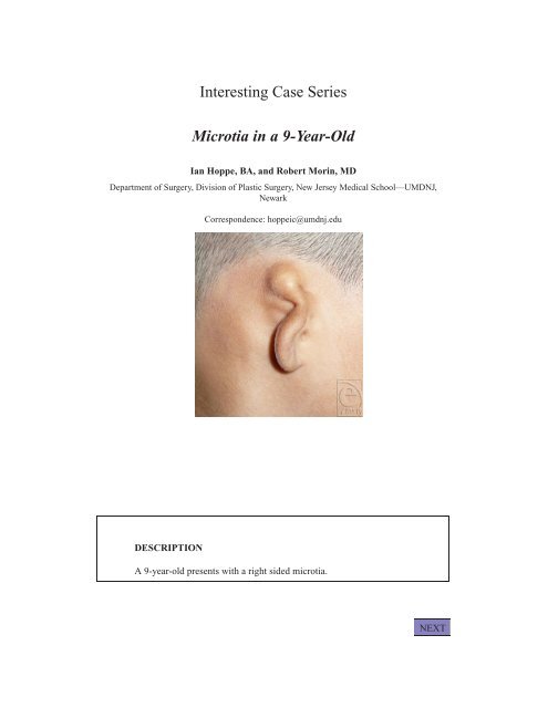
DESCRIPTION<br />
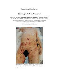
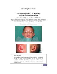
A 9-year-old presents with a right sided microtia.<br />
NEXT
QUESTIONS<br />
1. What are the embryological orig<strong>in</strong>s of the external ear?<br />
2. Does this child likely have hear<strong>in</strong>g capability <strong>in</strong> this ear?<br />
3. With what syndrome is bilateral microtia likely?<br />
4. What surgical approaches are available and what is the optimal age for<br />
repair?<br />
5. What life-threaten<strong>in</strong>g complication can result from surgery to correct this<br />
defect?<br />
BACK<br />
NEXT
DISCUSSION<br />
The auricle is derived from the first and second branchial arches. The<br />
tragus, helical crus, and superior helix are derived from anterior hillocks<br />
of the first branchial arch and the antitragus, antihelix, and lobule are<br />
derived from posterior hillocks of the second branchial arch.<br />
(http://cwx.prenhall.com/bookb<strong>in</strong>d/pubbooks/mart<strong>in</strong>i10/<br />
chapter18/medialib/developmentofexternalear.html).<br />
Reconstruction of microtia can take place as early as 2 years of age,<br />
because the reconstructed ear usually grows normally. However, it is<br />
recommended to wait until the child is 4 to 5 years of age because this<br />
is when the ear is with<strong>in</strong> 5 to 7 mm of its f<strong>in</strong>al vertical height.<br />
Most microtic ears have a conductive hear<strong>in</strong>g loss due to an atretic<br />
external auditory canal and tympanic membrane. The middle ear ossicles<br />
(especially the malleus and <strong>in</strong>cus) are often deformed. The <strong>in</strong>ner ear<br />
develops separately and should be normal. It is generally unnecessary<br />
to perform a tympanoplasty to improve hear<strong>in</strong>g because the hear<strong>in</strong>g <strong>in</strong><br />
the nonmicrotic ear is adequate. If tympanoplasty is performed it should<br />
be done after the external ear reconstruction is complete.<br />
BACK<br />
NEXT
<strong>Microtia</strong> most commonly presents unilaterally with right to left to<br />
bilateral ratios of 5:3:1. It is twice as common <strong>in</strong> males as it is <strong>in</strong> females.<br />
Hemifacial microsomia is a common associated deformity. Goldenhar<br />
syndrome commonly presents with bilateral microtia.<br />
Surgical treatment consists of the creation of a cartilag<strong>in</strong>ous framework<br />
by harvest<strong>in</strong>g costal cartilage. Portions of the 6th through 9th<br />
costal cartilages are used, particularly the synchondroses and float<strong>in</strong>g<br />
rib. In the first stage of reconstruction the framework is <strong>in</strong>serted <strong>in</strong>to an<br />
auricular sk<strong>in</strong> pocket after careful dissection. Other procedures such as<br />
lobule transposition, tragal construction, conchal excavation, and helical<br />
rim elevation generally take place dur<strong>in</strong>g later surgeries. An alternative<br />
approach <strong>in</strong>volves placement of a porous polyethylene implant with immediate<br />
coverage with a temporoparietal flap and sk<strong>in</strong> graft.<br />
Dur<strong>in</strong>g the harvest<strong>in</strong>g of the costal cartilage there is a risk of lifethreaten<strong>in</strong>g<br />
pneumothorax. Before clos<strong>in</strong>g the chest donor site, sal<strong>in</strong>e<br />
should be used to fill the wound cavity and positive pressure ventilation<br />
should be performed. If an air leak is noted, immediate repair is<br />
warranted. If the leak persists, a chest tube should be <strong>in</strong>serted.<br />
REFERENCES<br />
Thomson H, W<strong>in</strong>slow J. <strong>Microtia</strong> reconstruction: does the cartilage framework grow? Plast Reconstr Surg.<br />
1989;84(6):908–5.<br />
Firm<strong>in</strong> F. Ear reconstruction <strong>in</strong> cases of typical microtia. Personal experience based on 352 microtic ear<br />
corrections. Scand J Plast Reconstr Surg Hand Surg. 1998;32:35–47.<br />
Meyer R, de Goumöens R, Derder S. Comb<strong>in</strong>ed aesthetic and functional treatment of microtia. Aesthetic<br />
Plast Surg. 1997;21:159–167.<br />
Bauer BS. Congenital and acquired deformities of the ear. In: Bentz ML, Bauer BS, Zuker RM, eds.<br />
Pr<strong>in</strong>ciples and Practice of Pediatric Plastic Surgery. Vol 2. St Louis: Quality Medical Publish<strong>in</strong>g;<br />
2008:801–50.<br />
Re<strong>in</strong>isch J. <strong>Microtia</strong> reconstruction us<strong>in</strong>g a polyethylene implant: an eight-year surgical experience. Paper<br />
presented at: The 78th Annual Meet<strong>in</strong>g the American Association of Plastic Surgeons; May 5,<br />
1999; Colorado Spr<strong>in</strong>gs, CO.<br />
Bauer B, Brent B, Re<strong>in</strong>isch J, Thorne C. Panel discussion—“ear reconstruction.” Plast Reconstr Surgery<br />
Abstract. Suppl 188(4); Suppl 32, September 6.<br />
BACK