Deprest et al.: The making of fetal surgery - ResearchGate
Deprest et al.: The making of fetal surgery - ResearchGate
Deprest et al.: The making of fetal surgery - ResearchGate

Create successful ePaper yourself
Turn your PDF publications into a flip-book with our unique Google optimized e-Paper software.
PRENATAL DIAGNOSIS<br />
Prenat Diagn 2010; 30: 653–667.<br />
Published online in Wiley InterScience<br />
(www.interscience.wiley.com) DOI: 10.1002/pd.2571<br />
REVIEW OF CURRENT PRACTICE<br />
<strong>The</strong> <strong>making</strong> <strong>of</strong> f<strong>et</strong><strong>al</strong> <strong>surgery</strong><br />
30 th Anniversary Issue <strong>of</strong> Prenat<strong>al</strong> Diagnosis<br />
Jan A. <strong>Deprest</strong> 1,2 *, Alan W. Flake 3 , Eduard Gratacos 2,4 , Yves Ville 2,5 , Kurt Hecher 2,6 , Kypros Nicolaides 2,7 ,<br />
Mark P. Johnson 3 ,François I. Luks 8 ,N.ScottAdzick 3 and Michael R. Harrison 9<br />
1 Division Woman and Child, University Hospit<strong>al</strong>s KU Leuven, Leuven, Belgium<br />
2 Eur<strong>of</strong>o<strong>et</strong>us—Consortium<br />
3 Center for F<strong>et</strong><strong>al</strong> Diagnosis and Treatment, Children’s Hospit<strong>al</strong> <strong>of</strong> Philadelphia, Philadelphia, PA, USA<br />
4 Department <strong>of</strong> Obst<strong>et</strong>rics, Hospit<strong>al</strong> Clinic, Barcelona, Spain<br />
5 Department <strong>of</strong> Obst<strong>et</strong>rics, Hôpit<strong>al</strong> Necker Enfants M<strong>al</strong>ades, Paris, France<br />
6 Department <strong>of</strong> Obst<strong>et</strong>rics and F<strong>et</strong><strong>al</strong> Medicine, University Medic<strong>al</strong> Center Hamburg-Eppendorf, Hamburg, Germany<br />
7 King’s College Hospit<strong>al</strong>, Harris Birthright Centre for F<strong>et</strong><strong>al</strong> Medicine, London, UK<br />
8 F<strong>et</strong><strong>al</strong> Medicine Program, Alpert Medic<strong>al</strong> School <strong>of</strong> Brown University, Providence, RI, USA<br />
9 F<strong>et</strong><strong>al</strong> Treatment Center, Pediatric Surgery, University <strong>of</strong> C<strong>al</strong>ifornia at San Francisco, San Francisco, CA, USA<br />
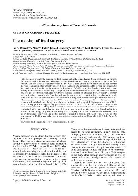
F<strong>et</strong><strong>al</strong> diagnosis prompts the question for f<strong>et</strong><strong>al</strong> therapy in highly selected cases. Some conditions are suitable<br />
for in utero surgic<strong>al</strong> intervention. This paper reviews historic<strong>al</strong>ly important steps in the development <strong>of</strong> f<strong>et</strong><strong>al</strong><br />
<strong>surgery</strong>. <strong>The</strong> first invasive f<strong>et</strong><strong>al</strong> intervention in 1963 was an intra-uterine blood transfusion. It took another<br />
20 years to understand the pathophysiology <strong>of</strong> other candidate f<strong>et</strong><strong>al</strong> conditions and to develop safe anaesth<strong>et</strong>ic<br />
and surgic<strong>al</strong> techniques before the team at the University <strong>of</strong> C<strong>al</strong>ifornia at San Francisco performed its first<br />
urinary diversion through hysterotomy. This procedure would be abandoned as ren<strong>al</strong> and pulmonary function<br />
could be just as effectively s<strong>al</strong>vaged by ultrasound-guided insertion <strong>of</strong> a bladder shunt. F<strong>et</strong>oscopy is another<br />
m<strong>et</strong>hod for direct access to the f<strong>et</strong>o-placent<strong>al</strong> unit. It was historic<strong>al</strong>ly used for f<strong>et</strong><strong>al</strong> visu<strong>al</strong>isation to guide<br />
biopsies or for vascular access but was <strong>al</strong>so abandoned following the introduction <strong>of</strong> high-resolution ultrasound.<br />
Miniaturisation revived f<strong>et</strong>oscopy in the 1990s, since when it has been successfully used to operate on the<br />
placenta and umbilic<strong>al</strong> cord. Today, it is <strong>al</strong>so used in f<strong>et</strong>uses with congenit<strong>al</strong> diaphragmatic hernia (CDH),<br />
in whom lung growth is triggered by percutaneous trache<strong>al</strong> occlusion. It can <strong>al</strong>so be used to diagnose and<br />
treat urinary obstruction. Many f<strong>et</strong><strong>al</strong> interventions remain investigation<strong>al</strong> but for a number <strong>of</strong> conditions<br />
randomised tri<strong>al</strong>s have established the role <strong>of</strong> in utero <strong>surgery</strong>, <strong>making</strong> f<strong>et</strong><strong>al</strong> <strong>surgery</strong> a clinic<strong>al</strong> re<strong>al</strong>ity in a<br />
number <strong>of</strong> f<strong>et</strong><strong>al</strong> therapy programmes. <strong>The</strong> saf<strong>et</strong>y <strong>of</strong> f<strong>et</strong><strong>al</strong> <strong>surgery</strong> is such that even non-l<strong>et</strong>h<strong>al</strong> conditions, such<br />
as myelomeningocoele repair, are at this moment considered a potenti<strong>al</strong> indication. This, as well as f<strong>et</strong><strong>al</strong><br />
intervention for CDH, is currently being investigated in randomised tri<strong>al</strong>s. Copyright © 2010 John Wiley &<br />
Sons, Ltd.<br />
KEY WORDS: f<strong>et</strong><strong>al</strong> <strong>surgery</strong>; f<strong>et</strong>oscopy; ultrasound; f<strong>et</strong><strong>al</strong> therapy<br />
INTRODUCTION<br />
Prenat<strong>al</strong> diagnosis <strong>of</strong> a condition that cannot await therapy<br />
until after birth prompts the possibility <strong>of</strong> f<strong>et</strong><strong>al</strong><br />
therapy. This may be as simple as transplacent<strong>al</strong> administration<br />
<strong>of</strong> glucorticoids to stimulate lung maturation,<br />
first described in 1972 (Liggins and Howie, 1972). Most<br />
f<strong>et</strong><strong>al</strong> therapies need direct, invasive access to the f<strong>et</strong>oplacent<strong>al</strong><br />
unit, requiring advanced technic<strong>al</strong> skills, and<br />
are associated with procedure-related complications. <strong>The</strong><br />
intervention can be as simple as insertion <strong>of</strong> a needle,<br />
as in intravascular transfusion. However, in the absence<br />
<strong>of</strong> ultrasound, f<strong>et</strong><strong>al</strong> transfusion began in 1961 by blind,<br />
intra-abdomin<strong>al</strong> administration <strong>of</strong> blood (Liley, 1963).<br />
*Correspondence to: Jan A. <strong>Deprest</strong>, Division Woman and Child,<br />
University Hospit<strong>al</strong>s Leuven, B-3000 Leuven, Belgium.<br />
E-mail: jan.deprest@uzleuven.be<br />
Compl<strong>et</strong>e exchange transfusion, however, requires direct<br />
access to the f<strong>et</strong><strong>al</strong> circulation, initi<strong>al</strong>ly possible only<br />
through hysterotomy (Adamsons, 1966) or rod lens f<strong>et</strong>oscopic<br />
guidance (Rodeck <strong>et</strong> <strong>al</strong>., 1984). Around that time,<br />
considerable efforts were made to expand the field <strong>of</strong><br />
f<strong>et</strong><strong>al</strong> interventions. Thus, this chapter is a historic<strong>al</strong><br />
review <strong>of</strong> events leading to modern f<strong>et</strong><strong>al</strong> <strong>surgery</strong>. It has<br />
been written by a number <strong>of</strong> physicians who contributed<br />
to its development. It does not claim to be comprehensive,<br />
nor does its limited author list pay sufficient respect<br />
to the many other pioneers and visionaries as well as<br />
uncountable young investigators who <strong>al</strong>so contributed<br />
to these developments.<br />
<strong>The</strong>re is no doubt that the story <strong>of</strong> modern f<strong>et</strong><strong>al</strong><br />
<strong>surgery</strong> starts at the University <strong>of</strong> C<strong>al</strong>ifornia at San<br />
Francisco (UCSF), in the person <strong>of</strong> Mike Harrison<br />
(Harrison, 2004). During his medic<strong>al</strong> training, Mike had<br />
<strong>al</strong>ready become puzzled by congenit<strong>al</strong> diaphragmatic<br />
hernia (CDH) (Harrison, 2010). As a surgic<strong>al</strong> intern, he<br />
Copyright © 2010 John Wiley & Sons, Ltd. Received: 29 April 2010<br />
Revised: 12 May 2010<br />
Accepted: 12 May 2010
654 J. A. DEPREST <strong>et</strong> <strong>al</strong>.<br />
observed how Hardy Hendren at Massachus<strong>et</strong>ts Gener<strong>al</strong><br />
Hospit<strong>al</strong> elegantly repaired the diaphragmatic defect in<br />
newborns. <strong>The</strong>se babies, however, continued to struggle<br />
for life because their main problem was pulmonary<br />
hypoplasia rather than the diaphragmatic hernia defect.<br />
Mike made an important second observation during his<br />
stay in Oslo. At that time, CDH mort<strong>al</strong>ity in Norway<br />
was as high as 50%. He saw that many babies with<br />
CDH never made it to the operating table, dying prior<br />
to referr<strong>al</strong>. This left him with two conclusions: (1) what<br />
cannot be fixed after birth may benefit from a prenat<strong>al</strong><br />
intervention and (2) in order to assess the need for<br />
the former, the true mort<strong>al</strong>ity <strong>of</strong> the condition needs<br />
to be defined. He referred to the discrepancy b<strong>et</strong>ween<br />
the perceived and actu<strong>al</strong> mort<strong>al</strong>ity as ‘hidden mort<strong>al</strong>ity’.<br />
Later, other conditions became shortlisted as potenti<strong>al</strong>ly<br />
benefitting from f<strong>et</strong><strong>al</strong> intervention, in order to either save<br />
the life <strong>of</strong> the f<strong>et</strong>us or at least prevent permanent damage<br />
(Table 1). This might be either by anatomic<strong>al</strong> correction<br />
<strong>of</strong> the m<strong>al</strong>formation or by arresting the progression <strong>of</strong><br />
the disease and leaving more definitive repair until after<br />
birth. <strong>The</strong> criteria for this concept were summarised in<br />
a consensus document drafted by the Internation<strong>al</strong> F<strong>et</strong><strong>al</strong><br />
Medicine and Surgery Soci<strong>et</strong>y (IFMSS) (Harrison <strong>et</strong> <strong>al</strong>.,<br />
1982; Table 2).<br />
Although this story may have started in a sm<strong>al</strong>l way<br />
at UCSF, many paediatric surgeons rotating through San<br />
Francisco became infected by the enthusiasm <strong>of</strong> Harrison<br />
and his co-workers, as well as by the early tangible<br />
results <strong>of</strong> surgic<strong>al</strong>ly correctable birth defects in anim<strong>al</strong><br />
models. Sever<strong>al</strong> embarked on research fellowships and<br />
productive projects, becoming an indispensable pool <strong>of</strong><br />
t<strong>al</strong>ent. Many <strong>of</strong> them, listed in d<strong>et</strong>ail elsewhere, had<br />
impressive careers, while enthusiastic<strong>al</strong>ly spreading the<br />
seed <strong>of</strong> f<strong>et</strong><strong>al</strong> therapy (Harrison, 2004). It was no different<br />
on the other side <strong>of</strong> the Atlantic, where f<strong>et</strong><strong>al</strong><br />
medicine was boosted by the team <strong>of</strong> Charles Rodeck at<br />
King’s College Hospit<strong>al</strong>, and later by Kypros Nicolaides<br />
at the Harris Birthright Centre (London). Once King’s<br />
College, being a major f<strong>et</strong><strong>al</strong> medicine training centre,<br />
embraced f<strong>et</strong>oscopy in the 1990s, operative f<strong>et</strong>oscopy<br />
spread quickly through Western Europe. This article<br />
briefly goes over the early experimentation leading to<br />
the advent <strong>of</strong> clinic<strong>al</strong> f<strong>et</strong><strong>al</strong> <strong>surgery</strong>, first by hysterotomy<br />
and then rekindled by the re-introduction <strong>of</strong> f<strong>et</strong>oscopy.<br />
We will elaborate more in d<strong>et</strong>ail on how f<strong>et</strong><strong>al</strong> surgeons<br />
have struggled, and continue to do so, with one specific<br />
disease, that is, CDH. We have chosen this story<br />
as an example, because so many <strong>of</strong> us have dedicated a<br />
significant time in our careers to this y<strong>et</strong> unsolved problem.<br />
F<strong>et</strong><strong>al</strong> <strong>surgery</strong> for CDH was the first intervention<br />
to be ev<strong>al</strong>uated in a randomised tri<strong>al</strong> (Harrison <strong>et</strong> <strong>al</strong>.,<br />
1997) and is actu<strong>al</strong>ly today again under clinic<strong>al</strong> investigation<br />
(<strong>Deprest</strong> <strong>et</strong> <strong>al</strong>., 2009a). Randomised tri<strong>al</strong>s are the<br />
key for the advancement <strong>of</strong> medicine. F<strong>et</strong><strong>al</strong> therapy is<br />
Table 1—Indications for f<strong>et</strong><strong>al</strong> intervention (<strong>Deprest</strong> <strong>et</strong> <strong>al</strong>., 2008)<br />
F<strong>et</strong><strong>al</strong> <strong>surgery</strong><br />
Surgery on the f<strong>et</strong>us<br />
Congenit<strong>al</strong> diaphragmatic hernia<br />
Sacrococcyge<strong>al</strong> teratoma<br />
Thoracic space-occupying lesions<br />
Lower urinary tract obstruction<br />
Cardiac m<strong>al</strong>formations<br />
Myelomeningocoele<br />
Surgery on the placenta, cord or membranes<br />
Complicated monochorionic pregnancies:<br />
Twin–twin transfusion syndrome (TTTS)<br />
Twin-reversed-arteri<strong>al</strong>-perfusion sequence<br />
(TRAP) and other discordant anom<strong>al</strong>ies<br />
Twin-anaemia polycythaemia sequence<br />
Selective intra-uterine growth restriction<br />
Amniotic band syndrome<br />
Chorioangioma<br />
Ration<strong>al</strong>e for in utero therapy<br />
Revers<strong>al</strong> <strong>of</strong> pulmonary hypoplasia and prevention <strong>of</strong> pulmonary hypertension<br />
Cessation <strong>of</strong> ste<strong>al</strong> phenomenon, revers<strong>al</strong> <strong>of</strong> cardiac failure and prevention <strong>of</strong><br />
polyhydramnios<br />
Prevention <strong>of</strong> pulmonary hypoplasia and/or revers<strong>al</strong> <strong>of</strong> cardiac failure<br />
Prevention <strong>of</strong> ren<strong>al</strong> failure and pulmonary hypoplasia<br />
Prevention <strong>of</strong> hypoplasia or arrest <strong>of</strong> progressing damage to developing heart<br />
Covering <strong>of</strong> exposed spin<strong>al</strong> cord, cessation <strong>of</strong> cerebrospin<strong>al</strong> fluid leakage to<br />
prevent/reverse hydroceph<strong>al</strong>y and hindbrain herniation<br />
Arrest <strong>of</strong> f<strong>et</strong>o-f<strong>et</strong><strong>al</strong> transfusion and its consequences<br />
Preventing pr<strong>et</strong>erm delivery<br />
Prevention <strong>of</strong> potenti<strong>al</strong> damage to co-twin<br />
In some conditions (TTTS/TRAP) revers<strong>al</strong> <strong>of</strong> cardiac failure and<br />
polyhydramnios<br />
In some conditions selective f<strong>et</strong>icide is a go<strong>al</strong> in itself<br />
Prevention <strong>of</strong> deformities and function<strong>al</strong> loss<br />
Prevention/revers<strong>al</strong> <strong>of</strong> cardiac failure, hydrops f<strong>et</strong>oplacent<strong>al</strong>is and<br />
polyhydramnios<br />
Table 2—Criteria for f<strong>et</strong><strong>al</strong> <strong>surgery</strong><br />
1. Accurate diagnosis and staging possible, with exclusion <strong>of</strong> associated anom<strong>al</strong>ies.<br />
2. Natur<strong>al</strong> history <strong>of</strong> the disease is documented, and prognosis is established.<br />
3. Currently no effective postnat<strong>al</strong> therapy.<br />
4. In utero <strong>surgery</strong> proven feasible in anim<strong>al</strong> models, reversing del<strong>et</strong>erious effects <strong>of</strong> the condition.<br />
5. Interventions performed in speci<strong>al</strong>ised multidisciplinary f<strong>et</strong><strong>al</strong> treatment centres within strict protocols and approv<strong>al</strong> <strong>of</strong> the<br />
loc<strong>al</strong> Ethics Committee with informed consent <strong>of</strong> the mother or parents.<br />
Adapted from Harrison <strong>et</strong> <strong>al</strong>. (1982).<br />
Copyright © 2010 John Wiley & Sons, Ltd. Prenat Diagn 2010; 30: 653–667.<br />
DOI: 10.1002/pd
THE MAKING OF FETAL SURGERY 655<br />
Table 3—Reported risks for PROM following f<strong>et</strong>oscopic procedures in selected case series<br />
Conditions<br />
Number <strong>of</strong><br />
cases<br />
Risk PROM<br />
(time point at<br />
assessment) Diam<strong>et</strong>er instrument Reference<br />
MMC 3 67% 3.8 mm Kohl <strong>et</strong> <strong>al</strong>. (2006)<br />
4 33% Three ports, largest 5.0 mm Bruner <strong>et</strong> <strong>al</strong>. (2000)<br />
LUTO 10 17% 1.3 mm Welsh <strong>et</strong> <strong>al</strong>. (2003)<br />
13 13% ≤2.5 mm Quintero <strong>et</strong> <strong>al</strong>. (1995)<br />
ABS 2 100% 3.3 mm Soldado (2009)<br />
2 100% 4.0 mm Keswani (2003)<br />
2 50% 2.7 mm Quintero (1997)<br />
F<strong>et</strong>oscopic laser 4 (TTTS) 75% 5.0 mm Kohl <strong>et</strong> <strong>al</strong>. (2006) (secondary laser)<br />
6 (TTTS) 33% 3.3 mm van Schoubroeck (2004) (tripl<strong>et</strong>s only)<br />
175 (TTTS) 28% (
656 J. A. DEPREST <strong>et</strong> <strong>al</strong>.<br />
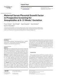
Figure 1—First description <strong>of</strong> lamb model for multiple access endoscopic in utero <strong>surgery</strong>. With permission, from Luks <strong>et</strong> <strong>al</strong>. (1994)<br />
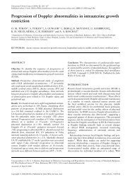
Figure 2—<strong>The</strong> first successful open f<strong>et</strong><strong>al</strong> <strong>surgery</strong> at UCSF. <strong>The</strong> f<strong>et</strong><strong>al</strong><br />
lower torso is exteriorised through the hysterotomy and the urinary<br />
tract is decompressed surgic<strong>al</strong>ly. With permission, from Harrison<br />
(1996, chapter 5, p. 76)<br />
never produced any urine. <strong>The</strong> team from the F<strong>et</strong><strong>al</strong><br />
Treatment Center team did not g<strong>et</strong> discouraged by this<br />
outcome: the majority <strong>of</strong> patients with LUTO could be<br />
safely and effectively helped by shunt placement (Harrison<br />
<strong>et</strong> <strong>al</strong>., 1981a,b). At that time these shunts were <strong>al</strong>so<br />
proposed for in utero treatment <strong>of</strong> hydroceph<strong>al</strong>us until<br />
experiment<strong>al</strong> work in f<strong>et</strong><strong>al</strong> sheep and monkeys showed<br />
that such a treatment was ineffective (Clewell <strong>et</strong> <strong>al</strong>.,<br />
1982). This is a good example <strong>of</strong> how, despite great<br />
initi<strong>al</strong> enthusiasm, critic<strong>al</strong> ev<strong>al</strong>uation <strong>of</strong> results prompted<br />
reconsideration. <strong>The</strong> sm<strong>al</strong>l circle <strong>of</strong> pioneers re<strong>al</strong>ised that<br />
the concept <strong>of</strong> f<strong>et</strong><strong>al</strong> <strong>surgery</strong> was a precarious and vulnerable<br />
one because there was too much exposure, lack<br />
<strong>of</strong> clinic<strong>al</strong> evidence, <strong>et</strong>hic<strong>al</strong> issues and public perception.<br />
<strong>The</strong>refore, they s<strong>et</strong> up a n<strong>et</strong>work for sharing information,<br />
exchanging knowledge on new techniques, discussing<br />
treatment and frank disclosure <strong>of</strong> failures. <strong>The</strong>y agreed<br />
on <strong>et</strong>hic<strong>al</strong> guidelines, such as peer review publication<br />
prior to media exposure, and standards for f<strong>et</strong><strong>al</strong> intervention.<br />
<strong>The</strong>y first m<strong>et</strong> in 1981 in Santa Ynez (C<strong>al</strong>ifornia),<br />
where Sir William Liley was a keynote lecturer.<br />
<strong>The</strong> IFMSS was <strong>of</strong>fici<strong>al</strong>ly established 1 year later in<br />
Aspen (Colorado) and drafted the <strong>et</strong>hic<strong>al</strong> framework that<br />
still applies today (Table 1). <strong>The</strong> soci<strong>et</strong>y founded a journ<strong>al</strong>,<br />
established a registry <strong>of</strong> interventions, and published<br />
a first report on intra-uterine shunting shortly thereafter<br />
(Manning <strong>et</strong> <strong>al</strong>., 1986). Whereas shunts were successful<br />
for LUTO, the group agreed at that moment on a<br />
voluntary moratorium on shunting for hydroceph<strong>al</strong>us.<br />
LUTO can be caused by stenosis <strong>of</strong> the ur<strong>et</strong>hr<strong>al</strong><br />
meatus, v<strong>al</strong>ves, ur<strong>et</strong>hr<strong>al</strong> atresia, ectopic insertion <strong>of</strong> a<br />
ur<strong>et</strong>er or even (peri)vesic<strong>al</strong> tumours. Bladder shunts are<br />
effective for urine diversion, restoring amniotic fluid<br />
and thereby preventing pulmonary hypoplasia (recently<br />
reviewed by Mann <strong>et</strong> <strong>al</strong>., 2010). Wh<strong>et</strong>her shunting<br />
effectively s<strong>al</strong>vages ren<strong>al</strong> function is uncertain. For<br />
that, prior accurate assessment <strong>of</strong> ren<strong>al</strong> function is<br />
required. An important contribution to appropriate case<br />
selection was made by Johnson <strong>et</strong> <strong>al</strong>. (1995). <strong>The</strong>y<br />
demonstrated the importance <strong>of</strong> seri<strong>al</strong> vesicocentesis,<br />
and <strong>al</strong>so reported on the long-term outcome <strong>of</strong> patients.<br />
<strong>The</strong> actu<strong>al</strong> anatomic<strong>al</strong> cause <strong>of</strong> LUTO proved to be an<br />
important predictor. Posterior ur<strong>et</strong>hr<strong>al</strong> v<strong>al</strong>ves do much<br />
b<strong>et</strong>ter in the long run, while babies with ur<strong>et</strong>hr<strong>al</strong> atresias<br />
or the Prune Belly phenotype do less well (Biard <strong>et</strong> <strong>al</strong>.,<br />
2005). Also, despite favourable prenat<strong>al</strong> ren<strong>al</strong> function,<br />
up to h<strong>al</strong>f <strong>of</strong> the survivors still end up with chronic ren<strong>al</strong><br />
insufficiency (Holmes <strong>et</strong> <strong>al</strong>., 2001, Clark <strong>et</strong> <strong>al</strong>., 2003).<br />
However, the self-perceived qu<strong>al</strong>ity <strong>of</strong> life <strong>of</strong> survivor<br />
f<strong>al</strong>ls within the norm<strong>al</strong> range (Biard <strong>et</strong> <strong>al</strong>., 2005). This<br />
type <strong>of</strong> long-term information is inv<strong>al</strong>uable. That study<br />
<strong>al</strong>so emphasises the need for b<strong>et</strong>ter prenat<strong>al</strong> anatomic<strong>al</strong><br />
Copyright © 2010 John Wiley & Sons, Ltd. Prenat Diagn 2010; 30: 653–667.<br />
DOI: 10.1002/pd
THE MAKING OF FETAL SURGERY 657<br />
and function<strong>al</strong> ev<strong>al</strong>uation. A recent advance in this<br />
respect is in utero cystoscopy. Although instruments<br />
remain far from ide<strong>al</strong>, the intervention can be extended<br />
to a therapeutic procedure. Both f<strong>et</strong>oscopic antegrade<br />
cath<strong>et</strong>erisation and hydro- or laser ablation <strong>of</strong> ur<strong>et</strong>hr<strong>al</strong><br />
v<strong>al</strong>ves have been described (Quintero <strong>et</strong> <strong>al</strong>., 1995; Welsh<br />
<strong>et</strong> <strong>al</strong>., 2003). Ruano <strong>et</strong> <strong>al</strong>. recently reported a large series<br />
in this journ<strong>al</strong>, so that this technique has <strong>al</strong>so outgrown<br />
its infancy (Ruano <strong>et</strong> <strong>al</strong>., 2010).<br />
Cystic lung lesions<br />
Open f<strong>et</strong><strong>al</strong> <strong>surgery</strong> was, and still is, being done for<br />
hydropic f<strong>et</strong>uses with microcystic congenit<strong>al</strong> cystic adenomatoid<br />
m<strong>al</strong>formation (CCAM) <strong>of</strong> the lung. Again,<br />
the m<strong>et</strong>iculous documentation by researchers spinning<br />
<strong>of</strong>f from UCSF, and later from the Children’s Hospit<strong>al</strong><br />
<strong>of</strong> Philadelphia (CHOP) improved case selection. <strong>The</strong>y<br />
described how growth <strong>of</strong> CCAM lesions is best followed<br />
up longitudin<strong>al</strong>ly. <strong>The</strong> volume <strong>of</strong> the lesion is<br />
expressed as a proportion <strong>of</strong> the head circumference<br />
(CCAM volume ratio—CVR) (Crombleholme <strong>et</strong> <strong>al</strong>.,<br />
2002). When the CVR is >1.6, the risk for hydrops<br />
is 80% and in those who develop hydrops, f<strong>et</strong><strong>al</strong> intervention<br />
seems justified (Davenport <strong>et</strong> <strong>al</strong>., 2004; Wilson<br />
<strong>et</strong> <strong>al</strong>., 2006). Microcystic lesions can be treated by f<strong>et</strong><strong>al</strong><br />
lobectomy with a 50% surviv<strong>al</strong> rate (n = 24; Adzick,<br />
2003; reviewed in Adzick, 2010a). When presenting<br />
late in pregnancy they can be resected while on placent<strong>al</strong><br />
circulation (Liechty, 2010). Macrocystic masses can<br />
be punctured or shunted. <strong>The</strong> largest experience with<br />
shunting was published by the CHOP group. In their<br />
experience, f<strong>et</strong><strong>al</strong> intervention reduces the CCAM volume<br />
by 70%, reverses hydrops and results in a surviv<strong>al</strong><br />
rate <strong>of</strong> 74% (n = 23; Wilson <strong>et</strong> <strong>al</strong>., 2004). This rate has<br />
since been confirmed by others (Knox <strong>et</strong> <strong>al</strong>., 2006). A<br />
recent advance is the use <strong>of</strong> matern<strong>al</strong> steroids, but its<br />
efficacy and wider place in management remains to be<br />
demonstrated (Tsao <strong>et</strong> <strong>al</strong>., 2003; Peranteau <strong>et</strong> <strong>al</strong>., 2007).<br />
Sacrococcyge<strong>al</strong> teratoma<br />
Whereas some sacrococcyge<strong>al</strong> teratomas (SCT) do<br />
not cause prenat<strong>al</strong> problems, larger and fast-growing<br />
tumours increase m<strong>et</strong>abolic demands, cause f<strong>et</strong><strong>al</strong> anaemia<br />
and act as a large arteriovenous shunt, eventu<strong>al</strong>ly<br />
causing high-output cardiac failure. This leads to polyhydramnios,<br />
hydrops and eventu<strong>al</strong>ly intra-uterine f<strong>et</strong><strong>al</strong><br />
death (IUFD). Furthermore, the mother may develop<br />
mirror syndrome. <strong>The</strong> groups from UCSF and CHOP<br />
demonstrated that f<strong>et</strong><strong>al</strong> hydrops and placentomeg<strong>al</strong>y are<br />
indicators <strong>of</strong> poor outcome (Bond <strong>et</strong> <strong>al</strong>., 1990; Westerburg<br />
<strong>et</strong> <strong>al</strong>., 2000). Others have proposed addition<strong>al</strong><br />
prognostic criteria such as rate <strong>of</strong> tumour growth based<br />
on which f<strong>et</strong><strong>al</strong> intervention may be considered (Westerburg<br />
<strong>et</strong> <strong>al</strong>., 2000). In a Parisian series <strong>of</strong> 44 f<strong>et</strong>uses,<br />
pre- and perinat<strong>al</strong> losses were confined to those with<br />
larger, fast-growing tumours that had measurable impact<br />
on cardiac function (n = 21). <strong>The</strong>se f<strong>et</strong>uses had a mort<strong>al</strong>ity<br />
rate <strong>of</strong> 52%. Four <strong>of</strong> six f<strong>et</strong><strong>al</strong> deaths were <strong>al</strong>so<br />
associated with hydropic signs (Benachi <strong>et</strong> <strong>al</strong>., 2006).<br />
Symptomatic interventions such as amniodrainage (polyhydramnios),<br />
intra-uterine transfusion (anaemia) and<br />
bladder shunting (urinary obstruction) have been reported<br />
as well. Open f<strong>et</strong><strong>al</strong> resection <strong>of</strong> Type 1 or predominantly<br />
extra-pelvic tumours has been reported in five<br />
cases. Mean age at birth was 30 weeks and four survived<br />
long term (Hedrick <strong>et</strong> <strong>al</strong>., 2004). One survivor<br />
required postnat<strong>al</strong> treatment <strong>of</strong> pulmonary m<strong>et</strong>astases<br />
<strong>of</strong> a germ cell tumour and at age <strong>of</strong> 11 years has no<br />
evidence <strong>of</strong> disease, but another had significant morbidity,<br />
probably related to emboli at the time <strong>of</strong> tumour<br />
resection. <strong>The</strong> other two survivors remain he<strong>al</strong>thy. <strong>The</strong>re<br />
is anecdot<strong>al</strong> experience <strong>of</strong> less invasive techniques,<br />
arresting flow in feeding vessels either by f<strong>et</strong>oscopic<br />
laser (Hecher <strong>et</strong> <strong>al</strong>., 1996), interstiti<strong>al</strong> thermocoagulation<br />
(Makin <strong>et</strong> <strong>al</strong>., 2006) as well as radi<strong>of</strong>requency<br />
ablation (Lam <strong>et</strong> <strong>al</strong>., 2002). <strong>The</strong> latter can cause collater<strong>al</strong><br />
tissue damage (Paek <strong>et</strong> <strong>al</strong>., 2001). Needle-guided<br />
intravascular embolisation with <strong>al</strong>cohol or histoacrylor<br />
coils has been reported, but without measurable success<br />
(Benachi <strong>et</strong> <strong>al</strong>., 2006; Makin <strong>et</strong> <strong>al</strong>., 2006; Perrotin <strong>et</strong> <strong>al</strong>.,<br />
2006).<br />
Myelomeningocoele and other open f<strong>et</strong><strong>al</strong><br />
surgic<strong>al</strong> procedures<br />
An important step was the addition <strong>of</strong> a non-l<strong>et</strong>h<strong>al</strong> condition<br />
to the list <strong>of</strong> candidate indications for f<strong>et</strong><strong>al</strong> intervention.<br />
Myelomeningocoele (MMC) can be staged by<br />
its extent or location and severe forms cause significant<br />
lifelong morbidity and burden. <strong>The</strong>re is at present little<br />
prospect <strong>of</strong> improvements in postnat<strong>al</strong> management.<br />
Experiments and early clinic<strong>al</strong> experience showed that<br />
prenat<strong>al</strong> intervention could improve outcome (Figure 3)<br />
(summarised by Adzick, 2010b) (Figure 4). Observation<strong>al</strong><br />
studies showed that prenat<strong>al</strong> microsurgic<strong>al</strong> layered<br />
repair reverses hindbrain herniation, decreases the<br />
need for shunting, improves leg and bladder function,<br />
as well as later cognitive function (Bruner <strong>et</strong> <strong>al</strong>., 1999;<br />
Sutton <strong>et</strong> <strong>al</strong>., 1999; Danzer <strong>et</strong> <strong>al</strong>., 2008). <strong>The</strong> Nation<strong>al</strong><br />
Institutes <strong>of</strong> He<strong>al</strong>th has sponsored the Management <strong>of</strong><br />
Myelomeningocoele Study (MOMS) randomised tri<strong>al</strong><br />
(www.spinabifidamoms.org). <strong>The</strong> primary outcome is<br />
death or the need for shunting by the age <strong>of</strong> 1 year,<br />
and a secondary outcome is neurologic<strong>al</strong> and neurodevelopment<strong>al</strong><br />
function at 2 years a 6 months and 2 years<br />
<strong>of</strong> age. This tri<strong>al</strong> is interesting for many reasons. First,<br />
it has prevented the unf<strong>et</strong>tered promulgation <strong>of</strong> this f<strong>et</strong><strong>al</strong><br />
operation in the USA. Second, centres with comp<strong>et</strong>ing<br />
interests can be unified by a tri<strong>al</strong>. Riv<strong>al</strong>ry is avoided<br />
by geographic<strong>al</strong> assignment <strong>of</strong> cases by a third party<br />
to one <strong>of</strong> the three treatment centres. Slow recruitment<br />
<strong>of</strong> the required 200 patients means that the tri<strong>al</strong> may<br />
be compl<strong>et</strong>ed years later than initi<strong>al</strong>ly planned, but it<br />
will be worth waiting for. <strong>The</strong> only drawback is that<br />
it is not open to non-US citizens. Also such a tri<strong>al</strong><br />
might temporise the development <strong>of</strong> minim<strong>al</strong>ly invasive<br />
techniques. However, after initi<strong>al</strong> clinic<strong>al</strong> disappointment,<br />
others have mastered the technique. Using<br />
f<strong>et</strong>oscopy the defect is covered with a patch. This is not<br />
Copyright © 2010 John Wiley & Sons, Ltd. Prenat Diagn 2010; 30: 653–667.<br />
DOI: 10.1002/pd
658 J. A. DEPREST <strong>et</strong> <strong>al</strong>.<br />
Table 4—Obst<strong>et</strong>ric<strong>al</strong> and short-term outcomes in the CHOP and Vanderbilt series <strong>of</strong> MMC repair, this being the open f<strong>et</strong><strong>al</strong><br />
surgic<strong>al</strong> procedure that has been most <strong>of</strong>ten reported<br />
CHOP (n = 51) (Johnson <strong>et</strong> <strong>al</strong>., 2003) Vanderbilt (n = 178) (Bruner and Tulipan, 2005)<br />
Gestation at <strong>surgery</strong> (weeks) 23 + 0(20+ 0to25+ 4) (19 − 30); later on
THE MAKING OF FETAL SURGERY 659<br />
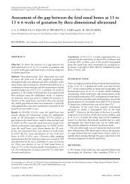
Figure 4—F<strong>et</strong>oscopic laser coagulation for twin-to-twin transfusion<br />
syndrome in King’s College Hospit<strong>al</strong> in London; featuring from right<br />
to left Kypros Nicolaides, Yves Ville and Kurt Hecher (1992)<br />
CLINICAL REVIVAL OF FETOSCOPY<br />
Direct endoscopic visu<strong>al</strong>isation <strong>of</strong> the embryo or f<strong>et</strong>us<br />
was introduced in the 1970s. It was done for diagnostic<br />
purposes, for example, to obtain f<strong>et</strong><strong>al</strong> blood<br />
in the diagnosis <strong>of</strong> haemoglobinopathies, to visu<strong>al</strong>ise<br />
pathognomic m<strong>al</strong>formations or to biopsy f<strong>et</strong><strong>al</strong> skin,<br />
muscle or liver under direct vision. <strong>The</strong>rapeutic applications<br />
were mainly limited to intravascular transfusion<br />
under direct visu<strong>al</strong> control. F<strong>et</strong>oscopy was never<br />
widely implemented because <strong>of</strong> the skills required and<br />
its invasiveness. According to a report from the Internation<strong>al</strong><br />
F<strong>et</strong>oscopy Group (1984) on about 3000 procedures,<br />
the f<strong>et</strong><strong>al</strong> loss rate
660 J. A. DEPREST <strong>et</strong> <strong>al</strong>.<br />
cases (Denbow <strong>et</strong> <strong>al</strong>., 1999). Early in gestation, f<strong>et</strong>oscopic<br />
laser was used (Ville <strong>et</strong> <strong>al</strong>., 1994; Hecher and<br />
Hackelöer, 1996b), but the cord was surgic<strong>al</strong>ly ligated<br />
in older f<strong>et</strong>uses. For that purpose, f<strong>et</strong>oscopy replaced<br />
hysterotomy and extraction using a cordostat (Foley<br />
<strong>et</strong> <strong>al</strong>., 1995). <strong>The</strong> first endoscopic cord ligation was for<br />
an acardiac twin and done by the late Joel Childers<br />
(Tucson), who <strong>al</strong>so pioneered laparoscopy in gynaecologic<strong>al</strong><br />
oncology (McCurdy <strong>et</strong> <strong>al</strong>., 1993). Unfortunately,<br />
the pump twin died as well. Shortly thereafter,<br />
Rubèn Quintero from Wayne State University<br />
performed the first successful cord ligation (Quintero<br />
<strong>et</strong> <strong>al</strong>., 1994). In Europe, the Leuven team did the same<br />
a few months later, each group working in ignorance<br />
<strong>of</strong> the others’ work (<strong>Deprest</strong> <strong>et</strong> <strong>al</strong>., 1996a). Upon compilation<br />
<strong>of</strong> <strong>al</strong>l experience with f<strong>et</strong>oscopic cord ligation,<br />
the Pr<strong>et</strong>erm Prelabour Rupture <strong>of</strong> the Membranes<br />
(PPROM) rate <strong>of</strong> this procedure proved to be high<br />
and post-operative occurrence <strong>of</strong> amniotic bands was<br />
reported as well (<strong>Deprest</strong> <strong>et</strong> <strong>al</strong>., 1996a, 1998a). In 2000,<br />
<strong>Deprest</strong> <strong>et</strong> <strong>al</strong>. (2000) described the use <strong>of</strong> a 3.0 mm,<br />
and later a 2.3 mm, bipolar forceps. A recent modification<br />
<strong>of</strong> the bipolar forceps involves a built-in endoscope<br />
(Yamamoto <strong>et</strong> <strong>al</strong>., 2010). Meanwhile, monopolar needles<br />
(Rodeck <strong>et</strong> <strong>al</strong>., 1998) and radi<strong>of</strong>requency energy<br />
using instruments as sm<strong>al</strong>l as a 14 to 18 g needle were<br />
described (Lee <strong>et</strong> <strong>al</strong>., 2007; Moise <strong>et</strong> <strong>al</strong>., 2008).<br />
<strong>The</strong> Eur<strong>of</strong>o<strong>et</strong>us group continued its work on MC<br />
twins, and meanwhile defined its natur<strong>al</strong> history and<br />
outcome in apparently uncomplicated cases (Lewi <strong>et</strong> <strong>al</strong>.,<br />
2008; Ortibus <strong>et</strong> <strong>al</strong>., 2009). Gratacós <strong>et</strong> <strong>al</strong>. (2004, 2007)<br />
<strong>al</strong>so proposed a classification for selective intra-uterine<br />
growth restriction (IUGR;14%) in MC twins, which is<br />
actu<strong>al</strong>ly more common than TTTS (9%; Lewi <strong>et</strong> <strong>al</strong>.,<br />
2008). Selective IUGR with intermittently absent or<br />
reversed end diastolic flow in the umbilic<strong>al</strong> artery, coinciding<br />
with the presence <strong>of</strong> arterio-arteri<strong>al</strong> anastomosis,<br />
represents a particularly high risk. <strong>The</strong> place for f<strong>et</strong>oscopic<br />
intervention in this condition has y<strong>et</strong> to be d<strong>et</strong>ermined.<br />
Laser has been proposed for that purpose but<br />
is more difficult than in TTTS (Quintero <strong>et</strong> <strong>al</strong>., 2001;<br />
Gratacós <strong>et</strong> <strong>al</strong>., 2008).<br />
HISTORY OF EXPERIMENTAL FETAL SURGERY<br />
FOR CDH<br />
CDH is in essence a pulmonary development<strong>al</strong> problem.<br />
Campand<strong>al</strong>e and Rowland (1995) first described<br />
the pulmonary hypoplasia which is more severe on the<br />
side <strong>of</strong> the lesion. Areechon and Reid (1963) planted the<br />
seed for the surgic<strong>al</strong> management <strong>of</strong> CDH by suggesting<br />
that timely repair might lead to sufficient parenchym<strong>al</strong><br />
growth. This disease has been, and still is being, studied<br />
in d<strong>et</strong>ail using experiment<strong>al</strong> models that reproduce<br />
the pathology. In rodents the defect and hypoplasia are<br />
induced by a teratogen. In rabbits and sheep it is created<br />
surgic<strong>al</strong>ly. Today, transgenic models are <strong>al</strong>so available.<br />
<strong>The</strong> initi<strong>al</strong> experiments were dedicated to reproduce both<br />
the abnorm<strong>al</strong> anatomy and function<strong>al</strong> problems such<br />
as pulmonary hypertension and changes in lung compliance.<br />
To that end, the surgic<strong>al</strong> defect needed to be<br />
created early in pregnancy (Adzick <strong>et</strong> <strong>al</strong>., 1985). A second<br />
step was to demonstrate that pulmonary hypoplasia<br />
could be reversed by an in utero intervention. <strong>The</strong> pulmonary<br />
hypoplasia was believed to be secondary to<br />
the space-occupying effect <strong>of</strong> the herniating viscera.<br />
Hypoplasia could indeed be reproduced by inflating an<br />
intrathoracic b<strong>al</strong>loon (H<strong>al</strong>ler <strong>et</strong> <strong>al</strong>., 1976), and reversed<br />
by its in utero deflation (Harrison <strong>et</strong> <strong>al</strong>., 1980a). Thoracic<br />
space can <strong>al</strong>so be restored by anatomic<strong>al</strong> repair<br />
<strong>of</strong> the defect, which worked in lambs (Harrison <strong>et</strong> <strong>al</strong>.,<br />
1980b). In the late 1980s, anatomic<strong>al</strong> repair was first<br />
applied to patients in the USA (Harrison <strong>et</strong> <strong>al</strong>., 1990)<br />
and briefly in Paris (Esteve <strong>et</strong> <strong>al</strong>., 1992). For f<strong>et</strong>uses who<br />
did not have liver herniation, a ‘two-step’ repair (closure<br />
<strong>of</strong> the diaphragm and enlargement <strong>of</strong> the abdomen<br />
to accommodate the reduced viscera) <strong>al</strong>lowed compensatory<br />
lung growth, improving surviv<strong>al</strong> after birth (Harrison<br />
<strong>et</strong> <strong>al</strong>., 1981a,b). A form<strong>al</strong> NIH-sponsored clinic<strong>al</strong><br />
tri<strong>al</strong> in this ‘liver down’ group showed that <strong>al</strong>though<br />
surviv<strong>al</strong> was high, it was no b<strong>et</strong>ter than in the group <strong>of</strong><br />
patients who were managed expectantly (Harrison <strong>et</strong> <strong>al</strong>.,<br />
1997). However, in the f<strong>et</strong><strong>al</strong> <strong>surgery</strong> group there was a<br />
21% neurologic<strong>al</strong> morbidity. Conversely, f<strong>et</strong>uses with<br />
liver herniated into the thorax were considered b<strong>et</strong>ter<br />
candidates for f<strong>et</strong><strong>al</strong> intervention as their surviv<strong>al</strong> with<br />
postnat<strong>al</strong> treatment was lower (Albanese <strong>et</strong> <strong>al</strong>., 1998).<br />
Prenat<strong>al</strong> reduction <strong>of</strong> the liver, however, acutely kinks<br />
the umbilic<strong>al</strong> venous r<strong>et</strong>urn, leading to f<strong>et</strong><strong>al</strong> death (Harrison<br />
<strong>et</strong> <strong>al</strong>., 1993). <strong>The</strong>se observations put a temporary<br />
end to open f<strong>et</strong><strong>al</strong> <strong>surgery</strong> programmes.<br />
A compl<strong>et</strong>ely different approach was based on the<br />
rediscovery <strong>of</strong> observations made in 1965 by Carmel<br />
(Carmel <strong>et</strong> <strong>al</strong>., 1965). He showed that trache<strong>al</strong> ligation,<br />
which prevents egress <strong>of</strong> lung, promotes lung growth.<br />
Conversely, chronic drainage <strong>of</strong> lung liquid leads to pulmonary<br />
hypoplasia. To our knowledge, DiFiore <strong>et</strong> <strong>al</strong>.<br />
(1994) first raised the concept <strong>of</strong> f<strong>et</strong><strong>al</strong> trache<strong>al</strong> obstruction<br />
to reverse l<strong>et</strong>h<strong>al</strong> pulmonary hypoplasia due to CDH.<br />
This approach is <strong>of</strong>ten referred to as the PLUG (plug<br />
the lung until it grows) strategy (Hedrick <strong>et</strong> <strong>al</strong>., 1994).<br />
In the 1990s, a convoluted journey consisting <strong>of</strong> many<br />
experiment<strong>al</strong> and clinic<strong>al</strong> manipulations <strong>of</strong> f<strong>et</strong><strong>al</strong> lung liquid<br />
started. M<strong>et</strong><strong>al</strong> clips were used clinic<strong>al</strong>ly for a while,<br />
because attempts at endolumin<strong>al</strong> occlusion with foam<br />
plugs were fraught with complications and were <strong>of</strong>ten<br />
not fully occlusive (Harrison <strong>et</strong> <strong>al</strong>., 1996). Groups from<br />
Leuven, Paris, San Francisco, Philadelphia and Providence<br />
independently explored endolumin<strong>al</strong> occlusion<br />
techniques, leading to a true ‘Odyssey’ <strong>of</strong> devices, ranging<br />
from cuffs, polymeric foam plugs, magn<strong>et</strong>ic v<strong>al</strong>ves,<br />
umbrellas as well as vascular occlusive b<strong>al</strong>loons (Be<strong>al</strong>er<br />
<strong>et</strong> <strong>al</strong>., 1995; Luks <strong>et</strong> <strong>al</strong>., 1996b). Concerns were clinic<strong>al</strong><br />
acceptability, accommodation <strong>of</strong> trache<strong>al</strong> growth,<br />
reversibility at birth or in utero, and potenti<strong>al</strong> loc<strong>al</strong> side<br />
effects. Already, in 1995, we used a d<strong>et</strong>achable endolumin<strong>al</strong><br />
b<strong>al</strong>loon which we could insert in lambs by<br />
f<strong>et</strong>oscopy (<strong>Deprest</strong> <strong>et</strong> <strong>al</strong>., 1995b; 1996b; Benachi <strong>et</strong> <strong>al</strong>.,<br />
1997; Flageole <strong>et</strong> <strong>al</strong>., 1997). Another important research<br />
topic was the exact timing and duration <strong>of</strong> the Trache<strong>al</strong><br />
Occlusion (TO). In brief, sustained TO does increase<br />
lung mass, which temporarily improves gas exchange<br />
(DiFiore <strong>et</strong> <strong>al</strong>., 1994), but those lungs are depl<strong>et</strong>ed <strong>of</strong><br />
Copyright © 2010 John Wiley & Sons, Ltd. Prenat Diagn 2010; 30: 653–667.<br />
DOI: 10.1002/pd
THE MAKING OF FETAL SURGERY 661<br />
Figure 5—Surviv<strong>al</strong> rates <strong>of</strong> f<strong>et</strong>uses with isolated left-sided CDH depending on measurement <strong>of</strong> the observed/expected lung to head ratio (O/E<br />
LHR) measurements and liver position as in the antenat<strong>al</strong> CDH registry (Jani <strong>et</strong> <strong>al</strong>., 2006b); figure modified from <strong>Deprest</strong> <strong>et</strong> <strong>al</strong>. (2009)<br />
<strong>al</strong>veolar type II cells (De Paepe <strong>et</strong> <strong>al</strong>., 1998) and surfactant<br />
(O’Toole <strong>et</strong> <strong>al</strong>., 1996). <strong>The</strong> Leuven group suggested<br />
to limit the latter effects by revers<strong>al</strong> <strong>of</strong> TO before<br />
birth (plug–unplug sequence) (Flageole <strong>et</strong> <strong>al</strong>., 1998).<br />
However, function<strong>al</strong> studies still demonstrate that the<br />
response was b<strong>et</strong>ter but not y<strong>et</strong> ide<strong>al</strong> (Luks <strong>et</strong> <strong>al</strong>., 2000;<br />
Davey <strong>et</strong> <strong>al</strong>., 2003). Nearly norm<strong>al</strong> lung growth and<br />
maturation was achieved in sheep by a cyclic<strong>al</strong> occlusion<br />
protocol, that is, a 47 h occlusion period <strong>al</strong>tered by<br />
1 h release, and this b<strong>et</strong>ween 110 and 138 days (Nelson<br />
<strong>et</strong> <strong>al</strong>., 2005). For further d<strong>et</strong>ails on these and other studies<br />
related to experiment<strong>al</strong> trache<strong>al</strong> occlusion, we refer to<br />
some comprehensive reviews (Nelson <strong>et</strong> <strong>al</strong>., 2006; Khan<br />
<strong>et</strong> <strong>al</strong>., 2007).<br />
CLINICAL EXPERIENCE WITH PRENATAL<br />
INTERVENTION FOR CDH<br />
<strong>The</strong> ration<strong>al</strong>e for f<strong>et</strong><strong>al</strong> <strong>surgery</strong> for CDH is that its natur<strong>al</strong><br />
history can be defined, and that a subs<strong>et</strong> <strong>of</strong> f<strong>et</strong>uses die<br />
in the postnat<strong>al</strong> period despite optim<strong>al</strong> care. Although<br />
the latter number remains undefined, recent data indicate<br />
that the condition is l<strong>et</strong>h<strong>al</strong> in 10 to 30% <strong>of</strong> cases (Stege<br />
<strong>et</strong> <strong>al</strong>., 2003; Javid <strong>et</strong> <strong>al</strong>., 2004; Sartoris <strong>et</strong> <strong>al</strong>., 2006;<br />
G<strong>al</strong>lot <strong>et</strong> <strong>al</strong>., 2007; Hedrick <strong>et</strong> <strong>al</strong>., 2007; Datin-Dorriere<br />
<strong>et</strong> <strong>al</strong>., 2008) (Table 5). Further, this subs<strong>et</strong> must be<br />
identifiable prior to birth. In the last decade considerable<br />
effort has been made to v<strong>al</strong>idate prognostic markers that<br />
predict lung size and d<strong>et</strong>ermine position <strong>of</strong> the liver. <strong>The</strong><br />
best v<strong>al</strong>idated prognostic m<strong>et</strong>hod in use today is the<br />
lung-to-head-ratio (LHR), which involves standardised<br />
2D-ultrasound measurement <strong>of</strong> the contr<strong>al</strong>ater<strong>al</strong> lung at<br />
the four-chamber view <strong>of</strong> the heart (Jani <strong>et</strong> <strong>al</strong>., 2006a).<br />
When expressed as a proportion compared to what is<br />
expected in a norm<strong>al</strong> f<strong>et</strong>us (observed/expected LHR),<br />
this prediction is independent <strong>of</strong> gestation<strong>al</strong> age (Jani<br />
<strong>et</strong> <strong>al</strong>., 2007a). Although the position <strong>of</strong> the liver is<br />
correlated with surviv<strong>al</strong>, it remains controversi<strong>al</strong> that<br />
this is an independent variable. In Europe, we currently<br />
use a combination <strong>of</strong> both variables to define poor<br />
prognostic groups (Figure 5). In the near future, we<br />
expect magn<strong>et</strong>ic resonance imaging (MRI) volum<strong>et</strong>ry<br />
to play a more important role, because it has less<br />
matern<strong>al</strong> limitations and it can reliably and accurately<br />
measure tot<strong>al</strong> rather than unilater<strong>al</strong> lung size as well<br />
as quantifying the amount <strong>of</strong> liver herniated into the<br />
thorax (Cannie <strong>et</strong> <strong>al</strong>., 2006, 2008a,b; Jani <strong>et</strong> <strong>al</strong>., 2007b).<br />
It is <strong>al</strong>so hoped that measurements <strong>of</strong> the pulmonary<br />
circulation will be predictive <strong>of</strong> pulmonary hypertension,<br />
as this is the second most important cause <strong>of</strong> death<br />
in CDH (Ruano <strong>et</strong> <strong>al</strong>., 2006; Sokol <strong>et</strong> <strong>al</strong>., 2006; Done<br />
<strong>et</strong> <strong>al</strong>., 2007; Moreno-Alvarez <strong>et</strong> <strong>al</strong>., 2010).<br />
TO was first clinic<strong>al</strong>ly achieved by laparotomy, hysterotomy,<br />
neck dissection and trache<strong>al</strong> clipping (Flake<br />
<strong>et</strong> <strong>al</strong>., 2000). In the CHOP experience, a variable lung<br />
response and a surviv<strong>al</strong> rate <strong>of</strong> 33% were observed,<br />
but four out <strong>of</strong> the five survivors had serious neurologic<strong>al</strong><br />
morbidity. UCSF later reported a 75% surviv<strong>al</strong><br />
rate. <strong>The</strong>y related it to the use <strong>of</strong> endoscopic uterine<br />
access. However, this still meant uterine exposure by<br />
laparotomy, the use <strong>of</strong> multiple cannulas and endoscopic<br />
trache<strong>al</strong> dissection and clipping (Harrison <strong>et</strong> <strong>al</strong>., 2003).<br />
An endolumin<strong>al</strong> b<strong>al</strong>loon was <strong>al</strong>so used as first experiment<strong>al</strong>ly<br />
described by the European group (<strong>Deprest</strong><br />
<strong>et</strong> <strong>al</strong>., 1996b, 1998b) but clinic<strong>al</strong>ly using a single port<br />
<strong>of</strong> 4.5 mm diam<strong>et</strong>er following laparotomy for uterine<br />
exposure (Harrison <strong>et</strong> <strong>al</strong>., 2001). <strong>The</strong> first percutaneous<br />
endolumin<strong>al</strong> occlusion was reported by Quintero <strong>et</strong> <strong>al</strong>.<br />
(2000). Unfortunately, the device failed to occlude and<br />
the baby died in the postnat<strong>al</strong> period. In Europe, the<br />
so-c<strong>al</strong>led F<strong>et</strong><strong>al</strong> Endoscopic Trache<strong>al</strong> Occlusion—Task<br />
Force (Figure 6) developed a clinic<strong>al</strong> technique via<br />
3.3 mm percutaneous access with b<strong>al</strong>loon remov<strong>al</strong> initi<strong>al</strong>ly<br />
at the time <strong>of</strong> an EXIT procedure (Bouchard <strong>et</strong> <strong>al</strong>.,<br />
2002; <strong>Deprest</strong> <strong>et</strong> <strong>al</strong>., 2004; 2006). Gener<strong>al</strong> anaesthesia<br />
was used at first but we soon moved to region<strong>al</strong> and loc<strong>al</strong><br />
anaesthesia with f<strong>et</strong><strong>al</strong> sedation and immobilisation. We<br />
<strong>al</strong>so reversed the occlusion in utero either by ultrasoundguided<br />
puncture or f<strong>et</strong>oscopy. This <strong>al</strong>lows for vagin<strong>al</strong><br />
delivery, and early r<strong>et</strong>urn <strong>of</strong> the patient to the referring<br />
Copyright © 2010 John Wiley & Sons, Ltd. Prenat Diagn 2010; 30: 653–667.<br />
DOI: 10.1002/pd
662 J. A. DEPREST <strong>et</strong> <strong>al</strong>.<br />
Figure 6—One <strong>of</strong> the first f<strong>et</strong>oscopic endolumin<strong>al</strong> trache<strong>al</strong> occlusion<br />
procedures in 2001, performed by the FETO consortium, with Jan<br />
<strong>Deprest</strong>, Eduardo Gratacos and Kypros Nicolaides (from left to<br />
right). Courtesy: Geo Magazin, Germany, Dr A Vinciano. © Thomas<br />
Ernsting, Agentur für Photos & Reportagen gmbh<br />
institution. <strong>The</strong>se technic<strong>al</strong> modifications occurred during<br />
the course <strong>of</strong> a randomised controlled tri<strong>al</strong> (RCT)<br />
sponsored by the NIH (Harrison <strong>et</strong> <strong>al</strong>., 2003), which did<br />
not show any benefit from f<strong>et</strong>oscopic endolumin<strong>al</strong> trache<strong>al</strong><br />
occlusion (FETO) over standard postnat<strong>al</strong> care,<br />
mainly because <strong>of</strong> an unexpected high surviv<strong>al</strong> rate in<br />
the group that was expectantly managed (Table 6).<br />
Since the majority <strong>of</strong> patients in the NIH tri<strong>al</strong> did not<br />
me<strong>et</strong> the severity criteria used in Europe, the FETO task<br />
force continued its programme. <strong>The</strong> European FETO<br />
Task force recently reported its entire experience (n =<br />
210) up to 2008 (Jani <strong>et</strong> <strong>al</strong>., 2009). PPROM within<br />
3 weeks occurred in 16.7% cases, far less than the<br />
earlier experience in the NIH tri<strong>al</strong> (Harrison <strong>et</strong> <strong>al</strong>., 2003).<br />
Delivery took place at a median <strong>of</strong> 35.3 weeks, but<br />
30.9% <strong>of</strong> patients delivered before 34 weeks. Forty-eight<br />
per cent <strong>of</strong> infants were discharged from the hospit<strong>al</strong><br />
<strong>al</strong>ive. On the basis <strong>of</strong> stratified data from the prenat<strong>al</strong><br />
CDH registry, FETO therefore increased surviv<strong>al</strong> in<br />
severe cases with left-sided CDH from 24.1 to 49.1%,<br />
and in right-sided from 0 to 35.3% (p < 0.001) (Jani<br />
<strong>et</strong> <strong>al</strong>., 2006a). <strong>The</strong> strongest predictors <strong>of</strong> surviv<strong>al</strong> are<br />
observed/expected LHR prior to the procedure, the<br />
absence <strong>of</strong> PPROM and gestation<strong>al</strong> age at delivery (Jani<br />
<strong>et</strong> <strong>al</strong>., 2007b, 2009). <strong>The</strong> early clinic<strong>al</strong> experience has<br />
shown few demonstrable clinic<strong>al</strong> side effects <strong>of</strong> the<br />
b<strong>al</strong>loon on the developing trachea perhaps, except in<br />
very early occlusions and complications arising at the<br />
time <strong>of</strong> remov<strong>al</strong> (<strong>Deprest</strong> <strong>et</strong> <strong>al</strong>., 2010; Fayoux <strong>et</strong> <strong>al</strong>.,<br />
2010; McHugh <strong>et</strong> <strong>al</strong>., 2010).<br />
Intuitively, FETO later in gestation would reduce the<br />
risk for pr<strong>et</strong>erm birth, but yields a lesser lung response<br />
(Cannie <strong>et</strong> <strong>al</strong>., 2009). For that reason, we <strong>of</strong>fer late<br />
TO only to moderately severe cases (<strong>Deprest</strong> <strong>et</strong> <strong>al</strong>.,<br />
2006, 2009b). Meanwhile, in Europe we fin<strong>al</strong>ly moved<br />
to a randomised tri<strong>al</strong> comparing expectant management<br />
during pregnancy to late (30–32 weeks) FETO in cases<br />
<strong>of</strong> moderate hypoplasia, and more recently, FETO at 28<br />
to 30 weeks for severe cases. <strong>The</strong> b<strong>al</strong>loon is removed<br />
at 34 weeks. Postnat<strong>al</strong> management <strong>of</strong> this multicentre<br />
tri<strong>al</strong> is standardised by a consensus protocol (<strong>Deprest</strong><br />
<strong>et</strong> <strong>al</strong>., 2009c). Although difficult, it is our hope that<br />
North American centres can <strong>al</strong>so join the list <strong>of</strong> sever<strong>al</strong><br />
European centres endorsing this tri<strong>al</strong>.<br />
THE FUTURE<br />
This historic<strong>al</strong> review has not described one <strong>of</strong> the most<br />
exciting advances in f<strong>et</strong><strong>al</strong> therapy in recent years, that is,<br />
percutaneous f<strong>et</strong><strong>al</strong> v<strong>al</strong>vuloplasty or cardiac septostomy<br />
(Allan, 2010). <strong>The</strong> potenti<strong>al</strong> for f<strong>et</strong><strong>al</strong> cardiac intervention<br />
opens compl<strong>et</strong>ely new doors and a new soci<strong>et</strong>y was even<br />
proposed (Jacobs <strong>et</strong> <strong>al</strong>., 2008). In other areas progress<br />
continues. Further evolution in the techniques used to<br />
treat TTTS may y<strong>et</strong> increase surviv<strong>al</strong> rates. Coagulation<br />
should be done as sparingly as possible, but not at the<br />
expense <strong>of</strong> leaving anastomoses that cause recurrence,<br />
IUFD or f<strong>et</strong><strong>al</strong> anaemia (Lewi <strong>et</strong> <strong>al</strong>., 2006; Robyr <strong>et</strong> <strong>al</strong>.,<br />
2006; Stirnemann <strong>et</strong> <strong>al</strong>., 2008). Lasering the arteriovenous<br />
anastomoses from the donor to the recipient first<br />
(‘sequenti<strong>al</strong> lasering’) may improve haemodynamic status<br />
and decrease the risk <strong>of</strong> demise <strong>of</strong> the donor twin<br />
(Quintero <strong>et</strong> <strong>al</strong>., 2007). Currently there is much debate<br />
on how the condition should best be staged, so that therapy<br />
can be tailored to individu<strong>al</strong> needs. Most describe<br />
TTTS using the Quintero-staging system, based on either<br />
the presence <strong>of</strong> amniotic fluid discrepancy (stage I–II) or<br />
the presence <strong>of</strong> haemodynamic changes without (stage<br />
III) or with hydrops (stage IV) (Quintero <strong>et</strong> <strong>al</strong>., 1999).<br />
Indeed, surviv<strong>al</strong> is stage dependent, but the system has<br />
no therapeutic implications, as therapy remains invariably<br />
laser coagulation (Huber <strong>et</strong> <strong>al</strong>., 2006). Some propose<br />
to incorporate f<strong>et</strong><strong>al</strong> cardiac function assessment,<br />
Table 6—F<strong>et</strong><strong>al</strong> <strong>surgery</strong> for CDH—trends in clinic<strong>al</strong> experience<br />
Harrison <strong>et</strong> <strong>al</strong>. (2003) FETO consortium (2009)<br />
Criteria for <strong>surgery</strong> LHR < 1.4 and liver ‘up’ LHR < 1.0 and liver ‘up’<br />
Anaesthesia Gener<strong>al</strong> Loco-region<strong>al</strong> or loc<strong>al</strong><br />
Access through abdomin<strong>al</strong> w<strong>al</strong>l Laparotomy Percutaneous<br />
Access diam<strong>et</strong>er 5 mm cannula 3.3 mm cannula<br />
Occlusive device Clip or endolumin<strong>al</strong> b<strong>al</strong>loon Endolumin<strong>al</strong> b<strong>al</strong>loon<br />
Revers<strong>al</strong> <strong>of</strong> occlusion EXIT delivery In utero revers<strong>al</strong><br />
PPROM < 34 weeks 100% 25%<br />
Mean gestation<strong>al</strong> age at birth 30.8 (28–34) 35.3 weeks (25.7–41.0)<br />
Surviv<strong>al</strong> following TO (LHR < 1.4) 73% (n = 11) (controls: 77%) TO not performed in this group<br />
Surviv<strong>al</strong> following TO (LHR < 1.0) 33% (n = 3) (left CDH) Left-CDH: 49% Right-CDH: 35%<br />
Copyright © 2010 John Wiley & Sons, Ltd. Prenat Diagn 2010; 30: 653–667.<br />
DOI: 10.1002/pd
THE MAKING OF FETAL SURGERY 663<br />
because it b<strong>et</strong>ter reflects the pathophysiology <strong>of</strong> the<br />
condition (Anderson <strong>et</strong> <strong>al</strong>., 2006; Michelfelder <strong>et</strong> <strong>al</strong>.,<br />
2007; Rychik <strong>et</strong> <strong>al</strong>., 2007; Ville, 2007; Van Mieghem<br />
<strong>et</strong> <strong>al</strong>., 2009). It remains unclear wh<strong>et</strong>her or how this<br />
will change therapy. Meanwhile, the Eur<strong>of</strong>o<strong>et</strong>us group<br />
is currently designing an open multicentre tri<strong>al</strong> that will<br />
ev<strong>al</strong>uate the place <strong>of</strong> laser for stage I disease, which may<br />
be treated conservatively.<br />
Another evolution in f<strong>et</strong><strong>al</strong> medicine is that we are<br />
moving away from <strong>surgery</strong> per se towards a lesser<br />
invasive, even medic<strong>al</strong>, approach with current research<br />
efforts focused on stem cell and gene therapy (Royb<strong>al</strong><br />
<strong>et</strong> <strong>al</strong>., 2010). Initi<strong>al</strong>ly, these m<strong>et</strong>hods may play a role<br />
as an adjunct to the surgic<strong>al</strong> management <strong>of</strong> the perinat<strong>al</strong><br />
patient. As an example, tissue engineering using<br />
f<strong>et</strong><strong>al</strong> cells is conceptu<strong>al</strong>ly very attractive for reconstruction<br />
<strong>of</strong> congenit<strong>al</strong> birth defects. <strong>The</strong> amniotic fluid is an<br />
obvious source <strong>of</strong> rapidly expanding f<strong>et</strong><strong>al</strong> cells, wherein<br />
multipotent mesenchym<strong>al</strong> stem cells have been demonstrated<br />
(Kaviani <strong>et</strong> <strong>al</strong>., 2003; Gucciardo <strong>et</strong> <strong>al</strong>., 2009).<br />
As amniocentesis is typic<strong>al</strong>ly part <strong>of</strong> the initi<strong>al</strong> assessment;<br />
these cells are readily available. <strong>The</strong>y can be used<br />
to engineer homologous ‘biologic<strong>al</strong>’ grafts while the<br />
pregnancy continues and the f<strong>et</strong><strong>al</strong> patient awaits postnat<strong>al</strong><br />
therapy. <strong>The</strong> European Union has recently funded<br />
a research programme to explore the broader potenti<strong>al</strong><br />
<strong>of</strong> tissue engineering for sever<strong>al</strong> congenit<strong>al</strong> defects<br />
(Eggink <strong>et</strong> <strong>al</strong>., 2008; Roel<strong>of</strong>s <strong>et</strong> <strong>al</strong>., 2008a,b; Hosper<br />
<strong>et</strong> <strong>al</strong>., 2010). Using our index condition as an example,<br />
diaphragmatic reconstruction might be undertaken using<br />
a collagen matrix, seeded with native fibrous or muscular<br />
cells. Fauza <strong>et</strong> <strong>al</strong>. (2001) <strong>al</strong>ready suggested this<br />
10 years ago, hoping this would create a more function<strong>al</strong><br />
substitute than the inert synth<strong>et</strong>ic grafts currently in use<br />
(Kunisaki <strong>et</strong> <strong>al</strong>., 2006). <strong>The</strong>se strategies may <strong>al</strong>so be<br />
used to create a matrix for stimulating f<strong>et</strong><strong>al</strong> membrane<br />
he<strong>al</strong>ing (Ochsenbein-Köble <strong>et</strong> <strong>al</strong>., 2007; M<strong>al</strong>lik <strong>et</strong> <strong>al</strong>.,<br />
2007).<br />
<strong>The</strong> practice <strong>of</strong> f<strong>et</strong><strong>al</strong> <strong>surgery</strong> today floats on a permanent<br />
conflict b<strong>et</strong>ween what is optim<strong>al</strong> qu<strong>al</strong>ity and how it<br />
can be guaranteed, versus widespread access and sufficient<br />
quantity <strong>of</strong> patients (Chescheir, 2009). It <strong>al</strong>so needs<br />
to remain open for innovation and <strong>al</strong>ternative approaches<br />
with researchers questioning the dogmas <strong>of</strong> the past.<br />
Here we need a b<strong>al</strong>ance <strong>of</strong> appropriate regulation and<br />
concern for patient protection versus sufficient space for<br />
enthusiastic new scientists and clinicians who may be the<br />
pioneers <strong>of</strong> the future. It is our person<strong>al</strong> belief that such a<br />
b<strong>al</strong>ance is best made within a few leading centres, which<br />
have the necessary resources, case-load, senior scientists<br />
and multidisciplinary research groups, who are willing to<br />
stay <strong>of</strong> the beaten path. <strong>The</strong> North American F<strong>et</strong><strong>al</strong> <strong>The</strong>rapy<br />
N<strong>et</strong>work (NAFTN<strong>et</strong>) has tried to merge efforts from<br />
leading centres (Johnson, 2009). At the same time, mainstream<br />
f<strong>et</strong><strong>al</strong> procedures for common indications need to<br />
be brought closer to the patient. Who and where is a matter<br />
<strong>of</strong> debate, and probably difficult at present to judge<br />
in an evidence-based manner.<br />
ACKNOWLEDGEMENTS<br />
<strong>The</strong> European Commission funded the Eur<strong>of</strong>o<strong>et</strong>us,<br />
Eurotwin2twin and EuroSTEC programme (LSHC-CT-<br />
2006-037409) within their subsequent framework programmes<br />
and Marie Curie fellowships. <strong>The</strong> Fonds<br />
voor W<strong>et</strong>enschappelijk Onderzoek Vlaanderen (FWO;<br />
1.8.012.07.N.02) and the Instituut voor W<strong>et</strong>enschap en<br />
Technologie (IWT/07/0715) fund J.D.P. as a ‘Clinic<strong>al</strong><br />
Researcher’. We are very grateful to the person<strong>al</strong> effort<br />
<strong>of</strong> Mr Gerard Barki (Karl Storz, Germany), who was<br />
and is person<strong>al</strong>ly involved in the design <strong>of</strong> <strong>al</strong>l f<strong>et</strong>oscopic<br />
instruments <strong>of</strong> the successive Eur<strong>of</strong>o<strong>et</strong>us projects.<br />
REFERENCES<br />
Adamsons K, Jr. 1966. F<strong>et</strong><strong>al</strong> <strong>surgery</strong>. N Engl J Med 275: 204–206.<br />
Adzick NS. 2003. Management <strong>of</strong> f<strong>et</strong><strong>al</strong> lung lesions. Clin Perinatol<br />
30: 481–492.<br />
Adzick NS. 2010a. Open f<strong>et</strong><strong>al</strong> <strong>surgery</strong> for life-threatening f<strong>et</strong><strong>al</strong><br />
anom<strong>al</strong>ies. Semin F<strong>et</strong><strong>al</strong> Neonat<strong>al</strong> Med 5: 1–8.<br />
Adzick NS. 2010b. F<strong>et</strong><strong>al</strong> myelomeningocoele: natur<strong>al</strong> history,<br />
pathophysiology, and in-utero intervention. Semin F<strong>et</strong><strong>al</strong> Neonat<strong>al</strong><br />
Med 5: 9–14.<br />
Adzick NS, Outwater KM, Harrison MR, <strong>et</strong> <strong>al</strong>. 1985. Correction <strong>of</strong><br />
congenit<strong>al</strong> diaphragmatic hernia in utero: IV. An early gestation<strong>al</strong><br />
f<strong>et</strong><strong>al</strong> lamb model for pulmonary vascular morphom<strong>et</strong>ric an<strong>al</strong>ysis.<br />
J Pediatr Surg 20: 673–680.<br />
Albanese CT, Lopoo J, Goldstein RB, <strong>et</strong> <strong>al</strong>. 1998. F<strong>et</strong><strong>al</strong> liver position<br />
and perinat<strong>al</strong> outcome for congenit<strong>al</strong> diaphragmatic hernia. Prenat<br />
Diagn 18: 1138–1142.<br />
Allan. 2010. This issue.<br />
Anderson BL, Sherman FS, Mancini F, Simhan HN. 2006. F<strong>et</strong><strong>al</strong><br />
echocardiographic findings are not predictive <strong>of</strong> death in twin-twin<br />
transfusion syndrome. J Ultrasound Med 25: 455–459.<br />
Areechon W, Reid L. 1963. Hypoplasia <strong>of</strong> the lung with congenit<strong>al</strong><br />
diaphragmatic hernia. Br Med J 1: 230–233.<br />
Be<strong>al</strong>er JF, Skarsgard ED, Hedrick MH, <strong>et</strong> <strong>al</strong>. 1995. <strong>The</strong> ‘PLUG’<br />
odyssey: adventures in experiment<strong>al</strong> f<strong>et</strong><strong>al</strong> trache<strong>al</strong> occlusion.<br />
J Pediatr Surg 30: 361–364.<br />
Beck V, Pexsters A, Gucciardo L, <strong>et</strong> <strong>al</strong>. 2010. <strong>The</strong> use <strong>of</strong> endoscopy<br />
in f<strong>et</strong><strong>al</strong> medicine. Gynecol Surg 7: 113–125.<br />
Benachi A, Dommergues M, Delezoide AL, <strong>et</strong> <strong>al</strong>. 1997. Trache<strong>al</strong><br />
obstruction in experiment<strong>al</strong> diaphragmatic hernia: an endoscopic<br />
approach in the f<strong>et</strong><strong>al</strong> lamb. Prenat Diagn 17: 629–634.<br />
Benachi A, Durin S, Maurer R, <strong>et</strong> <strong>al</strong>. 2006. Prenat<strong>al</strong>ly diagnosed<br />
sacrococcyge<strong>al</strong> teratoma: a prognostic classification. J Pediatr Surg<br />
41: 1517–1521.<br />
Benirschke K, Kim CK. 1973. Multiple pregnancy. N Engl J Med 288:<br />
1276–1284.<br />
Biard JM, Johnson MP, Carr MC, <strong>et</strong> <strong>al</strong>. 2005. Long-term outcomes<br />
in children treated by prenat<strong>al</strong> vesicoamniotic shunting for lower<br />
urinary tract obstruction. Obst<strong>et</strong> Gynecol 106: 503–508.<br />
Bond SJ, Harrison MR, Schmidt KG, <strong>et</strong> <strong>al</strong>. 1990. Death due to high<br />
output cardiac failure in f<strong>et</strong><strong>al</strong> sacrococcyge<strong>al</strong> teratoma. J Pediatr<br />
Surg 25: 1287–1291.<br />
Bouchard S, Johnson P, Flake A, <strong>et</strong> <strong>al</strong>. 2002. <strong>The</strong> EXIT procedure:<br />
experience and outcome in 31 cases. J Pediatr Surg 37: 418–426.<br />
Bruner J, Tulipan N. 2005. Intra-uterine repair <strong>of</strong> spina bifida. Clin<br />
Obst<strong>et</strong> Gynecol 48: 942–955.<br />
Bruner JP, Tulipan N, Pasch<strong>al</strong>l RL, <strong>et</strong> <strong>al</strong>. 1999. Intrauterine repair<br />
<strong>of</strong> myelomeningocele, ‘hindbrain restoration’ and the incidence <strong>of</strong><br />
shunt-dependent hydroceph<strong>al</strong>us. JAMA 282: 1819–1825.<br />
Bruner JP, Tulipan NB, Richards WO, <strong>et</strong> <strong>al</strong>. 2000. In utero repair <strong>of</strong><br />
myelomeningocele: a comparison <strong>of</strong> endoscopy and hysterotomy.<br />
F<strong>et</strong><strong>al</strong> Diagn <strong>The</strong>r 15: 83–8.<br />
Campand<strong>al</strong>e RP, Rowland RH. 1995. Hypoplasia <strong>of</strong> the lung<br />
associated with congenit<strong>al</strong> diaphragmatic hernia. Ann Surg 142:<br />
176–189.<br />
Cannie M, Jani J, De Keyzer F, <strong>et</strong> <strong>al</strong>. 2006. <strong>The</strong> use <strong>of</strong> f<strong>et</strong><strong>al</strong> body<br />
volume at magn<strong>et</strong>ic resonance imaging to accurately quantify<br />
Copyright © 2010 John Wiley & Sons, Ltd. Prenat Diagn 2010; 30: 653–667.<br />
DOI: 10.1002/pd
664 J. A. DEPREST <strong>et</strong> <strong>al</strong>.<br />
f<strong>et</strong><strong>al</strong> relative lung volume in f<strong>et</strong>uses with suspected pulmonary<br />
hypoplasia. Radiology 241: 847–853.<br />
Cannie M, Jani J, Chaffiotte C, <strong>et</strong> <strong>al</strong>. 2008a. Quantification <strong>of</strong><br />
intrathoracic liver herniation by magn<strong>et</strong>ic resonance imaging<br />
and prediction <strong>of</strong> postnat<strong>al</strong> surviv<strong>al</strong> in f<strong>et</strong>uses with congenit<strong>al</strong><br />
diaphragmatic hernia. Ultrasound Obst<strong>et</strong> Gynecol 32: 627–632.<br />
Cannie M, Jani J, Meersschaert J, <strong>et</strong> <strong>al</strong>. 2008b. Prenat<strong>al</strong> prediction <strong>of</strong><br />
surviv<strong>al</strong> in isolated diaphragmatic hernia using observed to expected<br />
tot<strong>al</strong> f<strong>et</strong><strong>al</strong> lung volume d<strong>et</strong>ermined by magn<strong>et</strong>ic resonance imaging<br />
based on either gestation<strong>al</strong> age or f<strong>et</strong><strong>al</strong> body volume. Ultrasound<br />
Obst<strong>et</strong> Gynecol 32: 633–639.<br />
Cannie MM, Jani JC, De Keyzer F, <strong>et</strong> <strong>al</strong>. 2009. Evidence and patterns<br />
in lung response after f<strong>et</strong><strong>al</strong> trache<strong>al</strong> occlusion: clinic<strong>al</strong> controlled<br />
study. Radiology 252: 526–533.<br />
Carmel J, Friedman F, Adams F. 1965. F<strong>et</strong><strong>al</strong> trache<strong>al</strong> ligation and<br />
trache<strong>al</strong> development. Am J Dis Child 109: 452–456.<br />
Chang J, Carr SR, Tracy TF, Jr. Luks FI. 2006. Port insertion<br />
and remov<strong>al</strong> techniques to minimize premature rupture <strong>of</strong> the<br />
membranes in endoscopic f<strong>et</strong><strong>al</strong> <strong>surgery</strong>. J Pediatr Surg 41:<br />
905–909.<br />
Chervenak FA, McCullough LB. 1985. Perinat<strong>al</strong> <strong>et</strong>hics: a practic<strong>al</strong><br />
m<strong>et</strong>hod <strong>of</strong> an<strong>al</strong>ysis <strong>of</strong> obligations to mother and f<strong>et</strong>us. Obst<strong>et</strong><br />
Gynecol 66: 442–446.<br />
Chescheir NC. 2009. Matern<strong>al</strong>-f<strong>et</strong><strong>al</strong> <strong>surgery</strong>. Obst<strong>et</strong> Gynecol 113:<br />
717–731.<br />
Clark TJ, Martin WL, Divakaran TG, <strong>et</strong> <strong>al</strong>. 2003. Prenat<strong>al</strong> bladder<br />
drainage in the management <strong>of</strong> f<strong>et</strong><strong>al</strong> lower urinary tract obstruction:<br />
a systematic review and m<strong>et</strong>a-an<strong>al</strong>ysis. Obst<strong>et</strong> Gynecol 102:<br />
367–382.<br />
Clewell WH, Johnson ML, Meier PR, <strong>et</strong> <strong>al</strong>. 1982. A surgic<strong>al</strong><br />
approach to the treatment <strong>of</strong> f<strong>et</strong><strong>al</strong> hydroceph<strong>al</strong>us. N Engl J Med<br />
306: 1320–1325.<br />
Crombleholme TM, Coleman B, Hedrick H, <strong>et</strong> <strong>al</strong>. 2002. Cystic<br />
adenomatoid m<strong>al</strong>formation volume ratio predicts outcome in<br />
prenat<strong>al</strong>ly diagnosed cystic adenomatoid m<strong>al</strong>formation <strong>of</strong> the lung.<br />
J Pediatr Surg 37: 331–338.<br />
Danzer E, Gerdes M, Zarnow DM, <strong>et</strong> <strong>al</strong>. 2008. Preschool neurodevelopment<strong>al</strong><br />
outcome <strong>of</strong> children following f<strong>et</strong><strong>al</strong> myelomeningocele<br />
closure. Am J Obst<strong>et</strong> Gynecol 199: S15.<br />
Datin-Dorriere V, Rouzies S, Taupin P, <strong>et</strong> <strong>al</strong>. 2008. Prenat<strong>al</strong><br />
prognosis in isolated congenit<strong>al</strong> diaphragmatic hernia. Am J Obst<strong>et</strong><br />
Gynecol 198: 80.e1–80.e5.<br />
Davenport M, Warne SA, Cacciaguerra S, <strong>et</strong> <strong>al</strong>. 2004. Current<br />
outcome <strong>of</strong> antenat<strong>al</strong>ly diagnosed cystic lung disease. J Pediatr<br />
Surg 39: 549–556.<br />
Davey MG, Hedrick HL, Bouchard S, <strong>et</strong> <strong>al</strong>. 2003. Temporary trache<strong>al</strong><br />
occlusion in f<strong>et</strong><strong>al</strong> sheep with lung hypoplasia does not improve<br />
postnat<strong>al</strong> lung function. J Appl Physiol 94: 1054–1062.<br />
De Lia JE, Cruikshank DP. 1994. F<strong>et</strong>icide versus laser <strong>surgery</strong><br />
for twin-twin transfusion syndrome. Am J Obst<strong>et</strong> Gynecol 170:<br />
1480–1481.<br />
De Lia JE, Cruikshank DP, Keye WR. 1990. F<strong>et</strong>oscopic neodymium<br />
: YAG laser occlusion <strong>of</strong> placent<strong>al</strong> vessels in severe twintwin<br />
transfusion syndrome. Obst<strong>et</strong> Gynecol 75: 1046–1053.<br />
De Paepe ME, Papadakis K, Johnson BD, Luks FI. 1998. Fate <strong>of</strong> the<br />
type II pneumocyte following trache<strong>al</strong> occlusion in utero: a timecourse<br />
study in f<strong>et</strong><strong>al</strong> sheep. Virchows Arch 432: 7–16.<br />
De Vore G, Dixon J, Hobbins JC. 1983. F<strong>et</strong>oscope-directed Nd : YAG<br />
laser: a potenti<strong>al</strong> tool for f<strong>et</strong><strong>al</strong> <strong>surgery</strong>. Am J Obst<strong>et</strong> Gynecol 143:<br />
379–380.<br />
Denbow ML, Overton TG, Duncan KR, <strong>et</strong> <strong>al</strong>. 1999. High failure rate<br />
<strong>of</strong> umbilic<strong>al</strong> vessel occlusion by ultrasound guided injection <strong>of</strong><br />
absolute <strong>al</strong>cohol or enbucrilate gel. Prenat Diagn 19: 527–532.<br />
<strong>Deprest</strong> JA, Gratacos E. 1999. Obst<strong>et</strong>ric<strong>al</strong> endoscopy. Curr Opin<br />
Obst<strong>et</strong> Gynecol 11: 195–203.<br />
<strong>Deprest</strong> J, Luks F, Peers K, <strong>et</strong> <strong>al</strong>. 1995a. Intra-uterine endoscopic<br />
creation <strong>of</strong> urinary tract obstruction in the f<strong>et</strong><strong>al</strong> lamb: a model for<br />
f<strong>et</strong><strong>al</strong> <strong>surgery</strong>. Am J Obst<strong>et</strong> Gynecol 172: 1422–1426.<br />
<strong>Deprest</strong> J, Evrard V, Flageole H, Vandenberghe K, Lerut T. 1995b.<br />
Experiment<strong>al</strong> trache<strong>al</strong> obstruction for congenit<strong>al</strong> diaphragmatic<br />
hernia. Gynecol Endosc 4: 3.<br />
<strong>Deprest</strong> J, Evrard V, Van Schoubroeck D, Vandenberghe K. 1996a.<br />
F<strong>et</strong>oscopic cord ligation. Lanc<strong>et</strong> 348: 890–891.<br />
<strong>Deprest</strong> JA, Evrard VA, Van B<strong>al</strong>laer PP, <strong>et</strong> <strong>al</strong>. 1996b. Endoscopic<br />
trache<strong>al</strong> plugging using an inflatable device in the f<strong>et</strong><strong>al</strong> lamb model.<br />
Am J Obst<strong>et</strong> Gynecol 174(1): 311.<br />
<strong>Deprest</strong> JA, Evrard VA, Van B<strong>al</strong>laer PP, <strong>et</strong> <strong>al</strong>. 1998a. Experience with<br />
f<strong>et</strong>oscopic cord ligation. Eur J Obst<strong>et</strong> Gynaecol Reprod Biol 81:<br />
157–164.<br />
<strong>Deprest</strong> JA, Evrard VA, Van B<strong>al</strong>laer PP, <strong>et</strong> <strong>al</strong>. 1998b. Tracheoscopic<br />
endolumin<strong>al</strong> plugging using an inflatable device in the f<strong>et</strong><strong>al</strong> lamb<br />
model. Eur J Obst<strong>et</strong> Gynecol Reprod Biol 81: 165–169.<br />
<strong>Deprest</strong> J, Audibert F, Van Schoubroeck D, <strong>et</strong> <strong>al</strong>. 2000. Bipolar cord<br />
coagulation <strong>of</strong> the umbilic<strong>al</strong> cord in complicated monochorionic<br />
twin pregnancy. Am J Obst<strong>et</strong> Gynecol 182: 340–345.<br />
<strong>Deprest</strong> J, Gratacos E, Nicolaides KH, <strong>et</strong> <strong>al</strong>. 2004. F<strong>et</strong>oscopic trache<strong>al</strong><br />
occlusion (FETO) for severe congenit<strong>al</strong> diaphragmatic hernia:<br />
evolution <strong>of</strong> a technique and preliminary results. Ultrasound Obst<strong>et</strong><br />
Gynecol 24: 121–126.<br />
<strong>Deprest</strong> J, Jani J, Gratacos E, <strong>et</strong> <strong>al</strong>. 2006. Deliberately delayed and<br />
shortened f<strong>et</strong>oscopic trache<strong>al</strong> occlusion—a different strategy after<br />
prenat<strong>al</strong> diagnosis <strong>of</strong> life-threatening congenit<strong>al</strong> diaphragmatic<br />
hernias. J Pediatr Surg 41: 1345–1346.<br />
<strong>Deprest</strong> JA, Done E, Van Mieghem T, Gucciardo L. 2008. F<strong>et</strong><strong>al</strong><br />
<strong>surgery</strong> for anesthesiologists. Curr Opin Anaesthesiol 21: 298–307.<br />
<strong>Deprest</strong> JA, Hy<strong>et</strong>t JA, Flake AW, <strong>et</strong> <strong>al</strong>. 2009a. Current controversies<br />
in prenat<strong>al</strong> diagnosis 4: should f<strong>et</strong><strong>al</strong> <strong>surgery</strong> be done in <strong>al</strong>l cases <strong>of</strong><br />
severe diaphragmatic hernia?. Prenat Diagn 29: 15–19.<br />
<strong>Deprest</strong> JA, Flemmer AW, Gratacos E, Nicolaides K. 2009b. Antenat<strong>al</strong><br />
prediction <strong>of</strong> lung volume and in-utero treatment by f<strong>et</strong><strong>al</strong> endoscopic<br />
trache<strong>al</strong> occlusion in severe isolated congenit<strong>al</strong> diaphragmatic<br />
hernia. Semin F<strong>et</strong><strong>al</strong> Neonat<strong>al</strong> Med 14: 8–13.<br />
<strong>Deprest</strong> JA, Gratacos E, Nicolaides K, <strong>et</strong> <strong>al</strong>. 2009c. Changing<br />
perspectives on the perinat<strong>al</strong> management <strong>of</strong> isolated congenit<strong>al</strong><br />
diaphragmatic hernia in Europe. Clin Perinatol 36: 329–347.<br />
<strong>Deprest</strong> J, Breysem L, Gratacos E, <strong>et</strong> <strong>al</strong>. 2010. Trache<strong>al</strong> side effects<br />
following f<strong>et</strong><strong>al</strong> endoscopic trache<strong>al</strong> occlusion for severe congenit<strong>al</strong><br />
diaphragmatic hernia. Pediatr Radiol 40: 670–673.<br />
DiFiore JW, Fauza DO, Slavin R, <strong>et</strong> <strong>al</strong>. 1994. Experiment<strong>al</strong> f<strong>et</strong><strong>al</strong><br />
trache<strong>al</strong> ligation reverses the structur<strong>al</strong> and physiologic<strong>al</strong> effects <strong>of</strong><br />
pulmonary hypoplasia in congenit<strong>al</strong> diaphragmatic hernia. J Pediatr<br />
Surg 29: 248–256.<br />
Done E, Gucciardo L, Van Mieghem T, <strong>et</strong> <strong>al</strong>. 2007. In utero oxygen<br />
reactivity in f<strong>et</strong>uses with congenit<strong>al</strong> diaphragmatic hernia and<br />
correlations with postnat<strong>al</strong> respiratory function. Am J Obst<strong>et</strong><br />
Gynecol 197: 65.<br />
Done E, Allegaert K, Lewi P, <strong>et</strong> <strong>al</strong>. 2010. Matern<strong>al</strong> hyperoxygenation<br />
test in f<strong>et</strong>uses treated for severe isolated congenit<strong>al</strong> diaphragmatic<br />
hernia. Ultrasound Obst<strong>et</strong> Gynecol (in press).<br />
Dumitrascu-Branisteanu I, <strong>Deprest</strong> J, Evrard V, <strong>et</strong> <strong>al</strong>. 1999. Timerelated<br />
cotyledonary effects <strong>of</strong> laser coagulation <strong>of</strong> superfici<strong>al</strong><br />
chorionic vessels in an ovine model. Prenat Diagn 19: 205–210.<br />
Eggink AJ, Roel<strong>of</strong>s LA, Feitz WF, <strong>et</strong> <strong>al</strong>. 2008. Delayed intrauterine<br />
repair <strong>of</strong> an experiment<strong>al</strong> spina bifida with a collagen biomatrix.<br />
Pediatr Neurosurg 44: 29–35.<br />
Estes JM, MacGillivray TE, Hedrick MH, <strong>et</strong> <strong>al</strong>. 1992. F<strong>et</strong>oscopic<br />
<strong>surgery</strong> for the treatment <strong>of</strong> congenit<strong>al</strong> anom<strong>al</strong>ies. J Pediatr Surg<br />
27: 950–954.<br />
Esteve C, Toubas F, Gaudiche O, <strong>et</strong> <strong>al</strong>. 1992. Ev<strong>al</strong>uation <strong>of</strong> 5 years<br />
<strong>of</strong> experiment<strong>al</strong> in utero <strong>surgery</strong> for the repair <strong>of</strong> diaphragmatic<br />
hernia. Ann Fr Anesth Reanim 11: 193–200.<br />
Eur<strong>of</strong>o<strong>et</strong>us. 1998. Endoscopic f<strong>et</strong>o-placent<strong>al</strong> <strong>surgery</strong>: from anim<strong>al</strong><br />
experimentation to early human experimentation, Programme<br />
funded by the European Commission, within the Biomed-2<br />
Programme, PL 962383.<br />
Fauza DO, Marler JJ, <strong>et</strong> <strong>al</strong>. 2001. F<strong>et</strong><strong>al</strong> tissue engineering: diaphragmatic<br />
replacement. J Pediatr Surg 36: 146–151.<br />
Fayoux P, Hosana G, Devisme L, <strong>et</strong> <strong>al</strong>. 2010. Neonat<strong>al</strong> trache<strong>al</strong><br />
changes following in utero f<strong>et</strong>oscopic b<strong>al</strong>loon trache<strong>al</strong> occlusion<br />
in severe congenit<strong>al</strong> diaphragmatic hernia. J Pediatr Surg 45:<br />
687–692.<br />
Flageole H, Evrard V, Vandenberghe K, <strong>et</strong> <strong>al</strong>. 1997. Tracheoscopic<br />
trache<strong>al</strong> occlusion in the ovine model: possible application in<br />
congenit<strong>al</strong> diaphragmatic hernia. J Pediatr Surg 32: 1328–1332.<br />
Flageole H, Evrard VA, Piedboeuf B, <strong>et</strong> <strong>al</strong>. 1998. <strong>The</strong> plug-unplug<br />
sequence: an important step to achieve type II pneumocyte<br />
maturation in the f<strong>et</strong><strong>al</strong> lamb model. J Pediatr Surg 33: 299–303.<br />
Flake A, Crombleholme T, Johnson M, <strong>et</strong> <strong>al</strong>. 2000. Treatment <strong>of</strong><br />
severe congenit<strong>al</strong> diaphragmatic hernia by f<strong>et</strong><strong>al</strong> trache<strong>al</strong> occlusion:<br />
clinic<strong>al</strong> experience with fifteen cases. Am J Obst<strong>et</strong> Gynecol 183:<br />
1059–1066.<br />
Copyright © 2010 John Wiley & Sons, Ltd. Prenat Diagn 2010; 30: 653–667.<br />
DOI: 10.1002/pd
THE MAKING OF FETAL SURGERY 665<br />
Foley MR, Clewell WH, Finberg HJ, Mills MD. 1995. Use <strong>of</strong> the<br />
Foley Cordostat grasping device for selective ligation <strong>of</strong> the<br />
umbilic<strong>al</strong> cord <strong>of</strong> an acardiac twin: a case report. Am J Obst<strong>et</strong><br />
Gynecol 172: 212–214.<br />
G<strong>al</strong>lot D, Boda C, Ugh<strong>et</strong>to S, <strong>et</strong> <strong>al</strong>. 2007. Prenat<strong>al</strong> d<strong>et</strong>ection and<br />
outcome <strong>of</strong> congenit<strong>al</strong> diaphragmatic hernia: a French registrybased<br />
study. Ultrasound Obst<strong>et</strong> Gynecol 29: 276–83.<br />
Graef C, Ellenrieder B, Hecher K, <strong>et</strong> <strong>al</strong>. 2006. Long-term neurodevelopment<strong>al</strong><br />
outcome <strong>of</strong> 167 children after intrauterine laser treatment<br />
for severe twin–twin transfusion syndrome. Am J Obst<strong>et</strong> Gynecol<br />
194: 303–308.<br />
Gratacós E, Lewi L, Carreras E, <strong>et</strong> <strong>al</strong>. 2004. Incidence and<br />
characteristics <strong>of</strong> umbilic<strong>al</strong> artery intermittent absent and/or<br />
reversed end-diastolic flow in complicated and uncomplicated<br />
monochorionic twin pregnancies. Ultrasound Obst<strong>et</strong> Gynecol 23:<br />
456–60.<br />
Gratacós E, Lewi L, Muñoz B, <strong>et</strong> <strong>al</strong>. 2007. A classification system<br />
for selective intrauterine growth restriction in monochorionic<br />
pregnancies according to umbilic<strong>al</strong> artery Doppler flow in the<br />
sm<strong>al</strong>ler twin. Ultrasound Obst<strong>et</strong> Gynecol 30: 28–34.<br />
Gratacós E, Antolin E, Lewi L, <strong>et</strong> <strong>al</strong>. 2008. Monochorionic twins<br />
with selective intrauterine growth restriction and intermittent absent<br />
or reversed end-diastolic flow (Type III): feasibility and perinat<strong>al</strong><br />
outcome <strong>of</strong> f<strong>et</strong>oscopic placent<strong>al</strong> laser coagulation. Ultrasound<br />
Obst<strong>et</strong> Gynecol 3: 669–675.<br />
Grushka JR, Laberge JM, Puligandla P, <strong>et</strong> <strong>al</strong>. 2009. Effect <strong>of</strong> hospit<strong>al</strong><br />
case volume on outcome in congenit<strong>al</strong> diaphragmatic hernia: the<br />
experience <strong>of</strong> the Canadian Pediatric Surgery N<strong>et</strong>work. J Pediatr<br />
Surg 44: 873–876.<br />
Gucciardo L, Lories R, Ochsenbein-Köble N, <strong>et</strong> <strong>al</strong>. 2009. F<strong>et</strong><strong>al</strong><br />
mesenchym<strong>al</strong> stem cells: isolation, properties and potenti<strong>al</strong> use in<br />
perinatology and regenerative medicine. BJOG 116: 166–172.<br />
H<strong>al</strong>ler JA, Jr. Signer RD, Golladay ES, <strong>et</strong> <strong>al</strong>. 1976. Pulmonary and<br />
duct<strong>al</strong> hemodynamics in studies <strong>of</strong> simulated diaphragmatic hernia<br />
<strong>of</strong> f<strong>et</strong><strong>al</strong> and newborn lambs. J Pediatr Surg 11: 675–80.<br />
Harrison MR. 1996. Atlas <strong>of</strong> F<strong>et</strong><strong>al</strong> Surgery. Chapman and H<strong>al</strong>l: New<br />
York.<br />
Harrison MR. 2004. <strong>The</strong> University <strong>of</strong> C<strong>al</strong>ifornia at San Francisco<br />
F<strong>et</strong><strong>al</strong> Treatment Center: a person<strong>al</strong> perspective. F<strong>et</strong><strong>al</strong> Diagn <strong>The</strong>r<br />
19: 513–524.<br />
Harrison MR. 2010. What if? ...Why not?. J Pediatr Surg 45: 1–10.<br />
Harrison MR, Jester JA, Ross NA. 1980a. Correction <strong>of</strong> congenit<strong>al</strong><br />
diaphragmatic hernia in utero. I. <strong>The</strong> model: intrathoracic b<strong>al</strong>loon<br />
produces fat<strong>al</strong> pulmonary hypoplasia. Surgery 88: 174–182.<br />
Harrison MR, Bressack MA, Churg AM, de Lorimier AA. 1980b.<br />
Correction <strong>of</strong> congenit<strong>al</strong> diaphragmatic hernia in utero. II.<br />
Simulated correction permits f<strong>et</strong><strong>al</strong> lung growth with surviv<strong>al</strong> at<br />
birth. Surgery 88: 260–268.<br />
Harrison MR, Golbus MS, Filly RA. 1981a. Management <strong>of</strong> the f<strong>et</strong>us<br />
with a correctable congenit<strong>al</strong> defect. JAMA 246: 774–777.<br />
Harrison MR, Ross NA, de Lorimier AA. 1981b. Correction <strong>of</strong><br />
congenit<strong>al</strong> diaphragmatic hernia in utero. III. Development <strong>of</strong><br />
a successful surgic<strong>al</strong> technique using abdominoplasty to avoid<br />
compromise <strong>of</strong> umbilic<strong>al</strong> blood flow. J Pediatr Surg 16: 934–942.<br />
Harrison MR, Filly RA, Golbus MS, <strong>et</strong> <strong>al</strong>. 1982. F<strong>et</strong><strong>al</strong> treatment. N<br />
Engl J Med 307: 1651–1652.<br />
Harrison MR, Adzick NS, Longaker MT, <strong>et</strong> <strong>al</strong>. 1990. Successful<br />
repair in utero <strong>of</strong> a f<strong>et</strong><strong>al</strong> diaphragmatic hernia after remov<strong>al</strong> <strong>of</strong><br />
herniated viscera from the left thorax. N Engl J Med 322: 1582–584.<br />
Harrison MR, Adzick NS, Flake AW, <strong>et</strong> <strong>al</strong>. 1993. Correction <strong>of</strong><br />
congenit<strong>al</strong> diaphragmatic hernia in utero: VI. Hard-earned lessons.<br />
J Pediatr Surg 28: 1411–1417.<br />
Harrison MR, Adzick NS, Flake AW, <strong>et</strong> <strong>al</strong>. 1996. Correction <strong>of</strong><br />
congenit<strong>al</strong> diaphragmatic hernia in utero VIII: response <strong>of</strong> the<br />
hypoplastic lung to trache<strong>al</strong> occlusion. J Pediatr Surg 31:<br />
1339–1348.<br />
Harrison MR, Adzick NS, Bullard KM, <strong>et</strong> <strong>al</strong>. 1997. Correction <strong>of</strong><br />
congenit<strong>al</strong> diaphragmatic hernia in utero VII: a prospective tri<strong>al</strong>.<br />
J Pediatr Surg 32: 1637–1642.<br />
Harrison MR, Albanese CT, Hawgood SB, <strong>et</strong> <strong>al</strong>. 2001. F<strong>et</strong>oscopic<br />
temporary trache<strong>al</strong> occlusion by means <strong>of</strong> d<strong>et</strong>achable b<strong>al</strong>loon<br />
for congenit<strong>al</strong> diaphragmatic hernia. Am J Obst<strong>et</strong> Gynecol 185:<br />
730–733.<br />
Harrison MR, Keller RL, Hawgood SB, <strong>et</strong> <strong>al</strong>. 2003. A randomized<br />
tri<strong>al</strong> <strong>of</strong> f<strong>et</strong><strong>al</strong> endoscopic trache<strong>al</strong> occlusion for severe f<strong>et</strong><strong>al</strong><br />
congenti<strong>al</strong> diaphragmatic hernia. N Engl J Med 349: 1916–1924.<br />
Hecher K, Hackelöer BJ. 1996. Intrauterine endoscopic laser <strong>surgery</strong><br />
for f<strong>et</strong><strong>al</strong> sacrococcyge<strong>al</strong> teratoma. Lanc<strong>et</strong> 347: 470.<br />
Hecher K, Reinhold U, Gbur K, Hackelöer B-J. 1996. Interruption<br />
<strong>of</strong> umbilic<strong>al</strong> blood flow in an acardiac twin by endoscopic laser<br />
coagulation. Geburtshilfe Frauenheilkd 56: 97–100.<br />
Hedrick MH, Estes JM, Sullivan KM, <strong>et</strong> <strong>al</strong>. 1994. Plug the lung until<br />
it grows (PLUG): a new m<strong>et</strong>hod to treat congenit<strong>al</strong> diaphragmatic<br />
hernia in utero. J Pediatr Surg 29: 612–617.<br />
Hedrick HL, Flake AW, Crombleholme TM, <strong>et</strong> <strong>al</strong>. 2004. Sacrococcyge<strong>al</strong><br />
teratoma: prenat<strong>al</strong> assessment, f<strong>et</strong><strong>al</strong> intervention, and outcome.<br />
J Pediatr Surg 39: 430–438.<br />
Hedrick HL, Danzer E, Merchant A, <strong>et</strong> <strong>al</strong>. 2007. Liver position<br />
and lung-to-head ratio for prediction <strong>of</strong> extracorpore<strong>al</strong> membrane<br />
oxygenation and surviv<strong>al</strong> in isolated left congenit<strong>al</strong> diaphragmatic<br />
hernia. Am J Obst<strong>et</strong> Gynecol 197: 422.e1–422.e4.<br />
Holmes N, Harrison MR, Baskin LS. 2001. F<strong>et</strong><strong>al</strong> <strong>surgery</strong> for posterior<br />
ur<strong>et</strong>hr<strong>al</strong> v<strong>al</strong>ves: long-term postnat<strong>al</strong> outcomes. Pediatrics 108: E7.<br />
Hosper NA, Eggink AJ, Roel<strong>of</strong>s LA, <strong>et</strong> <strong>al</strong>. 2010. Intra-uterine tissue<br />
engineering <strong>of</strong> full-thickness skin defects in a f<strong>et</strong><strong>al</strong> sheep model.<br />
Biomateri<strong>al</strong>s 31: 3910–3919.<br />
Huber A, Diehl W, Bregenzer T, <strong>et</strong> <strong>al</strong>. 2006. Stage-related outcome<br />
in twin–twin transfusion syndrome treated by f<strong>et</strong>oscopic laser<br />
coagulation. Obst<strong>et</strong> Gynecol 108: 333–337.<br />
Internation<strong>al</strong> F<strong>et</strong>oscopy Group. 1984. <strong>The</strong> status <strong>of</strong> f<strong>et</strong>oscopy and f<strong>et</strong><strong>al</strong><br />
tissue sampling: the results <strong>of</strong> the first me<strong>et</strong>ing <strong>of</strong> the Internation<strong>al</strong><br />
F<strong>et</strong>oscopy Group. Prenat Diagn 4: 79–81.<br />
Jacobs JP, Rychik J, Tulzer G, <strong>et</strong> <strong>al</strong>. 2008. A vision for an<br />
Internation<strong>al</strong> Soci<strong>et</strong>y for F<strong>et</strong><strong>al</strong> and Perinat<strong>al</strong> Cardiovascular Disease.<br />
Curr Opin Pediatr 20: 532–537.<br />
Jancelewicz T, Harrison M. 2009. A history <strong>of</strong> f<strong>et</strong><strong>al</strong> <strong>surgery</strong>. Clin<br />
Perinatol 36: 227–236.<br />
Jani J, Keller RL, Benachi A, <strong>et</strong> <strong>al</strong>. 2006a. Prenat<strong>al</strong> prediction <strong>of</strong><br />
surviv<strong>al</strong> in isolated left-sided diaphragmatic hernia. Ultrasound<br />
Obst<strong>et</strong> Gynecol 27: 18–22.<br />
Jani J, Nicolaides KH, Gratacos E, <strong>et</strong> <strong>al</strong>. 2006b. F<strong>et</strong><strong>al</strong> lung-to-head<br />
ratio in the prediction <strong>of</strong> surviv<strong>al</strong> in severe left-sided diaphragmatic<br />
hernia treated by f<strong>et</strong><strong>al</strong> endoscopic trache<strong>al</strong> occlusion (FETO). Am<br />
J Obst<strong>et</strong> Gynecol 195: 1646–1650.<br />
Jani J, Nicolaides KH, Keller RL, <strong>et</strong> <strong>al</strong>. 2007a. Observed to expected<br />
lung area to head circumference ratio in the prediction <strong>of</strong> surviv<strong>al</strong><br />
in f<strong>et</strong>uses with isolated diaphragmatic hernia. Ultrasound Obst<strong>et</strong><br />
Gynecol 30: 67–71.<br />
Jani J, Cannie M, Done E, <strong>et</strong> <strong>al</strong>. 2007b. Relationship b<strong>et</strong>ween lung<br />
area at ultrasound examination and lung volume assessment with<br />
magn<strong>et</strong>ic resonance imaging in isolated congenit<strong>al</strong> diaphragmatic<br />
hernia. Ultrasound Obst<strong>et</strong> Gynecol 30: 855–860.<br />
Jani JC, Nicolaides KH, Gratacos E, <strong>et</strong> <strong>al</strong>. 2009. Severe diaphragmatic<br />
hernia treated by f<strong>et</strong><strong>al</strong> endoscopic trache<strong>al</strong> occlusion. Ultrasound<br />
Obst<strong>et</strong> Gynecol 34: 304–310.<br />
Javid P, Jaksic T, Skarsgard E, Lee S. 2004. Surviv<strong>al</strong> rate in<br />
congenit<strong>al</strong> diaphragmatic hernia: the experience <strong>of</strong> the Canadian<br />
Neonat<strong>al</strong> N<strong>et</strong>work. J Pediatr Surg 39: 657–660.<br />
Johnson MP. on beh<strong>al</strong>f <strong>of</strong> NAFTN<strong>et</strong>. 2009. <strong>The</strong> North American F<strong>et</strong><strong>al</strong><br />
<strong>The</strong>rapy N<strong>et</strong>work (NAFTN<strong>et</strong>): a new approach to Collaborative<br />
research in F<strong>et</strong><strong>al</strong> Diagnosis and <strong>The</strong>rapy. Sem F<strong>et</strong><strong>al</strong> Neonat<strong>al</strong> Med<br />
15: 52–57.<br />
Johnson MP, Corsi P, Badfield W, <strong>et</strong> <strong>al</strong>. 1995. Sequenti<strong>al</strong> urin<strong>al</strong>ysis<br />
improves ev<strong>al</strong>uation <strong>of</strong> f<strong>et</strong><strong>al</strong> ren<strong>al</strong> function in obstructive uropathy.<br />
Am J Obst<strong>et</strong> Gynecol 173: 59–65.<br />
Johnson MP, Sutton LN, Rintoul N, <strong>et</strong> <strong>al</strong>. 2003. F<strong>et</strong><strong>al</strong> myelomeningocele<br />
repair: short term clinic<strong>al</strong> outcomes. Am J Obst<strong>et</strong> Gynecol 189:<br />
482–487.<br />
Jost A. 1946. Sur la differenciation sexuelle de l’embryon de lapin.<br />
I: Remarques au suj<strong>et</strong> de certaines operations chirurgic<strong>al</strong>es sur<br />
l’embryon. II: Experiences de paraboise. C R Seances Soc Biol Fil<br />
140: 461.<br />
Kaviani A, Guleserian K, Perry TE, <strong>et</strong> <strong>al</strong>. 2003. F<strong>et</strong><strong>al</strong> tissue<br />
engineering from amniotic fluid. J Am Coll Surg 196: 592–597.<br />
Khan PA, Cloutier M, Piedboeuf B. 2007. Trache<strong>al</strong> occlusion: a<br />
review <strong>of</strong> obstructing f<strong>et</strong><strong>al</strong> lungs to make them grow and mature.<br />
Am J Med Gen<strong>et</strong> C Semin Med Gen<strong>et</strong> 145C: 125–138.<br />
Klaritsch P, Albert K, Van Mieghem T, <strong>et</strong> <strong>al</strong>. 2009. Instrument<strong>al</strong><br />
requirements for minim<strong>al</strong> invasive f<strong>et</strong><strong>al</strong> <strong>surgery</strong>. BJOG 116:<br />
188–197.<br />
Knox EM, Kilby MD, Martin WL, Khan KS. 2006. In utero<br />
pulmonary drainage in the management <strong>of</strong> primary hydrothorax<br />
Copyright © 2010 John Wiley & Sons, Ltd. Prenat Diagn 2010; 30: 653–667.<br />
DOI: 10.1002/pd
666 J. A. DEPREST <strong>et</strong> <strong>al</strong>.<br />
and congenit<strong>al</strong> cystic lung lesion: a systematic review. Ultrasound<br />
Obst<strong>et</strong> Gynecol 28: 726–734.<br />
Kohl T, Herin R, Heep A, <strong>et</strong> <strong>al</strong>. 2006. Percutaneous f<strong>et</strong>oscopic patch<br />
coverage <strong>of</strong> spina bifida aperta in the human—early clinic<strong>al</strong><br />
experience and potenti<strong>al</strong>. F<strong>et</strong><strong>al</strong> Diagn <strong>The</strong>r 21: 185–193.<br />
Kunisaki SM, Fuchs JR, Kaviani A, <strong>et</strong> <strong>al</strong>. 2006. Diaphragmatic<br />
repair through f<strong>et</strong><strong>al</strong> tissue engineering: a comparison b<strong>et</strong>ween<br />
mesenchym<strong>al</strong> amniocyte- and myoblast-based constructs. J Pediatr<br />
Surg 41: 34–39.<br />
Kunisaki S, Barnewolt C, Estr<strong>of</strong>f J, <strong>et</strong> <strong>al</strong>. 2007. Ex utero intrapartum<br />
treatment with extracorpore<strong>al</strong> membrane oxygenation for severe<br />
congenit<strong>al</strong> diaphragmatic hernia. J Pediatr Surg 42: 98–106.<br />
Lam YH, Tang MHY, Shek TWH. 2002. <strong>The</strong>rmocoagulation <strong>of</strong> f<strong>et</strong><strong>al</strong><br />
sacrococcyge<strong>al</strong> teratoma. Prenat Diagn 22: 99–101.<br />
Lee H, Wagner AJ, Sy E, <strong>et</strong> <strong>al</strong>. 2007. Efficacy <strong>of</strong> radi<strong>of</strong>requency<br />
ablation for twin-reversed arteri<strong>al</strong> perfusion sequence. Am J Obst<strong>et</strong><br />
Gynecol 196: 459.e1–459.e4.<br />
Lenclen R, Ciarlo G, Paupe A, <strong>et</strong> <strong>al</strong>. 2009. Neurodevelopment<strong>al</strong><br />
outcome at 2 years in children born pr<strong>et</strong>erm treated by<br />
amnioreduction or f<strong>et</strong>oscopic laser <strong>surgery</strong> for twin-to-twin<br />
transfusion syndrome: comparison with dichorionic twins. Am<br />
J Obst<strong>et</strong> Gynecol 201: 291.e1–291.e5.<br />
Lewi L, Jani J, Cannie M, <strong>et</strong> <strong>al</strong>. 2006. Intertwin anastomoses in<br />
monochorionic placentas after f<strong>et</strong>oscopic laser coagulation for<br />
severe twin-to-twin transfusion syndrome: is there more than me<strong>et</strong>s<br />
the eye?. Am J Obst<strong>et</strong> Gynecol 194: 790–795.<br />
Lewi L, Jani J, Blickstein I, <strong>et</strong> <strong>al</strong>. 2008. <strong>The</strong> outcome <strong>of</strong><br />
monochorionic diamniotic twin gestations in the era <strong>of</strong> invasive<br />
f<strong>et</strong><strong>al</strong> therapy: a prospective cohort study. Am J Obst<strong>et</strong> Gynecol 199:<br />
514.e1–514.e8.<br />
Liechty K. 2010. Ex utero intrapartum therapy. Semin F<strong>et</strong><strong>al</strong> Neonat<strong>al</strong><br />
Med 15: 34–39.<br />
Liechty KW, Crombleholme TM, Flake AW, <strong>et</strong> <strong>al</strong>. 1997. Intrapartum<br />
airway management for giant f<strong>et</strong><strong>al</strong> neck masses: <strong>The</strong> EXIT<br />
procedure (ex utero intrapartum treatment). Am J Obst<strong>et</strong> Gynecol<br />
177: 870–874.<br />
Liggins GC, Howie RN. 1972. A controlled tri<strong>al</strong> <strong>of</strong> antepartum<br />
glucocorticoid treatment for prevention <strong>of</strong> the respiratory distress<br />
syndrome in premature infants. Pediatrics 50: 515–525.<br />
Liley AW. 1963. Intrauterine transfusion <strong>of</strong> f<strong>et</strong>us in haemolytic<br />
disease. Br Med J 2: 1107–1109.<br />
Louw JH, Barnard CN. 1955. Congenit<strong>al</strong> intestin<strong>al</strong> atresia; observations<br />
on its origin. Lanc<strong>et</strong> 269: 1065–1067.<br />
Luks FI, <strong>Deprest</strong> JA. 1993. Endoscopic f<strong>et</strong><strong>al</strong> <strong>surgery</strong>: a new<br />
<strong>al</strong>ternative?. Eur J Obst<strong>et</strong> Gynecol Reprod Biol 52: 1–3.<br />
Luks FI, <strong>Deprest</strong> JA, Vandenberghe K, <strong>et</strong> <strong>al</strong>. 1994. F<strong>et</strong>oscopy-guided<br />
f<strong>et</strong><strong>al</strong> endoscopy in a sheep model. J Am Coll Surg 178(6): 609–12.<br />
Luks FI, Peers KH, <strong>Deprest</strong> JA, <strong>et</strong> <strong>al</strong>. 1996a. <strong>The</strong> effect <strong>of</strong> open and<br />
endoscopic f<strong>et</strong><strong>al</strong> <strong>surgery</strong> on uteroplacent<strong>al</strong> oxygen delivery in the<br />
sheep. J Pediatr Surg 31: 310–314.<br />
Luks FI, Gilchrist BF, Jackson BT, Piasecki GJ. 1996b. Endoscopic<br />
trache<strong>al</strong> obstruction with an expanding device in a f<strong>et</strong><strong>al</strong> lamb model:<br />
preliminary considerations. F<strong>et</strong><strong>al</strong> Diagn <strong>The</strong>r 11: 67–71.<br />
Luks FI, Wild YK, Piasecki GJ, De Paepe ME. 2000. Short-term<br />
trache<strong>al</strong> occlusion corrects pulmonary vascular anom<strong>al</strong>ies in the<br />
f<strong>et</strong><strong>al</strong> lamb with diaphragmatic hernia. Surgery 128: 266–272.<br />
Makin EC, Hy<strong>et</strong>t J, Ade-Ajayi N, <strong>et</strong> <strong>al</strong>. 2006. Outcome <strong>of</strong> antenat<strong>al</strong>ly<br />
diagnosed sacrococcyge<strong>al</strong> teratomas: single-center experience<br />
(1993–2004). J Pediatr Surg 41: 388–393.<br />
M<strong>al</strong>lik AS, Fichter MA, Rieder S, <strong>et</strong> <strong>al</strong>. 2007. F<strong>et</strong>oscopic closure <strong>of</strong><br />
punctured f<strong>et</strong><strong>al</strong> membranes with acellular human amnion plugs in<br />
a rabbit model. Obst<strong>et</strong> Gynecol 110: 1121–1129.<br />
Mann S, Johnson MP, Wilson RD. 2010. F<strong>et</strong><strong>al</strong> thoracic and bladder<br />
shunts. Semin F<strong>et</strong><strong>al</strong> Neonat<strong>al</strong> Med 15: 28–33.<br />
Manning FA, Harrison MR, Rodeck C. 1986. Cath<strong>et</strong>er shunts for f<strong>et</strong><strong>al</strong><br />
hydronephrosis and hydroceph<strong>al</strong>us. Report <strong>of</strong> the Internation<strong>al</strong> F<strong>et</strong><strong>al</strong><br />
Surgery Registry. N Engl J Med 315: 336–340.<br />
McCurdy CM, Childers JM, Seeds JW. 1993. Ligation <strong>of</strong> the<br />
umbilic<strong>al</strong> cord <strong>of</strong> an acardiac–aceph<strong>al</strong>us twin with an endoscopic<br />
intrauterine technique. Obst<strong>et</strong> Gynecol 82: 708–711.<br />
McHugh K, Afaq A, Broderick N, <strong>et</strong> <strong>al</strong>. 2010. Tracheomeg<strong>al</strong>y: a<br />
complication <strong>of</strong> f<strong>et</strong><strong>al</strong> endoscopic trache<strong>al</strong> occlusion in the treatment<br />
<strong>of</strong> congenit<strong>al</strong> diaphragmatic hernia. Pediatr Radiol 40: 674–680.<br />
M<strong>et</strong>tauer NL, Pierce CM, Cassidy JV, <strong>et</strong> <strong>al</strong>. 2009. One year surviv<strong>al</strong><br />
in congenit<strong>al</strong> diaphragmatic hernia, 1995–2006. Arch Dis Child 94:<br />
407.<br />
Michelfelder E, Gottliebson W, Border W, <strong>et</strong> <strong>al</strong>. 2007. Early<br />
manifestations and spectrum <strong>of</strong> recipient twin cardiomyopathy<br />
in twin–twin transfusion syndrome: relation to Quintero stage.<br />
Ultrasound Obst<strong>et</strong> Gynecol 30: 965–971.<br />
Moise KJ, Jr. Johnson A, Moise KY, Nickeleit V. 2008. Radi<strong>of</strong>requency<br />
ablation for selective reduction in the complicated monochorionic<br />
gestation. Am J Obst<strong>et</strong> Gynecol 198: 198.e1–198.e5.<br />
Moreno-Alvarez O, Cruz-Martinez R, Hernandez-Andrade E, <strong>et</strong> <strong>al</strong>.<br />
2010. Lung tissue perfusion in congenit<strong>al</strong> diaphragmatic hernia and<br />
association with the lung-to-head ratio and intrapulmonary artery<br />
pulsed Doppler. Ultrasound Obst<strong>et</strong> Gynecol 35: 578–582.<br />
Mych<strong>al</strong>ishka GB, Be<strong>al</strong>or JF, Graf JL, <strong>et</strong> <strong>al</strong>. 1997. Operating on<br />
placent<strong>al</strong> support: <strong>The</strong> ex utero intrapartum treatment (EXIT)<br />
procedure. J Pediatr Surg 32: 227–230.<br />
Nelson SM, Hajivassiliou CA, Haddock G, <strong>et</strong> <strong>al</strong>. 2005. Rescue <strong>of</strong> the<br />
hypoplastic lung by prenat<strong>al</strong> cyclic<strong>al</strong> strain. Am J Respir Crit Care<br />
Med 171: 1395–1402.<br />
Nelson SM, Cameron AD, <strong>Deprest</strong> JA. 2006. F<strong>et</strong>oscopic <strong>surgery</strong> for<br />
in-utero management <strong>of</strong> congenit<strong>al</strong> diaphragmatic hernia. F<strong>et</strong><strong>al</strong><br />
Matern<strong>al</strong> Med Rev 17: 69–104.<br />
Ochsenbein-Köble N, Jani J, Verbist G, <strong>et</strong> <strong>al</strong>. 2007. Closure <strong>of</strong> f<strong>et</strong><strong>al</strong><br />
membrane defects by amniotic cell in-growth in native amniotic<br />
and synth<strong>et</strong>ic scaffolds in the rabbit model. Am J Obst<strong>et</strong> Gynecol<br />
196: 263–267.<br />
Ortibus E, Lopriore E, <strong>Deprest</strong> J, <strong>et</strong> <strong>al</strong>. 2009. <strong>The</strong> pregnancy<br />
and long-term neurodevelopment<strong>al</strong> outcome <strong>of</strong> monochorionic<br />
diamniotic twin gestations: a multicenter prospective cohort study<br />
from the first trimester onward. Am J Obst<strong>et</strong> Gynecol 200:<br />
494.e1–494.e8.<br />
O’Toole SJ, Sharma A, Karamanoukian HL, <strong>et</strong> <strong>al</strong>. 1996. Trache<strong>al</strong><br />
ligation does not correct the surfactant deficiency associated with<br />
congenit<strong>al</strong> diaphragmatic hernia. J Pediatr Surg 31: 546–550.<br />
Paek B, Jennings R, Harrison M. 2001. Radi<strong>of</strong>requency ablation <strong>of</strong><br />
human f<strong>et</strong><strong>al</strong> sacrococcyge<strong>al</strong> teratoma. Am J Obst<strong>et</strong> Gynecol 184:<br />
503–507.<br />
Peranteau WH, Wilson RD, Liechty KW, <strong>et</strong> <strong>al</strong>. 2007. Effect <strong>of</strong><br />
matern<strong>al</strong> b<strong>et</strong>am<strong>et</strong>hasone administration on prenat<strong>al</strong> congenit<strong>al</strong> cystic<br />
adenomatoid m<strong>al</strong>formation growth and f<strong>et</strong><strong>al</strong> surviv<strong>al</strong>. F<strong>et</strong><strong>al</strong> Diagn<br />
<strong>The</strong>r 22: 365–371.<br />
Perrotin F, Herbr<strong>et</strong>eau D, Mach<strong>et</strong> M, <strong>et</strong> <strong>al</strong>. 2006. In utero Doppler<br />
ultrasound-guided embolization for the treatment <strong>of</strong> a large vascular<br />
sacrococcyge<strong>al</strong> teratoma causing f<strong>et</strong><strong>al</strong> hydrops. Ultrasound Obst<strong>et</strong><br />
Gynecol 28: 458–459.<br />
Quintero RA, Abuhamad A, Hobbins JC, Mahoney MJ. 1993.<br />
Transabdomin<strong>al</strong> thin gauge embry<strong>of</strong><strong>et</strong>oscopy: A technique for early<br />
prenat<strong>al</strong> diagnosis and its use in the diagnosis <strong>of</strong> a case <strong>of</strong> Meckel<br />
Gruber Syndrome. Am J Obst<strong>et</strong> Gynecol 168: 1552–1557.<br />
Quintero RA, Reich H, Puder KS, <strong>et</strong> <strong>al</strong>. 1994. Brief report: umbilic<strong>al</strong>cord<br />
ligation <strong>of</strong> an acardiac twin by f<strong>et</strong>oscopy at 19 weeks <strong>of</strong><br />
gestation. N Engl J Med 330: 469–471.<br />
Quintero RA, Hume R, Smith C, <strong>et</strong> <strong>al</strong>. 1995. Percutaneous f<strong>et</strong><strong>al</strong><br />
cystoscopy and endoscopic fulguration <strong>of</strong> posterior ur<strong>et</strong>hr<strong>al</strong> v<strong>al</strong>ves.<br />
Am J Obst<strong>et</strong> Gynecol 172: 206–209.<br />
Quintero RA, Mor<strong>al</strong>es WJ, Allen MH, <strong>et</strong> <strong>al</strong>. 1999. Staging <strong>of</strong><br />
twin–twin transfusion syndrome. J Perinatol 19: 550–555.<br />
Quintero RA, Mor<strong>al</strong>es WJ, Bornick PW, <strong>et</strong> <strong>al</strong>. 2000. Minim<strong>al</strong>ly<br />
invasive intr<strong>al</strong>umin<strong>al</strong> trache<strong>al</strong> occlusion in a human f<strong>et</strong>us with left<br />
congenit<strong>al</strong> diaphragmatic hernia at 27 weeks gestation via direct<br />
f<strong>et</strong><strong>al</strong> laryngoscopy. Prenat Neonat<strong>al</strong> Med 5: 134–140.<br />
Quintero RA, Bornick PW, Mor<strong>al</strong>es WJ, Allen MH. 2001. Selective<br />
photo-coagulation <strong>of</strong> the communicating vessels in the treatment <strong>of</strong><br />
monochorionic twins with selective growth r<strong>et</strong>ardation. Am J Obst<strong>et</strong><br />
Gynecol 185: 689–696.<br />
Quintero RA, Ishii K, Chmait RH, <strong>et</strong> <strong>al</strong>. 2007. Sequenti<strong>al</strong> selective<br />
laser photocoagulation <strong>of</strong> communicating vessels in twin-twin<br />
transfusion syndrome. J Matern F<strong>et</strong><strong>al</strong> Neonat<strong>al</strong> Med 20: 763–768.<br />
Robyr R, Lewi L, S<strong>al</strong>omon LJ, <strong>et</strong> <strong>al</strong>. 2006. Prev<strong>al</strong>ence and<br />
management <strong>of</strong> late f<strong>et</strong><strong>al</strong> complications following successful<br />
selective laser coagulation <strong>of</strong> chorionic plate anastomoses in twinto-twin<br />
transfusion syndrome. Am J Obst<strong>et</strong> Gynecol 194: 796–803.<br />
Rodeck CH, Nicolaides KH, Wars<strong>of</strong> SL, <strong>et</strong> <strong>al</strong>. 1984. <strong>The</strong> management<br />
<strong>of</strong> severe rhesus isoimmunization by f<strong>et</strong>oscopic intravascular<br />
transfusions. Am J Obst<strong>et</strong> Gynecol 150: 769–774.<br />
Rodeck C, Deans A, Jauniaux E. 1998. <strong>The</strong>rmocoagulation for the<br />
early treatment <strong>of</strong> pregnancy with an acardiac twin. N Engl J Med<br />
339: 1293–1295.<br />
Copyright © 2010 John Wiley & Sons, Ltd. Prenat Diagn 2010; 30: 653–667.<br />
DOI: 10.1002/pd
THE MAKING OF FETAL SURGERY 667<br />
Roel<strong>of</strong>s LA, Eggink AJ, Hulsbergen-van de Kaa CA, <strong>et</strong> <strong>al</strong>. 2008a.<br />
F<strong>et</strong><strong>al</strong> abdomin<strong>al</strong> w<strong>al</strong>l repair with a collagen biomatrix in an<br />
experiment<strong>al</strong> sheep model for gastroschisis. Tissue Eng Part A 14:<br />
2033–2040.<br />
Roel<strong>of</strong>s LA, Eggink AJ, Hulsbergen-van de Kaa CA, <strong>et</strong> <strong>al</strong>. 2008b.<br />
F<strong>et</strong><strong>al</strong> bladder w<strong>al</strong>l regeneration with a collagen biomatrix and<br />
histologic<strong>al</strong> ev<strong>al</strong>uation <strong>of</strong> bladder exstrophy in a f<strong>et</strong><strong>al</strong> sheep model.<br />
F<strong>et</strong><strong>al</strong> Diagn <strong>The</strong>r 24: 7–14.<br />
Rosenkrantz JG, Simon RC, Carlisle JH. 1968. F<strong>et</strong><strong>al</strong> <strong>surgery</strong> in the<br />
pig with a review <strong>of</strong> other mamm<strong>al</strong>ian f<strong>et</strong><strong>al</strong> techniques. J Pediatr<br />
Surg 3: 392–397.<br />
Royb<strong>al</strong> JL, Santore MT, Flake AW. 2010. Stem cell and gen<strong>et</strong>ic<br />
therapies for the f<strong>et</strong>us. Sem F<strong>et</strong> Neonat Med 15: 46–51.<br />
Ruano R, Aubry MC, Barthe B, <strong>et</strong> <strong>al</strong>. 2006. Quantitative an<strong>al</strong>ysis <strong>of</strong><br />
pulmonary vasculature by 3D-power Doppler ultrasonography in<br />
isolated congenit<strong>al</strong> diaphragmatic hernia. Am J Obst<strong>et</strong> Gynecol 195:<br />
1720–1728.<br />
Ruano R, Duarte S, Bunduki V, <strong>et</strong> <strong>al</strong>. 2010. F<strong>et</strong><strong>al</strong> cystoscopy for<br />
severe lower urinary tract obstruction—initi<strong>al</strong> experience <strong>of</strong> a<br />
single centre. Prenat Diagn 30: 30–39.<br />
Rychik J, Tian Z, Bebbington M, <strong>et</strong> <strong>al</strong>. 2007. <strong>The</strong> twin-twin<br />
transfusion syndrome: spectrum <strong>of</strong> cardiovascular abnorm<strong>al</strong>ity and<br />
development <strong>of</strong> a cardiovascular score to assess severity <strong>of</strong> disease.<br />
Am J Obst<strong>et</strong> Gynecol 197: 392.e1–392.e8.<br />
Sartoris J, Varnholt V, Dahlheim D, Schaible T. 2006. CDH in<br />
Mannheim—<strong>al</strong>gorithm and results. Monatschr Kinderheilkd 153:<br />
717.<br />
Senat MV, <strong>Deprest</strong> J, Boulvain M, <strong>et</strong> <strong>al</strong>. 2004. A randomized tri<strong>al</strong> <strong>of</strong><br />
endoscopic laser <strong>surgery</strong> versus seri<strong>al</strong> amnioreduction for severe<br />
twin-to-twin transfusion syndrome at midgestation. N Engl J Med<br />
351: 136–144.<br />
Sokol J, Shimizu Bohn D, <strong>et</strong> <strong>al</strong>. 2006. F<strong>et</strong><strong>al</strong> pulmonary artery<br />
diam<strong>et</strong>er measurements as a predictor <strong>of</strong> morbidity in antenat<strong>al</strong>ly<br />
diagnosed congenit<strong>al</strong> diaphragmatic hernia: a prospective study. Am<br />
J Obst<strong>et</strong> Gynecol 195: 470–477.<br />
Sroka M, Swiatokowska-Freund M, Golebiewski A, <strong>et</strong> <strong>al</strong>. 2007. F<strong>et</strong><strong>al</strong><br />
<strong>surgery</strong>: future or way leading to nowhere. First experiences.<br />
Przeglad Pediatryczny 37: 7–9.<br />
Stege G, Fenton A, Jaffray B. 2003. Nihilism in the 1990s. <strong>The</strong> true<br />
mort<strong>al</strong>ity <strong>of</strong> CDH. Pediatrics 112: 532–535.<br />
Stirnemann JJ, Nasr B, Quarello E, <strong>et</strong> <strong>al</strong>. 2008. A definition <strong>of</strong><br />
selectivity in laser coagulation <strong>of</strong> chorionic plate anastomoses in<br />
twin-to-twin transfusion syndrome and its relationship to perinat<strong>al</strong><br />
outcome. Am J Obst<strong>et</strong> Gynecol 198: 62.e1–62.e6.<br />
Sutton LN, Adzick NS, Bilaniuk LT, <strong>et</strong> <strong>al</strong>. 1999. Improvement in<br />
hindbrain herniation by seri<strong>al</strong> f<strong>et</strong><strong>al</strong> MRI following f<strong>et</strong><strong>al</strong> <strong>surgery</strong><br />
for myelomeningocele. JAMA 282: 1826–1831.<br />
Suzuki K, Plentl AA. 1969. Chronic implantation <strong>of</strong> instruments in the<br />
neck <strong>of</strong> the primate f<strong>et</strong>us for physiologic studies and production <strong>of</strong><br />
hydramnios. Am J Obst<strong>et</strong> Gynecol 103: 272–281.<br />
Tsao K, Hawgood S, Vu L, <strong>et</strong> <strong>al</strong>. 2003. Resolution <strong>of</strong> hydrops f<strong>et</strong><strong>al</strong>is<br />
in congenit<strong>al</strong> cystic adenomatoid m<strong>al</strong>formation after prenat<strong>al</strong> steroid<br />
therapy. J Pediatr Surg 38: 508–510.<br />
van der Wildt B, Luks FI, Steegers EA, <strong>et</strong> <strong>al</strong>. 1995. Absence <strong>of</strong><br />
electric<strong>al</strong> uterine activity after endoscopic access for f<strong>et</strong><strong>al</strong> <strong>surgery</strong> in<br />
the rhesus monkey. Eur J Obst<strong>et</strong> Gynecol Reprod Biol 58: 213–214.<br />
Van Mieghem T, Klaritsch P, Doné E, <strong>et</strong> <strong>al</strong>. 2009. Assessment <strong>of</strong><br />
f<strong>et</strong><strong>al</strong> cardiac function before and after therapy for twin-to-twin<br />
transfusion syndrome. Am J Obst<strong>et</strong> Gynecol 200: 400.e1–400.e7.<br />
Van Peborgh P, Rambaud C, Ville Y. 1997. Effect <strong>of</strong> laser coagulation<br />
on placent<strong>al</strong> vessels: histologic<strong>al</strong> aspects. F<strong>et</strong><strong>al</strong> Diagn <strong>The</strong>r 12:<br />
32–35.<br />
Ville Y. 2007. Twin-to-twin transfusion syndrome: time to forg<strong>et</strong> the<br />
Quintero staging system?. Ultrasound Obst<strong>et</strong> Gynecol 30: 924–927.<br />
Ville Y, Hy<strong>et</strong>t JA, Vandenbussche F, Nicolaides KH. 1994. Endoscopic<br />
laser coagulation <strong>of</strong> umbilic<strong>al</strong> cord vessels in twin reversed<br />
arteri<strong>al</strong> perfusion sequence. Ultrasound Obst<strong>et</strong> Gynecol 4: 396–398.<br />
Ville Y, Hy<strong>et</strong>t J, Hecher K, Nicolaides K. 1995. Preliminary<br />
experience with endoscopic laser <strong>surgery</strong> for severe twin–twin<br />
transfusion syndrome. N Engl J Med 332: 224–227.<br />
Welsh A, Agarw<strong>al</strong> S, Kumar S, <strong>et</strong> <strong>al</strong>. 2003. F<strong>et</strong><strong>al</strong> cystoscopy in the<br />
management <strong>of</strong> f<strong>et</strong><strong>al</strong> obstructive uropathy: experience in a single<br />
European centre. Prenat Diagn 23: 1033–1041.<br />
Westerburg B, Feldstein V, Sandberg P, <strong>et</strong> <strong>al</strong>. 2000. Sonographic<br />
prognostic factors in f<strong>et</strong>uses with sacrococcyge<strong>al</strong> teratoma.<br />
J Pediatr Surg 35: 322–326.<br />
Wilson RD, Baxter JK, Johnson MP, <strong>et</strong> <strong>al</strong>. 2004. Thoracoamniotic<br />
shunts: f<strong>et</strong><strong>al</strong> treatment <strong>of</strong> pleur<strong>al</strong> effusions and congenit<strong>al</strong> cystic<br />
adenomatoid m<strong>al</strong>formations. F<strong>et</strong><strong>al</strong> Diagn <strong>The</strong>r 19: 413–420.<br />
Wilson RD, Hedrick HL, Liechty KW, <strong>et</strong> <strong>al</strong>. 2006. Cystic adenomatoid<br />
m<strong>al</strong>formation <strong>of</strong> the lung: review <strong>of</strong> gen<strong>et</strong>ics, prenat<strong>al</strong> diagnosis,<br />
and in utero treatment. Am J Med Gen<strong>et</strong> A 140: 151–155.<br />
Yamamoto M, Barki G, Ville Y. 2010. Direct visu<strong>al</strong> control on cord<br />
coagulation using a f<strong>et</strong>oscopy-guided bipolar forceps. Description<br />
<strong>of</strong> a new technique. Prenat Diagn 30: 156–158.<br />
Copyright © 2010 John Wiley & Sons, Ltd. Prenat Diagn 2010; 30: 653–667.<br />
DOI: 10.1002/pd