Preview

You also want an ePaper? Increase the reach of your titles
YUMPU automatically turns print PDFs into web optimized ePapers that Google loves.
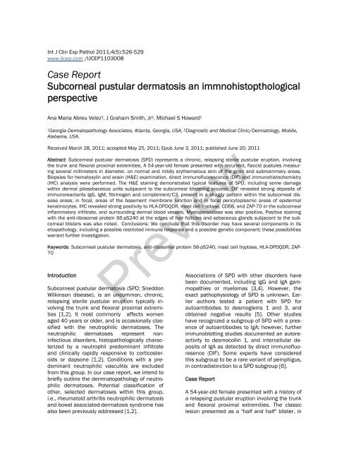
Int J Clin Exp Pathol 2011;4(5):526-529<br />
www.ijcep.com /IJCEP1103008<br />
Case Report<br />
Subcorneal pustular dermatosis an immnohistopthological<br />
perspective<br />
Ana Maria Abreu Velez 1 , J Graham Smith, Jr 2 , Michael S Howard 1<br />
1Georgia Dermatopathology Associates, Atlanta, Georgia, USA; 2 Diagnostic and Medical Clinic/Dermatology, Mobile,<br />
Alabama, USA.<br />
Received March 28, 2011; accepted May 25, 2011; Epub June 3, 2011; published June 20, 2011<br />
Abstract: Subcorneal pustular dermatosis (SPD) represents a chronic, relapsing sterile pustular eruption, involving<br />
the trunk and flexoral proximal extremities. A 54-year-old female presented with recurrent, flaccid pustules measuring<br />
several millimeters in diameter, on normal and mildly erythematous skin of the groin and submammary areas.<br />
Biopsies for hematoxylin and eosin (H&E) examination, direct immunofluorescence (DIF) and immunohistochemistry<br />
(IHC) analysis were performed. The H&E staining demonstrated typical features of SPD, including some damage<br />
within dermal pilosebaceous units subjacent to the subcorneal blistering process. DIF revealed strong deposits of<br />
immunoreactants IgG, IgM, fibrinogen and complement/C3, present in a shaggy pattern within the subcorneal disease<br />
areas; in focal, areas of the basement membrane junction and in focal pericytoplasmic areas of epidermal<br />
keratinocytes. IHC revealed strong positivity to HLA-DPDQDR, mast cell tryptase, CD68, and ZAP-70 in the subcorneal<br />
inflammatory infiltrate, and surrounding dermal blood vessels. Myeloperoxidase was also positive. Positive staining<br />
with the anti-ribosomal protein S6-pS240 at the edges of hair follicles and sebaceous glands subjacent to the subcorneal<br />
blisters was also noted. Conclusions: We conclude that this disorder may have several components in its<br />
etiopathology, including a possible restricted immune response and a possible genetic component; these possibilities<br />
warrant further investigation.<br />
Keywords: Subcorneal pustular dermatosis, anti-ribosomal protein S6-pS240, mast cell tryptase, HLA-DPDQDR, ZAP-<br />
70<br />
Introduction<br />
Subcorneal pustular dermatosis (SPD; Sneddon<br />
Wilkinson disease), is an uncommon, chronic,<br />
relapsing sterile pustular eruption typically involving<br />
the trunk and flexoral proximal extremities<br />
[1,2]. It most commonly affects women<br />
aged 40 years or older, and is occasionally classified<br />
with the neutrophilic dermatoses. The<br />
neutrophilic dermatoses represent noninfectious<br />
disorders, histopathologically characterized<br />
by a neutrophil predominant infiltrate<br />
and clinically rapidly responsive to corticosteroids<br />
or dapsone [1,2]. Conditions with a predominant<br />
neutrophilic vasculitis are excluded<br />
from this group. In our case report, we intend to<br />
briefly outline the dermnatopathology of neutrophilic<br />
dermatoses. Potential classification of<br />
other, selected dermatoses within this group,<br />
i.e., rheumatoid arthritis neutrophilic dermatosis<br />
and bowel associated-dermatosis syndrome has<br />
also been previously addressed [1,2].<br />
<strong>Preview</strong><br />
Associations of SPD with other disorders have<br />
been documented, including IgG and IgA gammopathies<br />
or myelomas [3,4]. However, the<br />
exact pathophysiology of SPD is unknown. Earlier<br />
authors tested a patient with SPD for<br />
autoantibodies to desmogleins 1 and 3, and<br />
obtained negative results [5]. Other studies<br />
have recognized a subgroup of SPD with a presence<br />
of autoantibodies to IgA; however, further<br />
immunoblotting studies documented an autoreactivity<br />
to desmocollin 1, and intercellular deposits<br />
of IgA as detected by direct immunofluoresence<br />
(DIF). Some experts have considered<br />
this subgroup to be a rare variant of pemphigus,<br />
in contradistinction to a SPD subgroup [6].<br />
Case Report<br />
A 54-year-old female presented with a history of<br />
a relapsing pustular eruption involving the trunk<br />
and flexoral proximal extremities. The classic<br />
lesion presented as a "half and half" blister, in
Subcorneal pustular dermatosis<br />
which purulent fluid seemed to accumulate in<br />
the lower half of the blister. The patient reported<br />
that some blisters had arisen within a<br />
few hours. Physical examination revealed individual<br />
pustular lesions in the flexoal areas of<br />
the extremities. Laboratory data included a normal<br />
complete blood count, and a normal erythrocyte<br />
sedimentation rate. Serum electrolytes,<br />
blood urea nitrogen, creatinine, liver function<br />
tests, urinalysis and chest radiographs were<br />
within normal limits. Biopsies of the lesions<br />
were performed, and submitted for hematoxylin<br />
and eosin (H&E) staining, multicolor direct immunofluorescence<br />
(DIF) and immunohistochemistry<br />
(IHC); technical procedures were followed<br />
as previously outlined [7,8]. We utilized the following<br />
Dako antibodies: HLA DPDQDR, mast cell<br />
tryptase, CD68, ZAP-70, anti-ribosomal protein<br />
S6-pS240 and myeloperoxidase.<br />
Microscopic examination<br />
H & E: Examination of the tissue sections demonstrated<br />
a subcorneal blistering disorder. Individual<br />
blisters were tense, and included serum<br />
scale crust. Within the blister lumens, numerous<br />
neutrophils were appreciated; lymphocytes and<br />
eosinophils were rare. Focal intraepidermal<br />
acantholysis was noted. No suprabasilar blistering<br />
was appreciated. Within the papillary dermis,<br />
a mild, superficial, perivascular infiltrate of<br />
lymphocytes, histiocytes, neutrophils and rare<br />
eosinophils was noted. No definitive evidence of<br />
an infectious, or a neoplastic process was seen.<br />
A PAS special stain was also reviewed; the positive<br />
control stained appropriately. The PAS special<br />
stain revealed no fungal organisms. Direct<br />
immunofluorescence (DIF) findings were as follows:<br />
IgG was seen as a sub-basement membrane<br />
zone(BMZ) reinforcement; specifically, a<br />
thin, linear band was appreciated under the<br />
BMZ of the dermal-epidermal junction. The pattern<br />
was distinctively different from the DIF patterns<br />
observed in bullous pemphigoid, dermatitis<br />
herpetiformis, epidermolysis bullosa acquisita<br />
and other subepidermal autoimmune blistering<br />
disorders. FITC conjugated IgE, IgA and<br />
fibrinogen were also seen in a pericytoplasmic<br />
and perinuclear pattern, in several patches<br />
within the epidermal stratum corneum (++).<br />
Other findings included IgM (+, intercellular epidermal<br />
stratum spinosum); IgD (+/-, focal BMZ<br />
cytoid bodies; complement/C1q (-); complement/C3<br />
(+, roof of subcorneal pustules); albumin<br />
(+, intercellular epidermal stratum spinosum);<br />
and fibrinogen (++, focally within papillary<br />
dermal tip areas, focally within the superficial<br />
corneal layer, and surrounding some upper and<br />
intermediate dermal blood vessels). The blister<br />
lumens were positive for IgG, IgA, IgM, IgE and<br />
fibrinogen (Figure 1). A Gram stain of the blister<br />
was negative for bacterial organisms. Immunohistochemisty<br />
(IHC) corroborated the DIF findings,<br />
and also revealed a strong positivity to<br />
mast cell tryptase (MCT), CD68 and HLA-<br />
DPDQDR surrounding upper dermal blood vessels<br />
under the inflammatory process (Figure 1).<br />
Myeloperoxidase was positive in the entire blister<br />
and in some of the corneal layers. We observed<br />
positive staining with HLA-DPDQDR,<br />
mast cell tryptase, CD68, and ZAP-70, antiribosomal<br />
protein S6-pS240 and myeloperoxidase<br />
Negative staining was noted with LAT, BAX,<br />
CD34, CD35, survivin and topoisomerase II alpha.<br />
Discussion<br />
Direct and indirect immunofluorescence results<br />
have previously been reported as negative in<br />
subcorneal pustular dermatosis. However, previous<br />
investigators utilized single antibody, single<br />
fluorochrome DIF techniques. In contradistinction,<br />
we have utilized simultaneous multiple<br />
antibody, multiple fluorochome techniques. IHC<br />
was further utilized to corroborate our DIF findings,<br />
which could have been interpreted in earlier<br />
reports as non-specific in nature. By IHC, we<br />
also found strong positivity to mast cell tryptase<br />
(MCT), CD68 and HLA-DPDQDR associated with<br />
upper dermal blood vessels subjacent to the<br />
blistering process. Myeloperoxidase was positive<br />
throughout the blister lumens, and focally<br />
within the corneal layer by IHC staining.<br />
<strong>Preview</strong><br />
Of interest, the upper corneal layer in proximity<br />
to the SPD blisters seems to be positive for IgG,<br />
IgM, IgA, IgE and fibrinogen. These findings suggest<br />
that SPD may display a delicate autoantibody<br />
response to localized “dysregulated cells”<br />
in the corneal layer. Very few studies have addressed<br />
the immunotypification of SPD. One of<br />
the few studies suggested that the accumulation<br />
of neutrophils in the subcorneal pustules<br />
indicated that the upper epidermis released<br />
chemoattractants.12 Further, the neutrophilic<br />
chemoattractants interleukin (IL)-1 beta, IL-6, IL-<br />
8, IL-10, leukotriene B4, and complement components<br />
C5a and C5a were found at increased<br />
levels in scale extracts from patients with SPD,<br />
527 Int J Clin Exp Pathol 2011;4(5):526-529
Subcorneal pustular dermatosis<br />
<strong>Preview</strong><br />
Figure 1. a. A group of clinical pustules near the axilla (black arrow). b. H & E stain, showing a subcorneal accumulation<br />
of neutrophils (blue arrow), within a subcorneal blister (black arrow). c. DIF showing localized deposits of FITC<br />
conjugated anti-human fibrinogen in multiple foci, including within a subcorneal blister (green staining; white arrow),<br />
within a dermal papillary tip (green staining; red arrow) and around upper dermal vessels (green staining; black arrow).<br />
Epidermal keratinocyte nuclei of are highlighted with 4',6-diamidino-2-phenylindole (Dapi) in blue d. Utilizing DIF<br />
and FITC conjugated fibrinogen in isolation, we were able to detect an identical reactivity pattern as observed in c<br />
(red arrows). e. IHC with anti-human fibrinogen antibody that corroborates our DIF findings of fibrinogen in a subcorneal<br />
blister (red arrow), within a dermal papillary tip (purple arrow) and around upper dermal blood vessels (black<br />
arrow). f. DIF showing staining between epidermal keratinocytes utilizing FITC conjugated anti-human IgG. The staining<br />
pattern resembles the intercellular staining pattern of pemphigus; however, the staining pattern observed in our<br />
patient appears more pericytoplasmic, and predominates in upper layers of the epidermis (white arrow). g. Similar to<br />
f, at higher magnification(white arrow) with epidermal keratinocyte nuclei counterstained with DAPI (light blue staining).<br />
h, i. Show similar DIF staining as f and g, but using FITC conjugated anti-human albumin. Please note enhanced<br />
staining in areas close to the epidermal corneal layer (red arrows). j and k, Show similar DIF staining as f and g, but<br />
using FITC conjugated anti-human-IgA. Please note enhanced staining in areas close to the epidermal corneal layer<br />
and the blister red arrows). l. Positive IHC staining with anti-human IgA within a subcorneal blister, and at multiple foci<br />
within the dermal extracellular matrix (brown staining, red arrows). m and n, IHCs demonstrating positive staining<br />
within a subcorneal blister and around upper dermal blood vessels with IgE in m, and with IgM in n (brown staining;<br />
red arrows). o. IHC, demonstrating strongly positive mast cell tryptase staining around the upper dermal blood vessels<br />
(brown staining). p, Strong IHC staining with anti-ribosomal protein S6-pS240.<br />
528 Int J Clin Exp Pathol 2011;4(5):526-529
Subcorneal pustular dermatosis<br />
compared with those of controls [9]. Also, tumor<br />
necrosis factor-alpha levels have been found to<br />
be significantly elevated in the serum and blister<br />
fluid of patients with SPD. Some authors<br />
have described the presence of epidermal IgA<br />
by DIF in what were considered SPD cases. The<br />
authors evaluated cases with clinical SPD-like<br />
disease from 29 patients, and intraepidermal<br />
IgA detected by DIF [9]. The disease pustules<br />
were considered to be subcorneal; intraepidermal.<br />
IgA deposits were usually found in the intercellular<br />
areas of the epidermis, although a<br />
subcorneal linear pattern was also described.<br />
The disease usually responded to dapsone. In<br />
six cases, a monoclonal IgA gammopathy was<br />
present [3,4].<br />
Our positive findings with HLA DPDQDR, mast<br />
cell tryptase, CD68, and ZAP-70, anti-ribosomal<br />
protein S6-pS240, and myeloperoxidase may<br />
indicate a restricted immune response, including<br />
the presence of T cell activation markers. In<br />
addition, the presence of ribosomal protein S6<br />
kinase (which plays a critical role in the regulation<br />
of cell growth and energy metabolism) may<br />
indicate alterations within the family of serine/<br />
threonine kinases, downstream effectors of the<br />
mTOR and PI3K signalling pathways [10, 11].<br />
Protein kinases are important regulators of intracellular<br />
signal transduction pathways, and<br />
thus play critical roles in diverse cellular functions.<br />
Previous reports have noted that steadystate<br />
levels of the S6K isoforms S6K1 and<br />
S6K2 are regulated by ubiquitination-mediated<br />
proteasomal degradation [10, 11]. Moreover,<br />
accumulating evidence indicates that the activation<br />
of protein kinases, in turn, commonly initiates<br />
their downregulation via the ubiquitin/<br />
proteasome pathway. Failure to regulate protein<br />
kinase activity or expression levels can elicit<br />
human disease. Thus, our findings documenting<br />
the presence of this antibody exclusively directed<br />
to focal areas of the hair follicle and sebaceous<br />
glands warrants further investigation.<br />
Acknowledgments<br />
Please address correspondence to: Ana Maria Abreu-<br />
Velez, MD, PhD, Georgia Dermatopathology Associates,<br />
1534 North Decatur Road. NE; Suite 206, Atlanta,<br />
GA 30307-1000, USA. Tel: (404) 371-0027,<br />
Fax: (404) 371-1900. Email: abreuvelez@yahoo.com<br />
References<br />
[1] Sneddon IB, Wilkinson DS. Subcorneal pustular<br />
dermatoses. Br J Dermatol. 1956; 68: 385-<br />
394.<br />
[2] Nischal KC, Khopkar U. An approach to the<br />
diagnosis of neutrophilic dermatoses: A histopathological<br />
perspective. Indian J Dermatol<br />
Venereol Leprol. 2007;73: 222-230.<br />
[3] Takata M, Inaoki M, Shodo M, Hirone T, Kaya<br />
H. Subcorneal pustular dermatosis associated<br />
with IgA myeloma and intraepidermal IgA deposits.<br />
Dermatology. 1994;189:111-114.<br />
[4] Scerri L, Zaki I, Allen BR. Pyoderma gangrenosum<br />
and subcorneal pustular dermatosis, without<br />
monoclonal gammopathy. Br J Dermatol.<br />
Mar 1994;130:398-399.<br />
[5] Bordignon M, Zattra E, Montesco MC, Alaibac<br />
M. Subcorneal pustular dermatosis (Sneddon-<br />
Wilkinson disease) with absence of desmoglein<br />
1 and 3 antibodies: case report and literature<br />
review. Am J Clin Dermatol. 2008;9:51-55.<br />
[6] Gruss C, Zillikens D, Hashinoto T, et. al. Rapid<br />
response of IgA pemphigus of subcorneal pustular<br />
dermatosis type to treatment with isotretinoin.<br />
J Am Acad Dermatol. 2000;43: 923-926.<br />
[7] Abreu Velez AM, Howard MS, Brown VM. Antibodies<br />
to piloerector (arrector pili) muscle in a<br />
case of lupus/lichen planus overlap syndrome.<br />
North Am J Med Sci. 2010; 2: 276-280<br />
[8] Abreu Velez AM, Klein AD, Howard MS. Autoreactivity<br />
to sweat glands and nerves in clinical<br />
scabies infection. North Am J Med Sci 2010; 2:<br />
422-425.<br />
[9] Grob JJ, Mege JL, Capo C, et al. Role of tumor<br />
necrosis factor-alpha in Sneddon-Wilkinson<br />
subcorneal pustular dermatosis. A model of<br />
neutrophil priming in vivo. J Am Acad Dermatol.<br />
1991;25:944-947.<br />
[10] Gwalter J, Wang ML. Gout I. The ubiquitination<br />
of ribosomal S6 kinases is independent from<br />
the mitogen-induced phosphorylation/<br />
activation of the kinase. Int J Biochem Cell<br />
Biol. 2009;4:828-833.<br />
[11] Lu Z, Hunter T. Degradation of activated protein<br />
kinases by ubiquitination. Annu Rev Biochem.<br />
2009;78:435-75.<br />
<strong>Preview</strong><br />
We would like to thank Jonathan S. Jones, HT<br />
(ASCP) for excellent technical work. All work was<br />
performed with funds from Georgia Dermatopathology<br />
Associates (GDA).<br />
529 Int J Clin Exp Pathol 2011;4(5):526-529