o_195ehsp3l1hu219e55rl1u798dna.pdf

Create successful ePaper yourself
Turn your PDF publications into a flip-book with our unique Google optimized e-Paper software.
www.najms.org North American Journal of Medical Sciences 2010 November, Volume 2. No. 11.<br />
Case Report<br />
OPEN ACCESS<br />
IgG/IgE bullous pemphigoid with<br />
CD45 lymphocytic reactivity to dermal<br />
blood vessels, nerves and eccrine sweat glands<br />
Ana Maria Abreu-Velez, MD., PhD. 1 , J. Graham Smith, Jr., MD. 2 , Michael S. Howard, MD. 1<br />
1 Georgia Dermatopathology Associates, Atlanta, Georgia, USA,<br />
2 Diagnostic and Medical Clinic/Dermatology, Mobile, Alabama, USA.<br />
Citation: Abreu-Velez AM, Smith JG, Howard MS. IgG/IgE bullous pemphigoid with CD45 lymphocytic reactivity to<br />
dermal blood vessels, nerves and eccrine sweat glands. North Am J Med Sci 2010; 2: 540-543.<br />
Doi: 10.4297/najms.2010.2540<br />
Availability: www.najms.org<br />
ISSN: 1947 – 2714<br />
Abstract<br />
Context: Bullous pemphigoid (BP), the most common autoimmune blistering disease, is mediated by autoantibodies. BP<br />
primarily affects the elderly and is characterized by the development of urticarial plaques surmounted by subepidermal<br />
blisters, and the deposition of immunoglobulins and complement at the basement membrane zone (BMZ) of the skin. BP is<br />
immunologically characterized by the development of autoantibodies targeting two structural proteins of the<br />
hemidesmosomes, BP180 (collagen XVII) and BP230 (BPAG1). Case Report: A 63 -year-old Caucasian female patient<br />
was evaluated for a 4 day history of several itching, erythematous blisters on her extremities. Biopsies for hematoxylin and<br />
eosin (H&E) examination, as well as Periodic acid-Schiff (PAS), immunohistochemistry (IHC) and direct<br />
immunofluorescence (DIF) analysis were performed. Results: H&E demonstrated a subepidermal blister, with partial<br />
re-epithelialization of the blister floor. Within the blister lumen, numerous neutrophils were present, with occasional<br />
eosinophils and lymphocytes also noted. Within the dermis, a mild, superficial, perivascular and periadnexal infiltrate of<br />
lymphocytes, histiocytes and occasional eosinophils was identified, with mild perivascular leukocytoclastic debris. The<br />
PAS stain was positive at the BMZ, and around selected blood vessels, nerves and sweat glands. DIF revealed linear<br />
deposits of IgG and Complement/C3 and fibrinogen at the BMZ, and around selected dermal nerves, blood vessels and<br />
sweat glands. Strong granular deposits of IgE were also observed, colocalizing with monoclonal antibodies to Collagen IV<br />
(CIV). By IHC, positive CD45 staining of lymphocytes was seen surrounding selected dermal blood vessels, eccrine sweat<br />
glands, and nerves. Conclusion: The patient displayed IgG, IgE, and fibrinogen autoantibodies against the BMZ, as well as<br />
around some dermal nerves and sweat glands; their binding in the skin could trigger complement activation. In addition,<br />
the role of the dermal CD45 positive lymphocytes warrants further investigation.<br />
Keywords: Bullous pemphigoid, IgE, nerves and sweat glands, autoantibodies.<br />
Correspondence to: Ana Maria Abreu-Velez, MD., PhD., Georgia Dermatopathology Associates, 1534 North Decatur Rd.<br />
NE; Suite 206, Atlanta, GA 30307-1000, USA. Tel.: (404) 3710027, Fax: (404) 3711900, Email: abreuvelez@yahoo.com<br />
Introduction<br />
Bullous pemphigoid (BP) is an autoimmune blistering<br />
disease primarily affecting elderly patients, and<br />
characterized by the development of urticarial plaques<br />
surmounted by subepidermal blisters and the deposition of<br />
both immunoglobulins and complement at the basement<br />
membrane zone (BMZ) of the skin. Immunologically, it is<br />
characterized by the development of autoantibodies<br />
540<br />
targeting two important proteins of the hemidesmosomes,<br />
BP180 (collagen XVII) and BP230 [1]. It is known that<br />
BP230 is an intracellular protein of the hemidesmosomal<br />
plaque, while BP180 is a transmembrane protein<br />
containing a collagenous extracellular domain. Significant<br />
experimental evidence indicates that BP180 is the primary<br />
target of the pathogenic autoantibodies. Autoantibodies are<br />
of both the IgG and IgE classes; their binding in the skin
www.najms.org North American Journal of Medical Sciences 2010 November, Volume 2. No. 11.<br />
triggers complement activation, mast cell degranulation<br />
and additional migration of eosinophils, mast cells, and<br />
neutrophils[1-4].Discharge of proteases from these<br />
inflammatory cells may result in cleavage of the BMZ and<br />
subsequent blister formation. However, the initial triggers<br />
of BP autoantibody production remain obscure.<br />
Case Report<br />
A 63 year old female patient was referred with a four day<br />
history of several, severely pruritic, erythematous blisters<br />
and crusts on her extremities (arms and thighs) (Fig. 1).<br />
Skin biopsies for hematoxylin and eosin (H & E), as well<br />
as for periodic acid-Schiff (PAS) and<br />
immunohistochemistry (IHC) were performed. In addition,<br />
direct immunofluorescence (DIF) and indirect<br />
immunofluorescence (IIF) with 0.1 M sodium chloride salt<br />
split skin were performed. In brief, DIF was performed<br />
utilizing skin cryosections, incubated with multiple<br />
fluorescein isothiocyanate (FITC)-conjugated secondary<br />
antibodies. The secondary antibodies were of rabbit origin,<br />
and included: a) anti-human IgG (γ chain), b) anti-human<br />
IgA (α chains), c) anti-human IgM (μ-chain), d)<br />
anti-human fibrinogen, and e) anti-human albumin (all<br />
used at 1:20 to 1:40 dilutions and obtained from Dako<br />
(Carpinteria, California, USA). We also utilized secondary<br />
antibodies of goat origin, including: a) anti-human IgE<br />
antiserum (Vector Laboratories, Bridgeport, New Jersey,<br />
USA) and b) anti-human C1q (Southern Biotech,<br />
Birmingham, Alabama, USA). Finally, monoclonal<br />
anti-mouse collagen IV (CIV) from Invitrogen(Carlsbad,<br />
California, USA) with a goat anti-mouse Texas red<br />
conjugated secondary antibody was utilized to highlight<br />
the basement membrane zone (BMZ). The slides were<br />
then counterstained with 4',6-diamidino-2-phenylindole<br />
(Dapi) (Pierce, Rockford, Illinois, USA) washed,<br />
coverslipped, and dried overnight at 4 o C. IHC staining<br />
using anti human CD45 was performed using a Dako<br />
automatized dual endogenous flex system, following Dako<br />
technical instructions.<br />
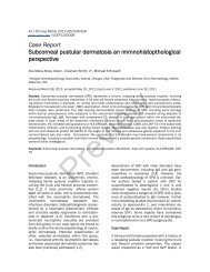
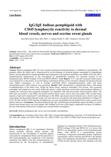
In Figure 1, we highlight our most significant H & E, PAS,<br />
DIF and IHC results, including localization of the BMZ<br />
autoantibodies utilizing the IIF sodium chloride split skin<br />
technique [4]. The DIF results in the basement membrane<br />
zone of the skin (BMZ) were as follows: IgG, linear, ++;<br />
IgE, granular, ++; C3, linear, ++; fibrinogen, linear, ++;<br />
Complement/C1q, linear, +/-; albumin, linear, +/-;<br />
Fibrinogen, linear, ++, and collagen IV, linear, ++. Within<br />
the dermis, we observed IgE, fibrinogen and C3 perineural<br />
and sweat gland reactivity (++). Indirect<br />
immunofluorescence(IIF) performed on 1M NaCl salt split<br />
skin displayed the following results: IgG (+, linear, blister<br />
roof); IgA (+, linear, blister roof); IgM (+, linear, blister<br />
roof); IgE (+, dermal perivascular and perineural);<br />
Complement/C1q(+, linear, blister roof); Complement/C3<br />
(+, linear, blister roof); Albumin(+, linear, blister roof);<br />
and Fibrinogen (+, linear, blister roof).<br />
Examination of the H&E tissue sections demonstrated a<br />
subepidermal, tense blister, with partial re-epithelialization<br />
541<br />
of the blister floor. No impetiginization of the epidermis<br />
was seen. Within the blister lumen, numerous neutrophils<br />
were present, with occasional eosinophils and lymphocytes<br />
also noted. Dermal papillary festoons were not observed<br />
(Fig. 1). Within the dermis, a mild, superficial, perivascular<br />
and periadnexal infiltrate of lymphocytes, histiocytes and<br />
occasional eosinophils was identified. Neutrophils were<br />
rare. Mild perivascular leukocytoclastic debris was present,<br />
but no frank vasculitis was appreciated. A PAS special stain<br />
displayed positivity at the basement membrane zone<br />
(BMZ), as well as around some dermal blood vessels,<br />
nerves and eccrine sweat glands (Fig. 1). The PAS special<br />
stain revealed no fungal organisms.<br />
Fig. 1a, H & E staining (100X) demonstrating the subepidermal<br />
blister (red arrow). 1b, DIF, positive staining with FITC<br />
conjugated anti-human IgE at the BMZ in a linear pattern (green<br />
staining, lower white arrow), colocalizing with the collagen<br />
IV(CIV) antibody (red staining, top yellow arrow). Please notice<br />
that CIV also stains the upper dermal blood vessel areas (lower<br />
yellow arrow). The epidermal corneal layer also shows some IgE<br />
reactivity (upper white arrow). 1c. DIF, showing destruction of<br />
epidermal keratinocytes, characterized by amorphous staining of<br />
cell nuclei with Dapi (blue staining, yellow arrows). The red<br />
arrow shows defragmented pieces of CIV in the blister lumen<br />
(red particles); the white arrows show positive staining with<br />
FITC conjugated antihuman fibrinogen, on both sides of the<br />
blister (epidermal and dermal). 1d, Shows a clinical blister<br />
(white arrow) and some adjacent crusts (blue arrows). 1e, DIF.<br />
Note the keratinocytes nuclei in blue (Dapi), and the mapping of<br />
the blister with CIV antibody (red staining, yellow arrows).<br />
Please note the delicate, trans-epidermal excretion of tiny<br />
fragments of CIV, seen as small red dots among the keratinocytes.<br />
1f. Periodic acid-Schiff (PAS) stain, displaying pink positivity at<br />
the BMZ (red arrow). 1g. Positive staining of a nerve with FITC<br />
conjugated anti-human fibrinogen (green staining; red arrows), as<br />
well as against eccrine sweat glands (yellow arrows). 1h. The<br />
identity of the nerve was confirmed by colocalizing, positive<br />
S-100 IHC staining (brown staining, red arrow). 1i, 1l and 1n. In<br />
addition, we utilized IHC to confirm a significant lymphocytic<br />
infiltrate around dermal neurovascular package and eccrine sweat
www.najms.org North American Journal of Medical Sciences 2010 November, Volume 2. No. 11.<br />
glands with CD45 (brown staining, red arrows). 1j. PAS positive,<br />
pink staining of a nerve sheath (red arrow). 1k, DIF positive<br />
staining of a blood vessel wall with FITC conjugated<br />
Complement/C3 (green-yellowish staining, yellow arrow),<br />
colocalizing with CIV antibody (red staining, red arrows). 1m. H<br />
& E stain, showing lymphocytes infiltrating around dermal<br />
nerves (black arrow), eccrine sweat glands (yellow arrow) and an<br />
eccrine sweat gland ductus (blue arrow). 1o. PAS positive<br />
staining of eccrine sweat gland peripheral membranes (pink<br />
staining, black arrows).<br />
Discussion<br />
Bullous pemphigoid and epidermolysis bullosa acquisita<br />
(EBA) may have indistinguishable clinical, histologic, and<br />
routine immunofluorescence features. Thus, these two<br />
diseases can be reliably distinguished 1) in seropositive<br />
cases by indirect immunoflrouescence (IIF) testing on salt<br />
lamina lucida split skin, 2) in research studies by direct<br />
immunoelectron microscopy and 3) in patients with<br />
circulating autoantibodies, by immunoblotting (IB)<br />
Western studies [4]. The use of these methods is limited by<br />
their availability and expense, and the requirement for<br />
circulating autoantibodies. In our case, following initial<br />
direct immunofluorescence of the biopsy specimen,<br />
additional salt split skin studies utilizing 1.0 mol/L sodium<br />
chloride were performed. In EBA, the IgG linear BMZ<br />
deposits classically appear on the dermal side of a split<br />
specimen; in BP, predominantly or exclusively in the<br />
epidermal side. In our case, the salt split skin pattern<br />
favored the diagnosis of BP, with the additional features of<br />
IgE and fibrinogen also present on both sides of the NaCl<br />
induced blister.<br />
Other differential diagnoses we considered included<br />
bullous impetigo, and a bullous arthropod bite reaction. In<br />
general, patient encounters with biting or stinging<br />
arthropods elicit localized reactions including pruritic<br />
macules, urticarial wheals and papular reactions [5, 6].<br />
Less frequently, localized bullous or hemorrhagic or<br />
disseminated papular reactions may be seen, particularly<br />
in children and immunologically naive adults [5, 6]. With<br />
the exclusion of bee and wasp venom allergies,<br />
immediate-type allergic reactions to arthropod stings and<br />
bites are uncommon [5, 6]. Thus, we considered a bullous<br />
arthropod bite reaction in our differential diagnosis, which<br />
could produce late-phase reactions including the<br />
development of sustained edema, vesicles, blisters, and/or<br />
intense pruritus [5, 6].<br />
Although IgG represents the most commonly documented<br />
immunoglobulin subclass in BP patients, several recent<br />
reports also highlight the importance of IgE; its binding in<br />
the skin triggers complement activation, and further<br />
accumulation of inflammatory cells [7-9]. Indeed, a<br />
correlation of IgE autoantibody with BP180 in a severe<br />
form of bullous pemphigoid has been described [8]. The<br />
case demonstrated positivity with IgG, IgE, fibrinogen and<br />
Complement/C3 against not only the BMZ, but also to<br />
dermal nerves, blood vessels and sweat glands.<br />
The best chartacterized BP antigens are associated with<br />
hemidesmosomes. The hemidesmosomes are present at the<br />
542<br />
BMZ (dermal/epidermal junction). The desmosomes are<br />
multi-protein complexes, promoting stable adhesion of<br />
epithelial cells to the underlying extracellular matrix [10].<br />
Interactions between different hemidesmosomal<br />
components with each other have been studied; these<br />
interactions have been studied utilizing double hybrid and<br />
cell transfection assays. The results demonstrated that:<br />
BP180 binds not only to BP230, but also to plectin [10].<br />
The interactions between these proteins are facilitated by<br />
the Y subdomain within the N-terminal plakin domains of<br />
BP230 and plectin, and residues 145-230 of the<br />
cytoplasmic domain of BP180. Relative to residues<br />
145-230, different, but overlapping, sequences on BP180<br />
mediate binding to integrin ß4 which, in turn, also<br />
associates with BP180 via its third fibronectin type III<br />
repeat. Further, sequences in the N-terminal extremity of<br />
BP230 mediate its binding to integrin ß4, which requires<br />
the C-terminal end of the connecting segment (up to the<br />
fourth fibronectin repeat of the integrin ß4 subunit) [10].<br />
Based on this knowledge, we speculate that, given our<br />
patient’s immunofluorescence positivity against the dermal<br />
sweat glands, possible epitope spreading between the<br />
BP180 and BP230 antigens could elicit reactivity to<br />
integrins and/or plectins expressed not only in multiple<br />
areas of the BMZ, but also in the dermal nerves, sweat<br />
glands and/or microvasculature [11]. In addition, serum<br />
from a patient with bullous pemphigoid was recently<br />
associated with neurological diseases, recognizing BP<br />
antigen in the skin and in the brain [12]. Eccrine<br />
syringofibroadenomatous hyperplasia has also been<br />
documented in some patients with BP [13, 14].<br />
We were able to demonstrate a direct correlation of our DIF<br />
and IIF/salt split skin positivity with positive PAS staining.<br />
Relative to patients who live in areas where DIF and IIF are<br />
not available, the PAS stain often shows a high correlation<br />
with the positivity of immunoglobulins and complement in<br />
autoimmune diseases, as shown in our case [15-17].<br />
Specifically, PAS is a staining method often used to detect<br />
glycogen in tissues. The reaction of periodic acid<br />
selectively oxidizes glucose residues, creating aldehydes;<br />
these then react with the Schiff reagent, yielding a<br />
purple-magenta color. Thus, PAS staining is often utilized<br />
for staining structures containing a high proportion of<br />
carbohydrate macromolecules (i.e., glycogen,<br />
glycoproteins, or proteoglycans), typically found in<br />
neutrophils, fungal organisms and basal laminae. Positive<br />
PAS staining may be found in diverse autoimmune<br />
diseases, including vasculitides, autoimmune bullous<br />
diseases, autoimmune nephritis and Goodpasture's<br />
syndrome [15-17].<br />
Finally, the significance in our case of abundant CD45<br />
positive lymphocytes around the dermal sweat glands,<br />
sweat ducts and neurovascular packages remains unknown.<br />
Further investigation of our case features of 1) BP<br />
IgE/IgG/fibrinogen linear deposits at the BMZ, 2)<br />
auto-reactivity to dermal sweat glands and nerves and 3)<br />
CD45 positive infiltrating dermal lymphocytes may lead to<br />
a better understanding of this disease.
www.najms.org North American Journal of Medical Sciences 2010 November, Volume 2. No. 11.<br />
Acknowledgement<br />
The study was supported by the funding from Georgia<br />
Dermatopathology Associates, Atlanta, GA, USA.<br />
References<br />
1. Wong MM, Giudice GJ, Fairley JA. Autoimmunity<br />
in bullous pemphigoid. G Ital Dermatol Venereol<br />
2009;144:411-421.<br />
2. Woźniak K, Kowalewski C. Alterations of basement<br />
membrane zone in autoimmune subepidermal bullous<br />
diseases. J Dermatol Sci 2005;40:169-175.<br />
3. Medenica L, Skiljević D. Diagnostic significance of<br />
immunofluorescent tests in dermatology. Med Pregl<br />
2009;62:539-546.<br />
4. Gammon WR, Kowalewski C, Chorzelski TP, Kumar<br />
V, Briggaman RA, Beutner EH. Direct<br />
immunofluorescence studies of sodium<br />
chloride-separated skin in the differential diagnosis<br />
of bullous pemphigoid and epidermolysis bullosa<br />
acquisita. J Am Acad Dermatol 1990;22:664-670.<br />
5. Leverkus M, Jochim RC, Schäd S, et al. Bullous<br />
allergic hypersensitivity to bed bug bites mediated by<br />
IgE against salivary nitrophorin. J Invest Dermatol<br />
2006; 126:91-96.<br />
6. Sarkisian EC, Boiko S. Acute localized bullous<br />
eruption in a boy. Bullous reaction to insect bites.<br />
Arch Dermatol. 1995;131:1329-1332.<br />
7. Dresow SK, Sitaru C, Recke A, Oostingh GJ,<br />
Zillikens D, Gibbs BF. IgE autoantibodies against the<br />
intracellular domain of BP180. Br J Dermatol 2009;<br />
160:429-432.<br />
8. Iwata Y, Komura K, Kodera M, et al. Correlation of<br />
IgE autoantibody to BP180 with a severe form of<br />
bullous pemphigoid. Arch Dermatol 2008;144:41-48.<br />
9. Messingham KA, Noe MH, Chapman MA, Giudice<br />
GJ, Fairley JA. A novel ELISA reveals high<br />
frequencies of BP180-specific IgE production in<br />
bullous pemphigoid. J Immunol Methods 2009; 346:<br />
18-25.<br />
10. Koster J, Geerts D, Favre B, Borradori L,<br />
Sonnenberg A. Analysis of the interactions<br />
between BP180, BP230, plectin and the integrin<br />
alpha6beta4 important for hemidesmosome assembly.<br />
J Cell Sci 2003;116:387-399.<br />
11. Hertle MD, Adams JC, Watt FM. Integrin expression<br />
during human epidermal development in vivo and in<br />
vitro. Development. 1991;112:193-206.<br />
12. Li L, Chen J, Wang B, Yao Y, Zuo Y. Sera from<br />
patients with bullous pemphigoid (BP) associated<br />
with neurological diseases; recognized BP antigen in<br />
the skin and brain. Br J Dermatol 2009;6:1343-1345.<br />
13. Nomura K, Kogawa T, Hashimoto I, Katabira Y.<br />
Eccrine syringofibroadenomatous hyperplasia in a<br />
patient with bullous pemphigoid: a case report and<br />
review of the literature. Dermatologica 1991; 182:<br />
59-62.<br />
14. Nomura K, Hashimoto I. Eccrine<br />
syringofibroadenomatosis in two patients with<br />
bullous pemphigoid. Dermatology 1997; 195:<br />
395-398.<br />
15. Sherber NS, Wigley FM, Scher RK. Autoimmune<br />
disorders: nail signs and therapeutic approaches.<br />
Dermatol Ther 2007;20:17-30.<br />
16. Horiguchi Y, Danno K, Ikai K, Imamura S. Colloid<br />
body formation in bullous pemphigoid. Arch<br />
Dermatol Res 1985;277:167-173.<br />
17. Danilewicz M, Wagrowska-Danilewicz M. The<br />
consequences for renal function of the glomerular<br />
deposition of PAS positive material in proliferative<br />
glomerulopathies. A quantitative study. Gen Diagn<br />
Pathol 1997;143:225-230.<br />
543