o_195ehlp5kdj51nurtqo1dmvroqa.pdf

Create successful ePaper yourself
Turn your PDF publications into a flip-book with our unique Google optimized e-Paper software.
Case Report<br />
DOI: 10.7241/ourd.20132.45<br />
IMMUNE RESPONSE IN A CUTANEOUS ALLERGIC DRUG<br />
REACTION SECONDARY TO IMIDAPRIL, BENAZAPRIL<br />
AND METFORMIN<br />
Ana Maria Abreu Velez 1 , Isabel Cristina Avila 2 , Michael S. Howard 1<br />
Source of Support:<br />
Georgia Dermatopathology<br />
Associates, Atlanta, Georgia, USA<br />
Competing Interests:<br />
None<br />
1<br />
Georgia Dermatopathology Associates, Atlanta, Georgia, USA<br />
2<br />
Ph.D. Candidate, University of Antioquia, Medellin, Colombia, SA.<br />
Corresponding author: Ana Maria Abreu Velez, MD PhD<br />
abreuvelez@yahoo.com<br />
Our Dermatol Online. 2013; 4(2): 192-195 Date of submission: 19.01.2013 / acceptance: 04.03.2013<br />
Abstract<br />
Introduction: Cutaneous drug reactions may be classified with regard to pathogenesis and clinical morphology. They may be mediated by<br />
both immunologic and non-immunologic mechanisms.<br />
Case report: A 56 year old female presented with widespread patches and macules, concentrated on her face, trunk and extremities. The<br />
lesions were pruritic, and temporally associated with intake of benzapril hydrochloride, imidapril and metformin.<br />
Methods: Biopsies for hematoxylin and eosin (H&E) examination, as well as for immunohistochemistry (IHC) and direct immunofluorescence<br />
(DIF) analysis were performed for diagnostic purposes, and also to evaluate the lesional immune response.<br />
Results: Hematoxylin and eosin staining demonstrated a histologically unremarkable epidermis. Within the dermis, a moderately florid,<br />
superficial and deep, perivascular infiltrate of lymphocytes, plasmacytoid lymphocytes, histiocytes and rare eosinophils was identified,<br />
consistent with an allergic drug reaction. DIF demonstrated deposits of IgE, Complement/C3 and fibrinogen around dermal blood vessels.<br />
IHC demonstrated positive staining with HAM-56 and myeloid/histoid antigen in the cell infiltrate around the upper dermal blood vessels.<br />
HLA-ABC was overexpressed around those vessels, as well as around dermal sweat glands. COX-2 demonstrated positive staining in both the<br />
epidermis and upper dermis.<br />
Conclusion: Drug reactions are significant causes of skin rashes. In the current case, we were able to identify multiple antigen presenting cells<br />
in the area of the main inflammatory process. The immunologic case findings suggest that allergic drug eruptions may represent complex<br />
processes. An allergic drug reaction should be suspected whenever dermal, perivascular deposits of fibrinogen, Complement/C3 and other<br />
markers such as IgE are identified via DIF.<br />
Key words: HLA-ABC; biomarkers; myeloid-histoid antigen; HAM-56; GFAP; COX-2<br />
Abbreviations and acronyms: Immunohistochemistry (IHC), hematoxylin and eosin (H&E), direct immunofluoresence (DIF)<br />
Cite this article:<br />
Ana Maria Abreu Velez, Isabel Cristina Avila, Michael S. Howard: Immune response in a cutaneous allergic drug reaction secondary to imidapril, benazapril and<br />
metformin. Our Dermatol Online. 2013; 4(2): 192-195<br />
Introduction<br />
Cutaneous drug reactions may be classified with respect<br />
to pathogenesis and clinical morphology [1-6]. They<br />
may be mediated by immunologic and non-immunologic<br />
mechanisms. Immunologic reactions involve the host<br />
immune response and may be mediated by IgE-dependent,<br />
immune complex-initiated, cytotoxic, or cellular immune<br />
mechanisms [1,2]. Non-immunologic reactions may occur<br />
via activation of effector pathways, overdoses, cumulative<br />
toxicity, side effects, interactions with other drugs, metabolic<br />
alterations or exacerbation of pre-existing dermatologic<br />
conditions [1-6]. Certain cutaneous clinical lesional<br />
patterns may be associated with cutaneous drug reactions.<br />
These include urticaria, photosensitive eruptions, erythema<br />
multiforme, disturbance of pigmentation, morbilliform<br />
reactions, fixed drug reactions, erythema nodosum, toxic<br />
epidermal necrolysis, vasculitides and bullous reactions<br />
[1-7]. In addition, certain drugs cause defined cutaneous<br />
syndromes. These include iodides and bromides,<br />
hydantoins, corticosteroids, antimalarial agents, gold,<br />
cancer chemotherapeutic agents, tetracyclines, thiazides and<br />
sulfonamides, nonsteroidal anti-inflammatory agents and<br />
coumarin.<br />
192 © Our Dermatol Online 2.2013<br />
www.odermatol.com
Case Report<br />
A 56 year old Caucasian female was evaluated after<br />
presenting suddenly with erythematous macules and patches<br />
following a one week regimen of imidapril, 5mg once a day<br />
with concurrent benzapril and metformin. These medications<br />
were prescribed for treatment of her hypertension and Type<br />
II diabetes mellitus.<br />
Methods<br />
Our histopathologic studies and hematoxylin and eosin<br />
staining were performed as previously described [3-8].<br />
Direct immunofluorescence (DIF) and<br />
immunohistochemistry (IHC):<br />
In brief, DIF skin cryosections were prepared, and incubated<br />
with multiple fluorochromes as previously reported [3-8].<br />
We utilized a normal skin negative control, obtained from<br />
patients undergoing aesthetic plastic surgery. To test the<br />
local immune response in lesional skin, we utilized the<br />
following markers: antibodies to immunoglobulins A, E, G,<br />
and M; Complement/C1q and C3; kappa and lambda light<br />
chains, and albumin and fibrinogen. All of these antibodies<br />
were either fluorescein isothiocyantate (FITC) or Texas<br />
red conjugated for the DIF testing, and obtained from<br />
Dako (Carpinteria, California, USA). We also utilized Cy3<br />
conjugated monoclonal anti-glial fibrillary acidic protein<br />
(GFAP) antibody from Sigma (Saint Louis, Missouri, USA).<br />
We studied the following markers via immunohistochemistry:<br />
Complement/C5b-9/MAC, HLA-ABC, monoclonal mouse<br />
anti-human myeloid/histiocyte antigen and COX-2, all<br />
also from Dako; and HAM-56 antibody from Cell Marque<br />
Corporation (Rocklin, California, USA). Our IHC studies<br />
were performed as previously described [3-8]. The IRB<br />
consent was obtained.<br />
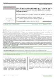
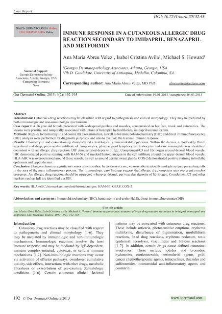
Figure 1. a and b. H&E staining at 100X and 200X respectively, showing a mixed inflammatory infiltrate along the upper and<br />
intermediate neurovascular plexuses of the skin(red arrows), as well as a strong infiltrate of lymphocytes, plasmacytoid lymphocytes,<br />
histiocytes and eosinophils near dermal hair follicles and sebaceous glands (blue arrow). c and d. At 100X and 400X magnification,<br />
respectively. DIF demonstrating positive staining with FITC conjugated IgE in the upper and intermediate neurovascular plexuses<br />
of the dermis (green staining; red arrows). e. HLA-ABC positive IHC staining around the hair follicular unit and blood vessels<br />
around this structure. f. DIF, demonstrating anti-human FITC conjugated kappa light chain antibody with positive staining around<br />
hair follicle areas (green staining; white arrow). Also note positive Texas red conjugated Complement/C3 staining inside the hair<br />
follicle (red staining; red arrow). g. Complement C5b-9/MAC positive IHC staining around the hair follicles and sweat glands(brown<br />
staining; red arrow). h. FITC conjugated Complement/C5b-9/MAC positive DIF staining around the hair follicles and eccrine<br />
glands(green staining; red arrow) i. IHC, demonstrating anti-human kappa light chain positive staining around dermal sweat<br />
glands(green staining; red arrows).<br />
© Our Dermatol Online 2.2013 193
Figure 2. a and b. Positive IHC staining in the cell infiltrate with myeloid/histiocyte antigen (brown staining; red arrows). c and d.<br />
Positive IHC staining with HAM-56 (brown staining; red arrows). e. Positive DIF staining with FITC conjugated Complement/C3<br />
to dermal blood vessels (green staining; white arrow). f. Positive Texas red conjugated Complement/C3 DIF staining in the isthmus<br />
of the hair follicle (red staining; white arrow).<br />
Results<br />
Examination of the H&E tissue sections demonstrated a<br />
histologically unremarkable epidermis. Within the dermis, a<br />
moderately florid, superficial and deep, perivascular infiltrate<br />
of lymphocytes, plasmacytoid lymphocytes and histiocytes<br />
was identified. Neutrophils and eosinophils were rare. Mild,<br />
deep dermal eccrine gland inflammation was also noted<br />
(Fig. 1). No dermal mucin deposition was seen. On DIF<br />
review, FITC conjugated anti-human IgE and Complement/<br />
C3 antibodies were positive around dermal blood vessels,<br />
especially those within the upper and intermediate dermal<br />
plexuses. Anti-human kappa light chain, Complement/C3,<br />
C1q and fibrinogen FITC conjugated antibody staining was<br />
positive within dermal eccrine sweat glands (Fig. 1, 2). On<br />
IHC review, the Complement/C5b-9/MAC complex antibody<br />
stained positive around dermal blood vessels, hair follicles<br />
and eccrine sweat glands (Fig. 1, 2). IHC also demonstrated<br />
positive staining via HAM-56 and myeloid/histoid antibodies<br />
in the cell infiltrate around the upper dermal blood vessels.<br />
HLA-ABC was overexpressed around those vessels, as well<br />
as around dermal sweat glands. COX-2 was positive in both<br />
the epidermis and upper dermis.<br />
Discussion<br />
Allergic reactions include 1) mild clinical events such<br />
as pruritus; 2) moderate events, including generalized skin<br />
eruptions and gastrointestinal and respiratory symptoms, and<br />
3) severe reactions such as anaphylaxis with cardiovascular<br />
complications; these reactions represent common clinical<br />
challenges [1-6]. Allergic reactions may develop to inhaled<br />
substances, food and food additives, and foreign substances<br />
(blood, latex, etc.). Many medications are documented<br />
causes of anaphylactic reactions, asthma, and generalized<br />
urticaria or angioedema [1-6]. Moreover, multiple skin<br />
reactions are induced by drugs via immune complexes,<br />
complement mediated reactions, direct histamine liberation<br />
and modulators of arachidonic acid metabolism. Notably, we<br />
found strong expression of COX-2 in both the epidermis and<br />
upper dermis. Finally, insect venom allergies may manifest<br />
with pain, disseminated exanthems and angioedema [1-9].<br />
The discovery of new associations between drug toxicities<br />
and specific HLA alleles has been facilitated by the use of<br />
DNA-based molecular techniques and the introduction of<br />
high-resolution HLA typing, which have replaced serologic<br />
typing in this field of study [10,11]. Drug toxicity/HLA<br />
associations have been best documented for immunologically<br />
mediated reactions, such as drug hypersensitivity reactions<br />
associated with the use of abacavir, and severe cutaneous<br />
adverse drug reactions, such as Stevens-Johnson syndrome<br />
and toxic epidermal necrolysis induced by carbamazepine<br />
and allopurinol use, respectively. The testing of HLA-ABC<br />
screening for the early diagnoses of potential drug reactions<br />
may thus be of interest in dermatologic practice for selected<br />
patients [10,11].<br />
In our results, we found multiple antigen presenting cells<br />
present within the inflammatory reaction around dermal<br />
blood vessels, hair follicles and sweat glands. Other authors<br />
have reported similar findings [11]. Other authors also<br />
reported that following neurotoxicity screening, a gliosis<br />
reaction represents a hallmark of many types of nervous<br />
system injury [12].<br />
194 © Our Dermatol Online 2.2013
Using a battery of neurotoxic agents, the authors showed that<br />
overexpression of the astroglial protein glial fibrillary acidic<br />
protein (GFAP) [12], could represent a skin biomarker of<br />
drug neurotoxicity. Qualitative and quantitative analysis of<br />
GFAP has shown this biomarker to be a sensitive and specific<br />
indicator of neurotoxic conditions. In our study, we found<br />
that GFAP was overexpressed and seemed to be associated<br />
with the hair follicular isthmus.<br />
In summary, we conclude that the in situ immune<br />
response to our selected drug eruption is complex; more<br />
cases are needed to understand its pathogenesis. Given the<br />
aging of the population in some countries, older patients are<br />
often prescribed multiple medications for multiple clinical<br />
issues. The vendors of metformin clearly state that either the<br />
doctors and/or the pharmacist should avoid using metformin<br />
simultaneously with nonprescription medications such us<br />
vitamins, nutritional supplements, and herbal products. They<br />
also recommend avoiding the following medications:<br />
1) acetazolamide (Diamox);<br />
2) amiloride (Midamor, in Moduretic);<br />
3) angiotensin-converting enzyme (ACE) inhibitors such<br />
as benazepril (Lotensin), captopril (Capoten), enalapril<br />
(Vasotec), fosinopril (Monopril), lisinopril (Prinivil,<br />
Zestril), moexipril (Univasc), perindopril (Aceon), quinapril<br />
(Accupril), ramipril (Altace), and trandolapril (Mavik);<br />
4) beta-blockers such as atenolol (Tenormin), labetalol<br />
(Normodyne), metoprolol (Lopressor, Toprol XL), nadolol<br />
(Corgard), and propranolol (Inderal);<br />
5) calcium channel blockers such as amlodipine (Norvasc),<br />
diltiazem (Cardizem, Dilacor, Tiazac, others), felodipine<br />
(Plendil), isradipine (DynaCirc), nicardipine (Cardene),<br />
nifedipine (Adalat, Procardia), nimodipine (Nimotop),<br />
nisoldipine (Sular), and verapamil (Calan, Isoptin, Verelan);<br />
6) cimetidine (Tagamet);<br />
7) digoxin (Lanoxin);<br />
8) other diuretics, including furosemide (Lasix);<br />
9) androgen and estrogen hormone replacement therapy;<br />
10) insulin and other medications for diabetes;<br />
11) isoniazid;<br />
12) medications for asthma and colds;<br />
13) medications for mental illness and nausea;<br />
14) medications for thyroid disease;<br />
15) morphine (MS Contin, others);<br />
16) niacin;<br />
17) oral contraceptives;<br />
18) other oral steroids such as dexamethasone (Decadron,<br />
Dexone), methylprednisolone (Medrol), and prednisone<br />
(Deltasone);<br />
19) phenytoin (Dilantin, Phenytek);<br />
20) procainamide (Procanbid);<br />
21) quinidine;<br />
22) quinine;<br />
23) ranitidine (Zantac);<br />
24) topiramate (Topamax);<br />
25) triamterene (Dyazide, Maxzide, others);<br />
26) trimethoprim (Primsol);<br />
27) vancomycin (Vancocin);<br />
28) zonisamide (Zonegran) [13].<br />
REFERENCES<br />
1. Wintroub BU, Stern RS: Cutaneous drug reactions: pathogenesis<br />
and clinical classification. Am Acad Dermatol. 1985;13:167-79.<br />
2. Stern RS, Wintroub BU: Adverse drug reactions: reporting and<br />
evaluating cutaneous reactions. Adv Dermatol. 1987;2:3-17.<br />
3. Abreu-Velez AM, Klein DA, Smoller BR, Howard MS: Bullous<br />
allergic drug eruption with presence of myeloperoxidase and<br />
reorganization of the dermal vessels observed by using CD34 and<br />
collagen IV antibodies N Am J Med Sci. 2011;3:82-4.<br />
4. Abreu-Velez AM, Howard MS, Smoller BR: Atopic dermatitis<br />
with possible polysensitization and monkey esophagus reactivity. N<br />
Am J Med Sci. 2010;2:336-40.<br />
5. Abreu-Velez AM, Pinto FJ Jr, Howard MS: Dyshidrotic eczema:<br />
relevance to the immune response in situ. N Am J Med Sci.<br />
2009;1:117-20.<br />
6. Abreu-Velez AM, Jackson BL, Howard MS: Deposition of<br />
immunoreactants in a cutaneous allergic drug reaction. N Am J Med<br />
Sci. 2009;1:180-3.<br />
7. Abreu-Velez AM, Smith JG Jr, Howard MS: Presence of neutrophil<br />
extracellular traps and antineutrophil cytoplasmic antibodies<br />
associated with vasculitides. N Am J Med Sci. 2009;1:309-13.<br />
8. Abreu-Velez AM, Loebl AM, Howard MS: Spongiotic dermatitis<br />
with a mixed inflammatory infiltrate of lymphocytes, antigen<br />
presenting cells, immunoglobulins and complement. N Dermatol<br />
Online. 2011;2:52-7.<br />
9. Abreu-Velez AM, Jackson BL, Howard MS: Salt and pepper<br />
staining patterns for LAT, ZAP-70 and MUM-1 in a vasculitic<br />
bullous allergic drug eruption. N Dermatol Online. 2011;2:104-7.<br />
10. Abreu-Velez, AM. Klein AD, Howard MS: LAT, EGFR -pY197,<br />
PCNL2, CDX2, HLA-DPDQDR, bromodeoxyuridine, JAM-A,<br />
and ezrin immunoreactants in a rubbed spongiotic dermatitis. Our<br />
Dermatol Online. 2011;2:211-5.<br />
11. Phillips EJ, Mallal SA: HLA and drug-induced toxicity. Curr<br />
Opin Mol Ther. 2009.11:231-42.<br />
12. O’Callaghan JP, Sriram K: Glial fibrillary acidic protein and<br />
related glial proteins as biomarkers of neurotoxicity. Expert Opin<br />
Drug Saf. 2005;4:433-42.<br />
13. http://www.ncbi.nlm.nih.gov/pubmedhealth/PMH0000974/<br />
Copyright by Ana Maria Abreu Velez, et al. This is an open access article distributed under the terms of the Creative Commons Attribution License,<br />
which permits unrestricted use, distribution, and reproduction in any medium, provided the original author and source are credited.<br />
© Our Dermatol Online 2.2013 195