o_195eh1omi1qffneg1lemtn01enua.pdf

You also want an ePaper? Increase the reach of your titles
YUMPU automatically turns print PDFs into web optimized ePapers that Google loves.
Original Article<br />
DOI: 10.7241/ourd.20133.67<br />
TISSUE INHIBITOR OF METALLOPROTEINASE 1,<br />
MATRIX METALLOPROTEINASE 9, ΑLPHA-1<br />
ANTITRYPSIN, METALLOTHIONEIN AND UROKINASE<br />
TYPE PLASMINOGEN ACTIVATOR RECEPTOR IN SKIN<br />
BIOPSIES FROM PATIENTS AFFECTED BY<br />
AUTOIMMUNE BLISTERING DISEASES<br />
Ana Maria Abreu Velez 1,2 , Maria Mercedes Yepes Naranjo 2,3 ,<br />
Isabel Cristina Avila 2 , Martha Luz Londoño 2 , Paul B. Googe Jr. 4 ,<br />
Jorge Enrique Velásquez Velez 5 , Ivan Dario Velez 6 ,<br />
Yulieth Alexandra Upegui 6 , Alejandra Jimenez-Echavarria 6 ,<br />
Natalia Regina Mesa-Herrera 7 , Hong Yi 8 , Juliana Calle-Isaza 9 ,<br />
Michael S. Howard 1<br />
Source of Support:<br />
Georgia Dermatopathology<br />
Associates, Atlanta, Georgia, USA<br />
(MSH, AMAV), University of<br />
Antioquia (AMAV) and Knoxville<br />
Dermatopathology Laboratory,<br />
Knoxville, Tennessee, USA (PBG),<br />
Mineros SA, Medellin, Colombia, SA<br />
and the Embassy of Japan in<br />
Colombia, Medellin, Colombia, SA.<br />
Competing Interests:<br />
None<br />
1<br />
Georgia Dermatopathology Associates, Atlanta, Georgia, U.S.A<br />
2<br />
Program of Endemic pemphigus and Autoimmune Blistering Diseases, University of<br />
Antioquia, Medellin Colombia<br />
3<br />
Susalud, Medellin, Colombia<br />
4<br />
Knoxville Dermatopathology Laboratory, Knoxville, Tennessee, U.S.A<br />
5<br />
HGM - Clinica CES - Clinica SOMER - Fundacion HUSVP, Rionegro, Colombia, SA<br />
6<br />
PECET, University of Antioquia, Medellin, Colombia, SA<br />
7<br />
Biochemistry and Molecular Genetics Group, Universidad de Antioquia, Medellin,<br />
Colombia, SA<br />
8<br />
Robert P. Apkarian Integrated Electron Microscopy Core, Emory University Medical<br />
Center, Atlanta, Georgia, USA<br />
9<br />
Hospital General de Medellin, Colombia, S.A<br />
Corresponding author: Ana Maria Abreu Velez, MD PhD<br />
abreuvelez@yahoo.com<br />
Our Dermatol Online. 2013; 4(3): 275-280 Date of submission: 16.03.2013 / acceptance: 19.04.2013<br />
Abstract<br />
Introduction: Proteinases and proteinase inhibitors have been described to play a role in autoimmune skin blistering diseases. We studied<br />
skin lesional biopsies from patients affected by several autoimmune skin blistering diseases for proteinases and proteinase inhibitors.<br />
Methods: We utilized immunohistochemistry to evaluate biopsies for α-1-antitrypsin, human matrix metalloproteinase 9 (MMP9), human<br />
tissue inhibitor of metalloproteinases 1 (TIMP-1), metallothionein and urokinase type plasminogen activator receptor (uPAR). We tested 30<br />
patients affected by endemic pemphigus, 30 controls from the endemic area, and 15 normal controls. We also tested 30 biopsies from patients<br />
with bullous pemphigoid (BP), 20 with pemphigus vulgaris (PV), 8 with pemphigus foliaceus, and 14 with dermatitis herpetiformis (DH).<br />
Results: Contrary to findings in the current literature, most autoimmune skin blistering disease biopsies were negative for uPAR and MMP9.<br />
Only some chronic patients with El Bagre-EPF were positive to MMP9 in the dermis, in proximity to telocytes. TIMP-1 and metallothionein<br />
were positive in half of the biopsies from BP patients at the basement membrane of the skin, within several skin appendices, in areas of dermal<br />
blood vessel inflammation and within dermal mesenchymal-epithelial cell junctions.<br />
Key words: endemic pemphigus foliaceus; autoimmune blistering skin diseases; matrix metalloproteinase 9; tissue inhibitor of<br />
metalloproteinases 1; urokinase type plasminogen activator receptor; α-1-antitrypsin<br />
Cite this article:<br />
Ana Maria Abreu Velez, Maria Mercedes Yepes Naranjo, Isabel Cristina Avila, Martha Luz Londoño, Paul B. Googe Jr., Jorge Enrique Velásquez Velez, Ivan<br />
Dario Velez, Yulieth Alexandra Upegui, Alejandra Jimenez- Echavarria, Natalia Regina Mesa-Herrera Zapata, Hong Yi, Juliana Calle-Isaza, Michael S. Howard:<br />
Tissue inhibitor of metalloproteinase 1, matrix metalloproteinase 9, αlpha-1 antitrypsin, metallothionein and urokinase type plasminogen activator receptor in<br />
skin biopsies from patients affected by autoimmune blistering diseases. Our Dermatol Online. 2013; 4(3): 275-280.<br />
www.odermatol.com<br />
© Our Dermatol Online 3.2013 275
Abbreviations and acronyms: Bullous pemphigoid (BP), immunohistochemistry (IHC), direct and indirect immunofluorescence (DIF, IIF),<br />
hematoxylin and eosin (H&E), basement membrane zone (BMZ), intercellular staining between keratinocytes (ICS), pemphigus vulgaris (PV),<br />
cicatricial pemphigoid (CP), autoimmune blistering skin disease (ABD), matrix metalloproteinase 9 (MMP9), tissue inhibitor of metalloproteinases<br />
1 (TIMP-1), extracellular matrix (ECM), urokinase type plasminogen activator receptor (u-Par).<br />
Introduction<br />
Multiple theories have been proposed regarding the<br />
pathophysiology of cutaneous autoimmune blistering skin<br />
diseases (ABDs). Some involve plasminogen activation,<br />
desmoglein compensation, acetylcholine receptor antibodies,<br />
and intracellular signal control of autoantibodies [1]. Moreover,<br />
human autoantibodies and the presence of complement are<br />
primary factors in producing the blisters of human autoimmune<br />
skin blistering diseases and are thought to exert their pathogenic<br />
effect via proteases [2,3].<br />
Few studies have tested for proteases and protease inhibitors in<br />
lesional skin from patients affected by ABDs [4,5]. We decided<br />
to investigate enzymes that could be modulated by ions that<br />
have been postulated as triggers for ABDs. We also aimed to<br />
investigate enzymes that are related to xenobiotics, based on our<br />
previous findings of metals and metalloids in skin biopsies of<br />
patients with a new variant of endemic pemphigus foliaceus in El<br />
Bagre, Colombia (El-Bagre-EPF) that are exposed to significant<br />
mercury pollution [5]. Thus, we utilized immunohistochemistry<br />
(IHC) to test for anti-human-α-1-antitrypsin, anti-human matrix<br />
metalloproteinase 9 (MMP9), anti-human tissue inhibitor of<br />
metalloproteinases 1 (TIMP1), urokinase type plasminogen<br />
activator receptor (uPAR) and for metallothionein in patients<br />
affected by autoimmune skin blistering diseases.<br />
Methods<br />
Subjects of study<br />
We tested 30 biopsies from El Bagre-EPF patients and 30<br />
controls from the endemic area. Of the 30 control biopsies,<br />
15 were taken from healthy first degree relatives and 15 from<br />
healthy, non-related persons. We also utilized 15 control skin<br />
biopsies from cosmetic surgery patients in the USA, taken<br />
from the chest and/or abdomen. The Bagre-EPF patients<br />
were previously diagnosed by us, fulfilling specific criteria as<br />
previously documented [6-10]. We also tested another group<br />
of ABD patients, whose skin biopsies were obtained from the<br />
archival files of two private dermatopathology laboratories in<br />
the USA. Most of the archival sample patients were not taking<br />
immunosuppressive therapeutic medications at the time of<br />
biopsy. We evaluated 34 biopsies from bullous pemphigoid<br />
(BP) patients, 20 from patients with pemphigus vulgaris (PV), 8<br />
from patients with non-endemic pemphigus foliaceus (PF) and<br />
4 from patients with dermatitis herpetiformis (DH). We also<br />
tested biopsies from heart, liver, kidney and lung tissue from<br />
4 autopsies of El Bagre-EPF patients. For all of the El Bagre<br />
area patients and controls we obtained written consents, as well<br />
as Institutional Review Board (IRB) permission. The archival<br />
biopsies were IRB exempt due to the lack of patient identifiers.<br />
Intensity of immunohistochemistry staining<br />
The staining intensity of the immunohistochemistry<br />
antibodies was evaluated 1) qualitatively by two independent<br />
observers, as well as 2) in a semiquantitative mode by automated<br />
computer image analysis (specifically designed to quantify<br />
immunohistochemistry staining in hematoxylin-counterstained<br />
histologic sections). For the image analysis, slides were scanned<br />
with a ScanScope CS scanning system (Aperio Technologies,<br />
Vista, California, USA), utilizing bright field imaging at 20× and<br />
40× magnifications. The strength of the staining was evaluated<br />
on a scale from 0 to 4, where 0 represented negative staining and<br />
4 the strongest staining.<br />
Immunohistochemistry staining<br />
We performed IHC using antibodies conjugated with<br />
horseradish peroxidase (HRP)-labelled secondary antibodies.<br />
We utilized multiple monoclonal and polyclonal antibodies,<br />
all from Dako (Carpinteria, California, USA). For all our IHC<br />
testing, we used a dual endogenous peroxidase blockage, with<br />
the addition of an Envision dual link (to assist in chromogen<br />
attachment). We applied the chromogen 3,3-diaminobenzidine,<br />
and counterstained with hematoxylin. The samples were run in<br />
a Dako Autostainer Universal Staining System, as previously<br />
described (13-15). Positive and negative controls were<br />
consistently performed. For IHC, we utilized Dako antibodies<br />
to polyclonal rabbit anti-human α-1-antitrypsin (cat. No.<br />
IR505, flex ready to use, antigen retrieval high pH), rabbit<br />
anti-human MMP9, (cat. No. A0150, dilution 1:75, antigen<br />
retrieval heat), monoclonal mouse anti-human TIMP-1 (cat.<br />
No. M0639, dilution 1:50, antigen retrieval heat), monoclonal<br />
mouse anti-human metallothionein (cat. No. M0639, dilution<br />
1:50, antigen retrieval high pH), and monoclonal mouse antihuman<br />
uPAR (cat. No M7294, dil, 1:25, antigen retrieval high<br />
pH). We also utilized control tissue from 4 non-El Bagre EPF<br />
patient autopsies (from the El Bagre EPF endemic area), to rule<br />
out false positive and false negative results due to spontaneous<br />
autolysis. The organs from the autopsies were taken within 12<br />
hours of patient death. The direct immunofluoresecent studies<br />
(DIF) were performed as previously described [6-10].<br />
Indirect immunoelectron microscopy (IEM)<br />
Performed as previously described [10]. In brief,<br />
postembedding immunogold labeling was performed on<br />
samples of El Bagre-EPF sera and controls. Rat skin was<br />
utilized as the substrate antigen; the tissue was dissected,<br />
fixed in 4% glutaraldehyde with 0.2% paraformaldehyde, and<br />
embedded in Lowicryl® resin. The tissue was then sectioned<br />
at 70 nm thickness. The samples were blocked with a solution<br />
from Aurion (Electron Microcopy Sciences/EMS, Hatfield,<br />
Pennsylvania, USA). Our tissue grids were then washed with<br />
PBS-BSAC (Aurion, EMS). The primary antibodies were<br />
incubated overnight at 4°C.<br />
The next day the grids were again washed; a secondary antibody<br />
solution, specifically 10nm gold-conjugated protein A PBS<br />
BSAC (Aurion, EMS) was applied. The samples were then<br />
double-stained with uranyl acetate and lead citrate. The samples<br />
were reviewed under a Hitachi H7500 transmission electron<br />
microscope. Images of immunogold particles displaying any<br />
pattern of positivity were recorded, and converted to TIF format.<br />
Statistical methods<br />
Differences in staining intensity and positivity were<br />
evaluated using a GraphPad Software statistical analysis system,<br />
and employing Student’s t-test.<br />
276 © Our Dermatol Online 3.2013
We considered a statistical significance to be present with a<br />
p value of 0.05 or less, assuming a normal distribution of the<br />
samples.<br />
Results<br />
In Table I and Figures 1 through 2 we summarize our<br />
primary results. We observed consistent patterns of IHC<br />
positivity relative to each autoimmune skin blistering disease.<br />
For example, α-1 antitrypsin was positive in all of the lesions<br />
of DH within the subepidermal blisters (p
Markers BP n=34 PV n=14 PF n=8 DH n=14 Normal<br />
skin<br />
controls<br />
n=15<br />
ά -1 anti-<br />
-trypsin<br />
In 4/34<br />
biopsies, some<br />
staining in<br />
blister, in dermis<br />
and around<br />
the eccrine<br />
ducts.<br />
Positive in the<br />
epidermis in<br />
spots, in some<br />
blister debris<br />
and<br />
subcorneal in<br />
8/14.<br />
Positive in the<br />
upper vessels<br />
and in the<br />
inflammatory<br />
dermis (3/4).<br />
Upper dermis,<br />
blister and under<br />
in 8/10. Positive<br />
in some<br />
fibroblastoid cells.<br />
Mostly<br />
negative.<br />
El Bagre EPF<br />
n=30<br />
Positive<br />
staining in some<br />
of the<br />
basal<br />
keratinocytes<br />
hair follicle,<br />
neurovascular<br />
supplies and<br />
subcutaneous<br />
fat.<br />
Control<br />
endemic area<br />
group n=15<br />
4/15 cases were<br />
positive, in<br />
neurovascular<br />
bundles of the<br />
skin and appendices<br />
and sweat<br />
glands.<br />
MMP-9<br />
Staining in the<br />
blister, dermal<br />
vessels METS<br />
matrix (4/34).<br />
Most cases<br />
negative.<br />
Negative.<br />
ICS like, dermis<br />
under the blister,<br />
neurovascular<br />
supplies of appendices<br />
(3/10).<br />
Mostly<br />
negative.<br />
9/30 of the chronic<br />
cases corneal<br />
layer, hair follicle<br />
some vessels.<br />
Positivity in the<br />
METS.<br />
Mostly<br />
negative.<br />
TIMP1<br />
Weak<br />
positive around<br />
the blisters,<br />
vessels and in<br />
the METs and<br />
fat (19/34).<br />
Positive in<br />
basal<br />
keratinocytes,<br />
blisters debris,<br />
dermal vessels<br />
and METs<br />
(7/14); linear<br />
deposition at<br />
the BMZ.<br />
Positive in 2/4<br />
cases around the<br />
blisters, upper<br />
vessels and<br />
METs.<br />
Positive in the blisters<br />
in the dermal<br />
papillae (3/10).<br />
Mostly<br />
negative.<br />
Corneal layer,<br />
some<br />
epidermis; also,<br />
dermal blood<br />
vessels and<br />
sweat glands.<br />
2/15 Positive<br />
sweat and<br />
sebaceous<br />
glands, vessels<br />
and extracellular<br />
fibroblastoid<br />
cells (8/15).<br />
Metallothionein<br />
Positive in the<br />
BMZ of the<br />
hair follicles,<br />
and some cells<br />
of the METs<br />
(19/34).<br />
Positive at the<br />
BMZ of the<br />
hair follicles<br />
and sebaceous<br />
glands, as well<br />
as in some areas<br />
in the MTEs<br />
matrix (9/14).<br />
Positive in 2/4<br />
cases around<br />
the blister and<br />
upper vessels<br />
and METs.<br />
Positive under the<br />
blisters (4/10).<br />
Mostly<br />
negative.<br />
Corneal, and<br />
cytoplasms of<br />
the keratinocytes<br />
of the spinous<br />
layer. Sebaceous<br />
glands in 23/30<br />
patients and<br />
some fat tissue.<br />
3/15 positive in<br />
the corneal layer.<br />
Positive staining<br />
to the sweat<br />
glands (8/15).<br />
uPAR Negative Negative Negative Negative Negative Negative Negative<br />
Table I. Summary of staining patterns of proteases and protease inhibitors in multiple autoimmune skin blistering diseases<br />
Several cases of DH were positive for α-1 antitrypsin. We<br />
previously described a fatal case of El Bagre-EPF with high<br />
levels of α-1 antitrypsin in a patient superinfected by varicellazoster<br />
virus [23]. The El-Bagre-EPF cases also stained positively<br />
for MMP9 only in chronic patients under treatment with<br />
prednisone for long periods. The MMP9 reactivity was located<br />
in areas of newly reported dermal cell junctions, including<br />
dermal METs [24] and telocytes [25]. Our finding may explain<br />
dermal histologic sclerodermoid changes occasionally noted in<br />
El Bagre EPF, with attendant loss of skin appendices.<br />
The fact that the ABDs improve with glucocorticoids may be<br />
related to our findings. Other studies of BP have shown altered<br />
expression of MMPs and TIMPs within lesional blisters [22].<br />
Conclusions<br />
We conclude that the observed profile expression of<br />
proteases, protease inhibitors and other enzymes in autoimmune<br />
skin blistering diseases seems to differ from many published<br />
animal models, thus highlighting significant immunologic<br />
differences between these animal models and the in vivo<br />
autoimmune skin blistering diseases of human patients. The<br />
TIMP1 and metallothionein seem to be expressed in an inverse<br />
correlation, suggestive of one enzyme attempting to repair<br />
collateral damage from the other enzyme. It is possible that the<br />
capacity to bind heavy metals on these enzymes may indicate<br />
some exposure to these xenobiotics in ABDs. The presence of<br />
enzymes and their inhibitors (not only in the blister areas, but<br />
also in the inflamed dermal vessels, skin appendices and METs)<br />
merits further testing of autoimmunity in these areas.<br />
Acknowledgements<br />
To assisting personnel at the Hospital Nuestra Senora del<br />
Carmen in El Bagre Colombia; and also to Jonathan S. Jones HT<br />
(ASCP) at Georgia Dermatopathology Associates for excellent<br />
technical assistance.<br />
278 © Our Dermatol Online 3.2013
REFERENCES<br />
1. Cotell S, Robinson ND, Chan LS: Autoimmune blistering skin<br />
diseases. Am J Emerg Med. 2000;18:288-99.<br />
2. Cirillo N, Dell’ Ermo A, Gombos F, Lanza A: The specific<br />
proteolysis hypothesis of pemphigus: does the song remain the<br />
same? Med Hypotheses. 2008;70,333-37.<br />
3. Shimanovich I, Mihai S, Oostingh GJ, Ilenchuk TT, Bröcker EB,<br />
Opdenakker G, et al: Granulocyte-derived elastase and gelatinase<br />
B are required for dermal-epidermal separation induced by<br />
autoantibodies from patients with epidermolysis bullosa acquisita<br />
and bullous pemphigoid. J Pathol. 2004;204;519-27.<br />
4. Kitajima Y, Aoyama Y, Seishima M: Transmembrane signaling<br />
for adhesive regulation of desmosomes and hemidesmosomes, and<br />
for cell-cell detachment induced by pemphigus IgG in cultured<br />
keratinocytes: involvement of protein kinase C. J Investig Dermatol<br />
Symp Proc. 1999;4;137-44.<br />
5. Abréu Vélez AM, Warfvinge G, Herrera WL, Abréu Vélez CE,<br />
Montoya M F, Hardy DM, et al: Detection of mercury and other<br />
undetermined materials in skin biopsies of endemic pemphigus<br />
foliaceus. Am J Dermatopathol. 2003;25:5:384-91.<br />
6. Abrèu-Velez AM, Hashimoto T, Bollag WB, Tobón Arroyave S,<br />
Abrèu-Velez CE, Londoño ML, et al: A unique form of endemic<br />
pemphigus in northern Colombia. J Am Acad Dermatol. 2003;49:599-<br />
608.<br />
7. Abrèu-Velez AM, Beutner EH, Montoya F, Bollag WB, Hashimoto<br />
T: Analyses of autoantigens in a new form of endemic pemphigus<br />
foliaceus in Colombia. J Am Acad Dermatol. 2003;49:609-14.<br />
8. Abreu-Velez AM, Robles EV, Howard MS: A new variant of<br />
endemic pemphigus foliaceus in El-Bagre, Colombia: the Hardy-<br />
Weinberg-Castle law and linked short tandem repeats. N Am J Med<br />
Sci. 2009;1:169-78.<br />
9. Howard MS, Yepes MM, Maldonado-Estrada JG, Villa-Robles E,<br />
Jaramillo A, Botero JH, et al: Broad histopathologic patterns of nonglabrous<br />
skin and glabrous skin from patients with a new variant of<br />
endemic pemphigus foliaceus-part 1. J Cutan Pathol. 2010;37:222-<br />
30.<br />
10. Abreu-Velez AM, Howard MS, Yi H, Gao W, Hashimoto<br />
T, Grossniklaus HE: Neural system antigens are recognized by<br />
autoantibodies from patients affected by a new variant of endemic<br />
pemphigus foliaceus in Colombia. J Clin Immunol. 2011;31:356-68.<br />
11. Asano S, Seishima M, Kitajima Y: Phosphatidylinositol-specificphospholipase<br />
C cleaves urokinase plasminogen activator receptor<br />
from the cell surface and leads to inhibition of pemphigus-IgGinduced<br />
acantholysis in DJM-1 cells, a squamous cell carcinoma<br />
line. Clin Exp Dermatol. 2001;26:289-95.<br />
12. Lo Muzio L, Pannone G, Staibano S, Mignogna MD, Rubini C,<br />
Farronato G, et al: Strict correlation between uPAR and plakoglobin<br />
expression in pemphigus vulgaris. J Cutan Pathol. 2002; 29:540-48.<br />
13. Verraes S, Hornebeck W, Polette M, Borradori L, Bernard<br />
P: Respective contribution of neutrophil elastase and matrix<br />
metalloproteinase 9 in the degradation of BP180 (type XVII collagen)<br />
in human bullous pemphigoid. J Invest Dermatol. 2001;117:1091-6.<br />
14. Liu Z, Li N, Diaz LA, Shipley M, Senior RM, Werb Z: Synergy<br />
between a plasminogen cascade and MMP-9 in autoimmune disease.<br />
J Clin Invest. 2005;115:879-87.<br />
15. Niimi Y, Pawankar R, Kawana S: Increased expression of<br />
matrix metalloproteinase-2, matrix metalloproteinase-9 and matrix<br />
metalloproteinase-13 in lesional skin of bullous pemphigoid. Int<br />
Arch Allergy Immunol. 2006;139:104-13.<br />
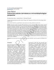
Figure 1. a ά-1 anti-trypsin DIF positive staining in a case of PV inside the blister (red arrow, dark brown staining) and some<br />
punctuate staining in upper dermal blood vessels (200x). b. Case of DH, demonstrating positive TIMP-1 IHC staining in the<br />
blister and punctuate staining in upper dermal blood vessels (red arrows, brown staining) (100x). c. Case of PV, with TIMP-1<br />
positive IHC staining in cells within and around the blister, and some in upper dermal blood vessels (red arrows, brown staining).<br />
d. BP case with positive IHC staining for metallothionein in the base of the blister and around a dermal sweat gland ductus (red<br />
arrows, brown staining). A few positive areas of punctate staining are also noted on upper dermal blood vessels. e. A BP patient<br />
with positive metallothionein IHC staining around dermal blood vessels (red arrows, brown staining). f. A DH case, with positive<br />
IHC staining in the blister with ά-1 anti-trypsin and on upper dermal blood vessels (red arrows, brown staining).<br />
© Our Dermatol Online 3.2013 279
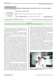
Figure 1. a IHC positive TIMP-1 staining of heart necropsy tissue from an El Bagre-EPF patient; and in b, the same patient in<br />
renal tubule tissue black arrows, red/brown staining). c. A BP case, with positive staining with TIMP1 over the blister BMZ and<br />
between dermal extracellular matrix fibers (red arrows, brown staining). d. The same case of BP as in c, using DIF with FITCI<br />
conjugated fibrinogen and showing that dermal staining is present, possibly related to dermal reactivity(red arrow, light<br />
yellow/green staining). e. A BP case, demonstrating IHC positive staining in the sweat glands with metallothionein (red<br />
arrows, dark staining). f. IHC positive to metallothionein in a patient with PV at the base membrane zone as well as in the<br />
dermal cell junctions and or the MET. g. DIF from an El Bagre-EPF patient using FITCI conjugated anti-human IgG antibody,<br />
and showing positive staining between the dermal fibers (red arrows, yellow/white staining). h. IEM, showing positive 10 nm<br />
Gold labeled anti-human IgG antibodies, positive to several cell junctions in the dermis (red arrows, black dots). i. An El Bagre-<br />
EPF patient, demonstrating positive IHC metallothionein staining in epidermal acantholytic cells and in the subjacent inflamed<br />
dermis (red arrows, brown staining).<br />
16. Salmela MT, Pender SL, Reunala T, MacDonald T, Saarialho-Kere<br />
U: Parallel expression of macrophage metalloelastase (MMP-12) in<br />
duodenal and skin lesions of patients with dermatitis herpetiformis.<br />
Gut. 2001;48, 496-52.<br />
17. Oikarinen A, Kylmäniemi M, Autio-Harmainen H, Autio P,<br />
Salo T: Demonstration of 72-kDa and 92-kDa forms of type IV<br />
collagenase in human skin: variable expression in various blistering<br />
diseases, induction during re-epithelialization, and decrease by<br />
topical glucocorticoids. J Invest Dermatol. 1993;101:205-10.<br />
18. Zebrowska A, Narbutt J, Sysa-Jedrzejowska A, Kobos J,<br />
Waszczykowska E: The imbalance between metalloproteinases and<br />
their tissue inhibitors is involved in the pathogenesis of dermatitis<br />
herpetiformis. Mediators Inflamm. 2005;6:373-9.<br />
19. Narbutt J, Waszczykowska E, Lukamowicz J, Sysa-Jedrzejowska<br />
A, Kobos J, Zebrowska A: Disturbances of the expression of<br />
metalloproteinases and their tissue inhibitors cause destruction of the<br />
basement membrane in pemphigoid. Pol J Pathol. 2006;57:71-6.<br />
20. Abreu-Velez AM, Yi H, Girard JG, Jiao Z, Duque-Ramírez M,<br />
Arias LF, et al: Dermatitis herpetiformis bodies and autoantibodies to<br />
noncutaneous organs and mitochondria in dermatitis herpetiformis.<br />
Our Dermatol Online. 2012;3:283-91.<br />
21. Gomez DE, Alonso DF, Yoshiji H, Thorgeirsson UP: Tissue<br />
inhibitors of metalloproteinases: structure, regulation and biological<br />
functions. Eur J Cell Biol. 1997;74:111-22.<br />
22. Simpkins CO: Metallothionein in human disease. Cell Mol Biol.<br />
2000; 46:465-88.<br />
23. Abreu-Velez AM, Smoller BR, Gao W, Grossniklaus HE, Jiao Z,<br />
Arias LF, et al: Varicella-zoster virus (VZV), and alpha 1 antitrypsin:<br />
a fatal outcome in a patient affected by endemic pemphigus foliaceus.<br />
Int J Dermatol. 2012;51:809-16.<br />
24. Franke WW, Rickelt S: Mesenchymal-epithelial transitions:<br />
spontaneous and cumulative syntheses of epithelial marker<br />
molecules and their assemblies to novel cell junctions connecting<br />
human hematopoietic tumor cells to carcinomatoid tissue structures.<br />
Int J Cancer. 2011;129,2588-99.<br />
25. Ceafalan L, Gherghiceanu M, Popescu LM, Simionescu O:<br />
Telocytes in human skin--are they involved in skin regeneration? J<br />
Cell Mol Med. 2012; 16,1405-20.<br />
Copyright by Ana Maria Abreu Velez, et al. This is an open access article distributed under the terms of the Creative Commons Attribution License,<br />
which permits unrestricted use, distribution, and reproduction in any medium, provided the original author and source are credited.<br />
280 © Our Dermatol Online 3.2013