

Comprehensive Ophthalmology Free Papers - aioseducation
Comprehensive Ophthalmology Free Papers - aioseducation
Comprehensive Ophthalmology Free Papers - aioseducation

You also want an ePaper? Increase the reach of your titles
YUMPU automatically turns print PDFs into web optimized ePapers that Google loves.
70th AIOC Proceedings, Cochin 2012<br />
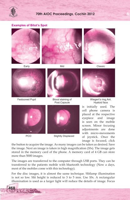
Examples of Bitot’s Spot<br />
Early Mid Classic<br />
is initially used. The<br />
cell phone camera is<br />
placed at the respective<br />
eyepiece and image<br />
PCO<br />
Slightly Displaced<br />
is seen on the mobile<br />
screen. Minor focusing<br />
adjustments are done<br />
with micro-movements<br />
of joystick. Once the<br />
image is focused, click<br />
the button to acquire the image. As many images can be taken as desired. Save<br />
the image. Next an image is taken in high magnification (10x). The image gets<br />
stored in the memory card of the phone. A memory card of 4 GB can store<br />
more than 5000 images.<br />
The images are transferred to the computer through USB ports. They can be<br />
transferred to the patients mobile with bluetooth technology (Now a days,<br />
most of the mobiles come with this technology).<br />
For the disc images, it is almost the same technique. Slitlamp illumination<br />
is not so low. Slit height is reduced to 3 to 5 mm. Use 10x. A rectangular<br />
illumination is used as a larger light will reduce the details of image. Focus<br />
468<br />
Festooned Pupil Blood staining of Wiegert’s ring,Ant.<br />
Post.Capsule<br />
Hyaloid face


