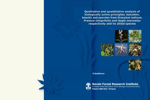
Qualitative and quantitative analysis of biologically active principles ...
Qualitative and quantitative analysis of biologically active principles ...
Qualitative and quantitative analysis of biologically active principles ...

Create successful ePaper yourself
Turn your PDF publications into a flip-book with our unique Google optimized e-Paper software.
KFRI Research Report No. 350 ISBN No. 09708103<br />
<strong>Qualitative</strong> <strong>and</strong> <strong>quantitative</strong> <strong>analysis</strong> <strong>of</strong><br />
<strong>biologically</strong> <strong>active</strong> <strong>principles</strong>, baicalein,<br />
luteolin <strong>and</strong> psoralen from oroxylum<br />
indicum, premna serratifolia, aegle<br />
marmelos <strong>and</strong> their allied species<br />
N Sasidharan<br />
K F R I<br />
Kerala Forest Research Institute<br />
Peechi 680 653, Thrissur<br />
2010
Title <strong>of</strong> the project : KFRI/486/05, <strong>Qualitative</strong> <strong>and</strong> <strong>quantitative</strong><br />
<strong>analysis</strong> <strong>of</strong> <strong>biologically</strong> <strong>active</strong> <strong>principles</strong>,<br />
Baicalein, Luteolin <strong>and</strong> Psoralen from<br />
Oroxylum indicum, Premna serratifolia,<br />
Objectives :<br />
Aegle marmelos <strong>and</strong> their allied species<br />
· Isolation <strong>and</strong> characterisation <strong>of</strong><br />
baicalein, luteolin <strong>and</strong> psoralen<br />
from Oroxylum indicum, Premna<br />
integrifolia <strong>and</strong> Aegle marmelos<br />
respectively through HPLC/GC.<br />
· Comparative <strong>analysis</strong> <strong>of</strong> the<br />
principle with its allied species.<br />
· Screening <strong>of</strong> biological activities<br />
by in vitro/in vivo assay system.<br />
Principal Investigator &<br />
Address<br />
: Dr N Sasidharan<br />
Head & Scientist, NTFP Department<br />
Kerala Forest Research Institute<br />
Peechi, Thrissur 680 653<br />
Research Associate : Dr. T.D. Babu<br />
Technical Assistant : Shri Willy George<br />
Date <strong>of</strong> commencement : April 2005<br />
Date <strong>of</strong> completion : April 2008
Contents<br />
Acknowledgements<br />
Abstract<br />
i<br />
1. Introduction 1<br />
2. Materials <strong>and</strong> methods 8<br />
2.1. Phytochemical <strong>analysis</strong> 8<br />
2.1.1. Screening for baicalein 8<br />
2.1.2. Screening for psoralen 12<br />
2.1.3. Screening for luteolin 15<br />
2.2. Biological property <strong>analysis</strong> 16<br />
2.3. Statistical <strong>analysis</strong> 20<br />
3. Results 20<br />
3.1. Detection <strong>of</strong> baicalein, psoralen <strong>and</strong> luteolin 20<br />
3.2. Biological properties 43<br />
4. Discussion 49<br />
5. References 53
Acknowledgements<br />
The guidance <strong>and</strong> the moral support <strong>and</strong> above all, the interest shown by<br />
Dr. K.V. Sankaran, Director <strong>and</strong> Dr. R. Gnanaharan, former Director, Kerala<br />
Forest Research Institute are acknowledged with due respect. The financial<br />
support for the study by the Kerala State Council for Science, Technology<br />
<strong>and</strong> Environment (KSCSTE) is gratefully acknowledged. I express my<br />
sincere thanks to Dr. Jose Padikkala, Pr<strong>of</strong>essor <strong>and</strong> Dr. Ramadasan Kuttan,<br />
Research Director, Amala Cancer Research Centre, Thrissur for providing<br />
facilities for carrying out biological property <strong>analysis</strong>. The editorial<br />
comments from Dr. E.M. Muralidharan, Dr. MP Sujatha <strong>and</strong> Dr. K.V. Bhat<br />
are acknowledged with thanks. I also place on record my sincere thanks to<br />
Dr. P. Sujanapal <strong>and</strong> Shri M.M. Roy for their help in carrying out laboratory<br />
studies <strong>and</strong> preparation <strong>of</strong> the report.
Abstract<br />
In the present study, analyses were made to detect the presence <strong>of</strong><br />
baicalein, in Scutellaria colebrookiana <strong>and</strong> S. violacea (Lamiaceae);<br />
Oroxylum indicum, Stereospermum colais, Stereospermum suaveolens,<br />
Dolich<strong>and</strong>rone arcuata, Radermachera xylocarpa <strong>and</strong> Millingtonia<br />
hortensis (Bignoniaceae). Premna serratifolia (Verbenaceae) was<br />
analysed for the presence <strong>of</strong> luteolin. Aegle marmelos, Clausena<br />
indica, Glycosmis pentaphylla, Murraya koenigii <strong>and</strong> Paramignya<br />
monophylla (Rutaceae) were also subjected to phytochemical<br />
<strong>analysis</strong> to detect the presence <strong>of</strong> Psoralen.<br />
Among the plants studied, the baicalein content was detected in<br />
Scutellaria colebrookiana, S. violacea <strong>and</strong> Oroxylum indicum. As per<br />
the procedure followed, luteolin was not detected in Premna<br />
serratifolia. Among the five Rutaceae species screened, psoralen was<br />
detected in Aegle marmelos <strong>and</strong> Murraya koenigii. The presence <strong>of</strong><br />
baicalein <strong>and</strong> psoralen was confirmed through Column<br />
chromatography, Spectrophotometry, Thin Layer Chromatography<br />
(TLC) <strong>and</strong> High Performance Liquid Chromatography (HPLC)<br />
analyses.<br />
Antioxidant properties <strong>of</strong> the selected species were analysed by<br />
scavenging the free radicals such as superoxide, hydroxyl radicals <strong>and</strong><br />
lipid peroxidation generated by in vitro assay systems. Among the<br />
four species studied for antioxidant properties, Scutellaria<br />
colebrookiana <strong>and</strong> S. violacea showed significant free radical<br />
scavenging activities. Further, the stable free radical 1,1Diphenyl2<br />
Picrylhydrazyl (DPPH) was effectively scavenged by chlor<strong>of</strong>orm <strong>and</strong><br />
acetone extracts <strong>of</strong> the roots. The data obtained from in vitro<br />
antioxidant studies indicate that Scutellaria colebrookiana <strong>and</strong> S.<br />
violacea possess significant antioxidant properties. This preliminary<br />
data suggests the wide spectrum biological potentials <strong>of</strong> the two<br />
Scutellaria species studied. Though luteolin content in Premna<br />
serratifolia was reported earlier, it was not detected in the present<br />
study as per the procedures followed. However, the plant extracts<br />
showed significant antioxidant properties in in vitro assays.<br />
i
1. Introduction<br />
India is enriched with vast resources <strong>of</strong> medicinal plants that have<br />
been extensively used in various Indian systems <strong>of</strong> medicine.<br />
Because <strong>of</strong> many adverse effects <strong>of</strong> synthetic products in therapy,<br />
the interest <strong>and</strong> usage <strong>of</strong> medicinal plants have increased<br />
tremendously. In most Indian systems <strong>of</strong> medicine, the source <strong>of</strong> a<br />
drug is <strong>of</strong>ten attributed to only one species, <strong>and</strong> its continuous<br />
extraction will lead to rarity (Patwardhan, 2000). In the absence <strong>of</strong><br />
the preferred species, the general trend is to meet the dem<strong>and</strong> with<br />
substitutes consisting <strong>of</strong> related or unrelated species without proper<br />
evaluation (Patwardhan et al., 2004). Hence, the identification <strong>of</strong><br />
plants, chemical constituents, <strong>and</strong> biological property <strong>analysis</strong> in<br />
the phytochemical industry are very important criteria.<br />
Recently, considerable attention has been paid to utilise eco or bi<strong>of</strong>riendly<br />
plant based products for the prevention <strong>and</strong> cure <strong>of</strong><br />
different human diseases. The identification <strong>of</strong> <strong>biologically</strong> <strong>active</strong><br />
compounds is an essential requirement in plantbased drug<br />
preparation. The compounds, responsible for biological activity, are<br />
secondary metabolites. Complete phytochemical investigations <strong>of</strong><br />
the medicinally important herbs have not been carried out so far.<br />
National Cancer Institute (NCI) has started a major screening<br />
programme to sort out the <strong>biologically</strong> <strong>active</strong> <strong>principles</strong> from natural<br />
resources since 1960. Over half a century after launching therapy<br />
for treatments, phytochemicals have become an important part <strong>of</strong><br />
drugs. Actually, 70% <strong>of</strong> drugs approved between 1940 <strong>and</strong> 2002 are<br />
either natural products or developed based on knowledge gained<br />
from natural products.<br />
The development <strong>of</strong> therapeutic drugs involves screening <strong>of</strong> natural<br />
products. During the past decades, a large number <strong>of</strong> compounds<br />
like various types <strong>of</strong> alkaloids, flavonoids glycosides, terpenes,<br />
phytoalexins, coumarins, polyphenols etc. were isolated, identified<br />
<strong>and</strong> their mechanism <strong>of</strong> action tested against various diseases.
Among plantderived chemicals, the flavonoids are the most<br />
effective group <strong>of</strong> compounds for general ailments because <strong>of</strong> their<br />
strong antioxidant efficacy. According to modern medicine, a number<br />
<strong>of</strong> the diseases are caused by the overproduction <strong>of</strong> free radicals.<br />
These highly re<strong>active</strong> radicals may damage the biological macro<br />
molecules including oxidation <strong>of</strong> enzymes, modification <strong>of</strong> amino<br />
acids, fragmentation <strong>of</strong> proteins, nucleic acids, etc. Even though the<br />
body system has several ‘safeguard’ mechanisms, these may not be<br />
sufficient to overcome the insult produced by excess stress. In such<br />
circumstances, supplementation <strong>of</strong> nontoxic antioxidants may<br />
have a chemoprotective role in the body (Logani et al., 1979).<br />
The development <strong>of</strong> molecular technique has led to the<br />
identification <strong>of</strong> key molecule that regulates normal biological<br />
process <strong>and</strong>, in some instances, has elucidated the cellular pathway<br />
responsible for diseases. This information is now exploited for<br />
molecular design <strong>of</strong> therapeutic drugs that specifically target the<br />
key pathways. Among these, a considerable interest has been<br />
focused on the biological efficacy <strong>of</strong> various alkaloids, flavonoids<br />
glycosides <strong>and</strong> polyophenolic groups. Most <strong>of</strong> these are widely<br />
distributed in nature mainly as secondary metabolites in all plants.<br />
Some <strong>of</strong> these flavonoids act as vital links in the electron transport<br />
chain playing important role in the biochemistry <strong>of</strong> energy<br />
production in their hosts. While many others show pronounced<br />
cytotoxic <strong>and</strong> allergenic actions that might enable the hosts to<br />
defend themselves against invading pathogen. A number <strong>of</strong> natural<br />
flavonoids as well as their synthetic analogues have been found to<br />
possess significant medicinal properties. Among these the 2<br />
flavonoids, baicalein, luteolin <strong>and</strong> a phytoalexin, psoralen are found<br />
to be considerable focus with its various biological activities.<br />
Baicalin (Figure 1), is a trihydroxy flavonoid glycoside, originally<br />
isolated from Scutellaria baicalensis. This herbaceous plant<br />
belonging to the family Lamiaceae, is well known in Chinese<br />
medicine. The plant is generally called ‘Skullcap’ <strong>and</strong> is widely used<br />
2
for the treatment <strong>of</strong> various diseases such as hepatitis, bronchitis<br />
as well as tumors, contemporarily. Based on Chinese medicine, this<br />
drug is considered as a detoxification <strong>and</strong> dampness removing<br />
agent <strong>and</strong> unblocks the lung <strong>and</strong> stomach meridians. Therefore, it<br />
can be used to treat the diseases related to respiratory <strong>and</strong> digestive<br />
systems. Various phytochemical as well as pharmacological studies<br />
have been reported its antioxidant, antitumour, antigenotoxic,<br />
antiinflammatory <strong>and</strong> antimutagenic properties. Apart from above<br />
properties, Scutellaria alone or in combination with other herbs has<br />
recently been shown to possess cytostatic effect on several cancer<br />
cell lines in vitro as well as in vivo (Yano et al., 1994). The bio<strong>active</strong><br />
components <strong>of</strong> Scutellaria have been confirmed to be flavones<br />
including wogonin, baicalin <strong>and</strong> its aglycon form baicalein. It is<br />
concluded that baicalein had the strongest biological properties. In<br />
particular, baicalein is a specific inhibitor <strong>of</strong> reverse transcriptase,<br />
the enzyme responsible for the multiplication <strong>of</strong> RNA <strong>of</strong> retroviruses<br />
including HIV. This glycoside can induce apoptosis in cancer cells via<br />
mitochondriamediated pathways <strong>and</strong> inhibit reverse transcriptase<br />
enzyme in retroviruses. Importantly, they show very little toxicity to<br />
normal epithelial <strong>and</strong> normal peripheral blood <strong>and</strong> myeloid cells (Li<br />
Weber, 2008). It also inhibits the proliferation <strong>of</strong> various transformed<br />
cell lines, induced apoptosis <strong>and</strong> inhibits COX2, which may occur<br />
via the downregulation <strong>of</strong> NFkB.<br />
The presence <strong>of</strong> flavonoid glycosides <strong>and</strong> its aglycon forms as well as<br />
some biological properties reported in Scutellaria baicalensis have<br />
also been reported from other species like S. lateriflora (American<br />
Skullcap), S. radix <strong>and</strong> S. barbata. The presence <strong>of</strong> baicalein, its<br />
variable glycoside <strong>and</strong> aglycon form have been recently reported<br />
from the unrelated Oroxylum indicum (Uddin et al., 2003). Gastroprotective<br />
effect (Zaveri <strong>and</strong> Jain, 2007), immunostimulant activity<br />
(Zaveri et al., 2006; Gohil et al., 2008), cytotoxic, antitumor effects (<br />
Roy et al., 2007), antiinflammatory, antimutagenesis, antibiosis,<br />
anticancer effects (Yin et al., 2007) <strong>of</strong> Oroxylum indicum are also<br />
3
eported. Considering the presence <strong>of</strong> baicalein in a number <strong>of</strong><br />
Scutelleria spp. <strong>and</strong> an unrelated Bignoniaceae species, Oroxylum<br />
indicum, the qualitative <strong>and</strong> <strong>quantitative</strong> analytical studies were<br />
carried out in Scutelleria spp. found in the Western Ghats <strong>of</strong> Kerala<br />
<strong>and</strong> allied species <strong>of</strong> Oroxylum indicum such as Stereospermum colais,<br />
S. suaveolens, Dolich<strong>and</strong>rone arcuata, Radermachera xylocarpa <strong>and</strong><br />
Millingtonia hortensis for the baicalein content.<br />
During aerobic metabolism, re<strong>active</strong> oxygen species (ROS) are<br />
constantly generated by cells as normal byproducts. Under physiologic<br />
conditions, free radicals such as superoxide (.O 2), hydroxyl radicals<br />
(.OH) <strong>and</strong> the nonradical hydrogen peroxide (H 2O 2,) are minor<br />
products <strong>of</strong> the mitochondrial oxidative respiratory chain. This<br />
occurs mostly in the form <strong>of</strong> .O2 which is metabolized to nonradical<br />
H2O2<br />
via a dismutation reaction, catalyzed by the superoxide<br />
oxidoreductase dismutase (SOD), a cytoplasmic enzyme that<br />
defends against oxidative stress (Curtin et al., 2002). Although ROS<br />
plays an important role in defense against pathogens <strong>and</strong> in the<br />
regulation <strong>of</strong> diverse cellular functions, including intracellular<br />
signaling, high levels <strong>of</strong> ROS induce oxidative stress that can lead to<br />
different types <strong>of</strong> diseases (Storz, 2005). Free radicals are chemically<br />
highly re<strong>active</strong> molecules that indiscriminately attack human tissue<br />
<strong>and</strong> cause all kinds <strong>of</strong> problems including ageing, inflammation,<br />
autoimmune diseases, cancer, cardiovascular diseases, chronic<br />
fatigue, depression, etc. Flavonoids belong to phenolic antioxidants<br />
(PhOH) which are capable to transfer electron free radicals <strong>and</strong> also<br />
act as chelators <strong>of</strong> redox<strong>active</strong> metal ions (Havsteen, 2002; Rice<br />
Evans et al., 1996; Valko et al., 2006). Thus, these compounds<br />
interfere with the oxidation <strong>of</strong> lipids <strong>and</strong> other molecules by the<br />
rapid donation <strong>of</strong> hydrogen atom to radicals ROO. + PhOH? ROOH<br />
+ PhO, <strong>and</strong> by take the redox<strong>active</strong> metal ions away. Unlike the<br />
hydroxyl (.OH) <strong>and</strong> superoxide (.O2) radicals, which are highly <strong>active</strong><br />
due to a very short halflife, the phenoxy radical intermediates are<br />
relatively stable. So they do not initiate (propagate) further radical<br />
4
eactions <strong>and</strong> thereby stopping the radical reaction chain. Thus, the<br />
association <strong>of</strong> flavonoids intake with reduced risks <strong>of</strong> cancer can be<br />
largely explained by the antiROS activities <strong>of</strong> these compounds.<br />
Investigation <strong>of</strong> the bioactivity <strong>and</strong> structure relationships <strong>of</strong><br />
different flavonoids has revealed that the antioxidant properties <strong>of</strong><br />
flavonoids involves the presence <strong>of</strong> 2,3unsaturation in conjugation<br />
with a 4oxo group in the Cring, the hydroxyl goups in the Bring<br />
<strong>and</strong> the 5hydroxy group in the Aring. Baicalein <strong>and</strong> baicalin<br />
possess the 2, 3unsaturation <strong>and</strong> the 4oxo in the Cring <strong>and</strong> 5<br />
hydroxyl group in the Aring.<br />
As in the case <strong>of</strong> baicalein, luteolin (Figure 2) is also an important<br />
flavonoid considering its antioxidant activities. Luteolin shows<br />
strong inhibitory activity in several models <strong>of</strong> oxidative stress <strong>and</strong> in<br />
Trolox test, it is twice stronger than that <strong>of</strong> vitamin E. Luteolin shows<br />
strong scavenging properties for superoxide radicals, singlet oxygen<br />
<strong>and</strong> inhibits single str<strong>and</strong> break in DNA induced by singlet oxygen.<br />
When ranked in order <strong>of</strong> potency, luteolin is more effective than vitamin<br />
C <strong>and</strong> seven other flavonoids in reducing DNA oxidative damage.<br />
The body regulates ROS by means <strong>of</strong> enzyme systems <strong>and</strong><br />
producing the antioxidant glutathione, superoxide dismutase <strong>and</strong><br />
catalase. Usually these enzymes are less efficient in neutralizing<br />
ROS as a body ages <strong>and</strong> as a result, the body needs dietary sources<br />
<strong>of</strong> antioxidants. Furthermore, the enzyme called Gamma Glutamal<br />
Transpeptidase (GGT) degrades glutathione <strong>and</strong> GGT is up regulated<br />
when the liver attempts to eliminate toxins from our diet, lifestyle,<br />
or infections. Dietary compounds that reduce GGT levels therefore<br />
increase glutathione <strong>and</strong> restore the proper levels <strong>of</strong> glutathione.<br />
These include bi<strong>of</strong>lavonoids such as luteolin <strong>and</strong> its analogs such<br />
as quercetin, silymarin, which decreased GGT levels up to 23.79%<br />
in a doubleblind placebo controlled study.<br />
Thus, luteolin is such a good antioxidant that can help the body to<br />
withst<strong>and</strong> the adverse effects <strong>of</strong> radiation <strong>and</strong> chemotherapy. In a<br />
5
study from Japan, researchers went looking for the factor in Rooibos<br />
tea that was protecting DNA from radiationinduced free radicals.<br />
They discovered that the protective factor is luteolin <strong>and</strong> gave dramatic<br />
protection to the bone marrow <strong>and</strong> spleen against radiation. Luteolin<br />
also provided dramatic protection against this druginduced free<br />
radical damage. Bone marrow peroxidation decreased 91% <strong>and</strong> CPK<br />
levels (an indication <strong>of</strong> heart damage) were normalized by luteolin.<br />
Importantly, luteolin did not interfere with the therapeutic effects <strong>of</strong><br />
doxorubicin, an anticancer flavonoid. Together, the actions <strong>of</strong><br />
luteolin <strong>and</strong> other bi<strong>of</strong>lavonoids can provide powerful cancerfighting<br />
benefits.<br />
The luteolin from the artichoke has shown to prevent LDLcholesterol<br />
oxidation, which may reduce risk <strong>of</strong> atherosclerosis (Morton et al.,<br />
2000). The antiinflammatory <strong>and</strong> anti allergic properties <strong>of</strong> luteolin<br />
are also reported (Ueda et al., 2002; Sartor et al., 2002). Luteolin in<br />
low micromolar range inhibits the proliferation <strong>of</strong> normal <strong>and</strong> tumor<br />
cells, as well as in vitro angiogenesis <strong>and</strong> inhibits tyrosinekinase, an<br />
enzyme involved in tumor cell proliferation (Chowdhury et al., 2002).<br />
Moreover, recent reports suggest that luteolin is strong chemopreventive<br />
agent <strong>and</strong> strong anticarcinogen (Bagli et al., 2004). Dietary<br />
supplementation with luteolin <strong>and</strong> 20methylcholanthrene showed<br />
significant reduction on tumor expression, up to 60%. The tumor<br />
growth <strong>and</strong> tumor volume decreased to 85% compared with the<br />
control at the end <strong>of</strong> the experimental period. The ability <strong>of</strong> luteolin<br />
to inhibit Nacetlytransferase is relevant also for its antineoplastic<br />
properties. Luteolin inhibited the arylamine Nacetyltransferase<br />
activity <strong>and</strong> DNA2amin<strong>of</strong>luorene adducts formation in human <strong>and</strong><br />
mouse leukemia cells for 24 h. Luteolin also displays potent<br />
antimutagenic action against some dietary carcinogens (e.g.<br />
polychlorinated biphenyl).<br />
The presence <strong>of</strong> luteolin is reported in various plants including<br />
parsley, artichoke leaves, celery, peppers, olive oil, rosemary, lemon,<br />
peppermint, sage, thyme etc. Recently luteolin content was detected<br />
6
in one <strong>of</strong> the Dasamoola plant, Premna integrifolia (Gokani et al.,<br />
2007; Dasgupta et al., 1984). Considering the presence <strong>of</strong> luteolin<br />
in a number <strong>of</strong> plants, qualitative <strong>analysis</strong> as well biological<br />
property studies were carried out in the allied species <strong>of</strong> Premna<br />
serratifolia (Figure 3).<br />
The phototoxic phytoalexin, psoralen an effective ingredient<br />
originally extracted from the Chinese herb, Psoralea corylifolia L., is<br />
used to increase melanin in the skin. It is also used in the<br />
treatment <strong>of</strong> vitiligo, bald patches <strong>of</strong> hair <strong>and</strong> psoriasis. Recent<br />
studies reported that psoralen induces interstr<strong>and</strong> cross links in<br />
DNA when activated by UV light (AshwoodSmith et al., 1980). The<br />
presence <strong>of</strong> psoralen content in Aegle marmelos has been reported<br />
recently (Dhalwal et al., 2007; 2008).<br />
Figure 1. Structure <strong>of</strong> baicalein<br />
Figure 2. Structure <strong>of</strong> luteolin<br />
Figure 3. Structure <strong>of</strong> psoralen<br />
7
2. Materials <strong>and</strong> methods<br />
2.1. Phytochemical <strong>analysis</strong><br />
2.1.1. Screening for baicalein<br />
Allied species <strong>of</strong> Oroxylum indicum such as Stereospermum colais, S.<br />
suaveolens, Dolich<strong>and</strong>rone arcuata, Radermachera xylocarpa <strong>and</strong><br />
Millingtonia hortensis (Bignoniaceae); Scutellaria colebrookiana <strong>and</strong><br />
S. violacea (Lamiaceae) were collected from various parts <strong>of</strong> Kerala.<br />
Selected species <strong>and</strong> their pr<strong>of</strong>ile<br />
Oroxylum indicum (L.) Benth. ex Kurz (Plate 1a)<br />
Local names: Aralu, Palakapayyani, Vellapathiri<br />
Mediumsized deciduous trees. Leaves opposite, compound, 23<br />
pinnate, pinnae 59; opposite; rachis 60100 cm, stout, glabrous;<br />
leaflets 35 in each pinnae,; lamina ovate, base cordate, oblique<br />
or truncate; apex acuminate. Flowers 10 cm long, in lax terminal<br />
racemes, reddishpurple outside, pinkishyellow within, racemes<br />
to 3050 cm long. Fruit a capsule 4075 x 58 cm, 2 valved,<br />
compressed, tapering at both end; seeds 56 cm long, winged all<br />
around except at base.<br />
Stereospermum colais (Buch.Ham. ex Dillw.) Mabb. (Plate 1b)<br />
Local names: Pathiri, Poopathiri<br />
Large deciduous trees. Leaves opposite, imparipinnate; rachis 10<br />
21 cm long,; leaflets 713, opposite; lamina ellipticlanceolate,<br />
ellipticoblong, ovate or obovate; base oblique, acute or obtuse;<br />
apex acuminate or caudate acuminate; margin entire or serrate,<br />
glabrous above <strong>and</strong> puberulent beneath. Flowers bisexual, yellow<br />
8
veined with red, 2 cm long, in terminal drooping panicles. Fruit a<br />
capsule, to 35 x 0.7 cm, subtetragonus, tapering at apex <strong>and</strong><br />
base, epicarp thin, spirally splitting; seeds 8 mm long, wings<br />
obtuse at both ends.<br />
Stereospermum suaveolens (G. Don) DC. (Plate 1c)<br />
Local names: Kalagari, Paadal<br />
Deciduous trees. Leaves opposite, compound, imparipinnate,<br />
rachis 150450 mm long, slender; leaflets 511, lamina elliptic or<br />
ovate; base oblique, acute or unequally rounded; apex<br />
acuminate; margin entire or serrulate on young trees, shiny <strong>and</strong><br />
glabrous above <strong>and</strong> pubescent beneath, coriaceous. Flowers<br />
bisexual, 34 cm long, dull crimson, in drooping pubescent<br />
panicles. Fruit a capsule, 3060 x 0.5 cm, nearly terete, grey,<br />
lenticellate; seeds 3 cm long, trigonus, wedge shaped with a<br />
membranous wing with obtuse ends.<br />
Dolich<strong>and</strong>rone arcuata (Wight) Clarke (Plate 1d)<br />
Mediumsized deciduous trees. Leaves compound, opposite,<br />
estipulate; rachis 720 cm long; leaflets 511, opposite; petiolule<br />
to 530 mm, slender, tomentose; lamina 47.5 x 24 cm,<br />
orbicular or ellipticovate; base oblique or acute; apex acute or<br />
obtuse. Flowers bisexual, white, few in terminal corymbs or<br />
panicles. Capsule 2 valved, upto 45 x 1.5 cm, linear, terete,<br />
pubescent, speckled with white dots, curved.<br />
Radermachera xylocarpa (Roxb.) K. Schum. (Plate 1e)<br />
Local names: Kattumuringa, Pannimuringa<br />
Medium sized deciduous trees. Leaves opposite, bipinnate or<br />
tripinnate; rachis 2450 cm long; pinnae 79, leaflets 39 in each<br />
<strong>of</strong> the pinnae; lamina ellipticlanceolate, ovatelanceolate or<br />
ovate; base oblique or acute; apex acute or acuminate; margin<br />
9
entire, membranous, glabrous. Flowers bisexual, 4.5 cm long,<br />
white, tinged with yellow, fragrant, in terminal panicled cymes.<br />
Fruit a capsule, elongate, 3075 x 1.53.7 cm, warty tubercled,<br />
woody, bivalved, dissepiments spongy, slightly curved; seeds 1.5<br />
cm long with membranous wings at both ends.<br />
Millingtonia hortensis L.f. (Plate 1f)<br />
Local names: Akasaveppu, Maramalli<br />
Mediumsized trees. Leaves opposite, 23 pinnate, rachis 4570<br />
cm long, stout; pinnae 1117 pairs; leaflets 35, opposite; lamina<br />
2.58 ×1.55 cm, ovate or ellipticovate, base oblique, truncate or<br />
acute, apex acuminate, margin entire or coarsely dentatecrenate,<br />
glabrous except midrib <strong>and</strong> lateral nerves beneath.<br />
Flowers bisexual, white, in terminal corymbose panicles; corolla<br />
2.5 cm across; tube narrow, cylindric, 7 cm. Fruit an elongated<br />
capsule, 30×2 cm, 2valved; seeds many, winged.<br />
Scutellaria colebrookiana Benth. (Plate 2a & b)<br />
Herbs, puberulus. Leaves opposite, 24 cm across, deltoid, apex<br />
acute, base truncate, dentate, nerves 4 pairs; petiole 2.5 cm long.<br />
Racemes to 12 cm long. Flowers paired, calyx 5 x 5 mm, upper lip<br />
<strong>and</strong> lower lip equal, white, glabrous; corolla 12 mm long, pale<br />
purple, lower lip broader; filaments glabrous.<br />
Scutellaraia violacea Heyne ex Benth. (Plate 2c)<br />
Local name: Kattuthulasi<br />
Erect herbs, stem hispid. Leaves opposite, 47 x 35 cm, ovate, apex<br />
acute, base cordate, crenate, hispid; petiole 45 cm long. Racemes<br />
to 15 cm long, gl<strong>and</strong>ular hispid; bracts ovate, 3 mm long. Flowers<br />
paired, longpedicelled; calyx glabrous, upper lobe saccate; corolla<br />
pale blue, 15 mm long, lateral lobes <strong>of</strong> lower lip shorter, glabrous;<br />
filaments fimbriate at base.<br />
10
Extraction <strong>of</strong> baicalein from Oroxylum indicum <strong>and</strong> related<br />
species<br />
The roots <strong>of</strong> the plants were dried <strong>and</strong> powdered. Approximately 15<br />
g <strong>of</strong> sample was extracted with 300 ml ethyl acetate with a soxhlet<br />
extraction system. The solvent <strong>of</strong> extract was evaporated in a rotary<br />
evaporator (Buchi) at 60 0 C. The ethyl acetate extract was again<br />
fractionated through medium pressure liquid silica gel (60 meshes)<br />
column chromatography system with successive elution with<br />
hexane, hexane <strong>and</strong> ethyl acetate (1:1) <strong>and</strong> ethyl acetate. The<br />
collected elutes were subjected to Thin Layer Chromatography (TLC)<br />
with various solvent system <strong>and</strong> the baicalein glycosides content<br />
was detected by spraying Benedict’s Reagent <strong>and</strong> by<br />
spectrophotometry.<br />
Solvent system used for chromatographic separation<br />
1. Chlor<strong>of</strong>orm: acetic acid (9:1)<br />
2. Ethyl acetate: benzene (9:11)<br />
3. Benzene: ethyl acetate (19:1)<br />
4. Petroleum benzene: acetone: methanol (4:4:2)<br />
5. Acetone: methanol: water (4:3:3)<br />
6. Petroleum benzene: acetone: methanol: water (2:4:3:1)<br />
Column chromatography<br />
Approximately 1 ml ethyl acetate was added in the sample, mixed<br />
<strong>and</strong> loaded on silica gel column (15 cm length) <strong>and</strong> eluted<br />
successively with 5074 ml each nhexane, nhexane & ethyl<br />
acetate (3:1), nhexane & ethyl acetate (2:2), nhexane & ethyl<br />
acetate (1:3) <strong>and</strong> ethyl acetate. Elutes 50 ml <strong>of</strong> each extract was<br />
collected <strong>and</strong> evaporated to dryness.<br />
11
Extraction from Scutellaria<br />
The plants were dried at 50 0 C <strong>and</strong> powdered. Approximately 10 g <strong>of</strong><br />
sample was successively extracted with 300 ml <strong>of</strong> petroleum<br />
benzene, chlor<strong>of</strong>orm, acetone <strong>and</strong> methanol with soxhlet extraction<br />
system. The extracts were recycled for 6 times. The samples were<br />
transferred to 500 ml beakers <strong>and</strong> allowed to evaporate at room<br />
temperature.<br />
Spectrophotometric <strong>analysis</strong><br />
Baicalein purchased from Sigma Chemical Co., USA was used as<br />
st<strong>and</strong>ard. The st<strong>and</strong>ard baicalein or isolated samples (1 mg/1 ml)<br />
were dissolved in ethanol <strong>and</strong> the absorbance from 200 nm to 900<br />
nm was measured using a spectrophotometer (P.G. Instruments,<br />
USA) <strong>and</strong> the data were analysed with UV win5 s<strong>of</strong>tware.<br />
HPLC <strong>analysis</strong><br />
The presence <strong>of</strong> baicalein in Scutellaria species detected by medium<br />
pressure liquid chromatography was confirmed by High Pressure<br />
Liquid Chromatography (HPLC, Shimadzu, Japan) using st<strong>and</strong>ard<br />
baicalein.<br />
2.1.2. Screening for psoralen<br />
Fruits <strong>of</strong> Murraya koenigii, Glycosmis pentaphylla <strong>and</strong> Aegle<br />
marmelos were collected from the plains <strong>and</strong> Paramignya<br />
monophylla <strong>and</strong> Clausena indica from forests <strong>of</strong> Sholayar in<br />
Thrissur district.<br />
12
Selected species <strong>and</strong> their pr<strong>of</strong>ile<br />
Aegle marmelos (L.) Correa (Plate 2d)<br />
Local names: Koovalam, Vilvam<br />
Small deciduous trees. Leaves alternate, trifoliate; leaflets<br />
membranous <strong>and</strong> glabrous. Flowers yellowishwhite in an axillary<br />
panicle, regular, actinomorphic <strong>and</strong> bisexual. Sepals 5, small.<br />
Petals 5, imbricate. Stamens numerous (6080) inserted round the<br />
disc, anthers elongate. Fruit a berry, 510 cm across; seeds<br />
numerous, embedded in fleshy pulp.<br />
Murraya koenigii (L.) Spreng. (Plate 3a)<br />
Local names: Kariveppila, Karivepu<br />
Shrubs or small trees. Leaves pinnately 715 foliolate; leaflets, ovate,<br />
acuminate. Flowers regular, actinomorphic, bisexual in axillary <strong>and</strong><br />
terminal cymes. Sepals ovate, acute. Petals greenishwhite, clawed,<br />
imbricate. Stamens 10; anthers ovate. Ovary bilocular. Fruit a<br />
berry, subglobose, 710 mm across, black when ripe.<br />
Glycosmis pentaphylla (Retz.) DC. (Plate 3b)<br />
Local names; Kuttipannel, Kurum panel, panal.<br />
Shrubs. Leaves alternate, imparipinnate, acute; leaflets 35,<br />
subopposite. Flowers creamy white, regular, actinomorphic,<br />
bisexual in racemose panicles. Sepals minute, ciliolate. Petals 45.<br />
Stamens 10. Ovary 23 carpellary. Fruit a berry, globose, 810<br />
across, pink when ripe; seeds 12.<br />
Clausena indica (Dalz.) Oliver (Plate 3c)<br />
Local names: Kattukariveppila<br />
Small trees. Leaves imparipinnate, 9 foliolate; leaflets alternate.<br />
Flowers regular, actinomorphic, bisexual in a panicle. Sepals ovate,<br />
acute. Petals greenish white, ovate, glabrous. Stamens10. Ovary<br />
13
tritetra carpellary. Fruit a berry, globose, 78 mm across, creamy<br />
yellow when ripe; seeds 1 or 2.<br />
Paramignya monophylla Wight (Plate 3d)<br />
Climbing spiny shrubs. Leaves alternate, entire. Flowers regular,<br />
axillary. Sepals cupular 45 lobed. Petals 45 free. Stamens 8, free.<br />
Ovary 35 celled, style elongate. Fruit globose or obovoid berry, 253<br />
cm long; seeds large.<br />
Extraction <strong>of</strong> psoralen<br />
The collected fruits were washed thoroughly with water <strong>and</strong> air<br />
dried for few days. The air dried fruits were further dried in the oven<br />
at 50 0 C. The dried fruits were powdered in a mixer grinder.<br />
Approximately 1 g fruit powder was dissolved in 5 ml <strong>of</strong> methanol<br />
<strong>and</strong> stirred for 1 hour by a magnetic stirrer. Approximately 1 g <strong>of</strong><br />
fruit powder was also dissolved in 5 ml <strong>of</strong> ethanol, chlor<strong>of</strong>orm <strong>and</strong><br />
acetone. All these were subjected to magnetic stirring for 1 hour<br />
<strong>and</strong> left overnight. Each extract was filtered using Whatman filter<br />
paper <strong>and</strong> this filtrate was used for further TLC <strong>analysis</strong>.<br />
TLC solvent systems used<br />
1. toluene: dioxin: acetic acid ( 6: 3.5: 0.5 )<br />
2. chlor<strong>of</strong>orm: acetone: formic acid ( 7.5 : 1.5 : 1 )<br />
3. chlor<strong>of</strong>orm: ethyl acetate ( 6 : 4 )<br />
4. hexane: ethyl acetate: methanol: water ( 2.5 : 2.5 : 3:2 )<br />
5. ethyl acetate: methanol: water ( 3.5 : 3 : 3.5 )<br />
6. chlor<strong>of</strong>orm: acetic acid ( 5.5 : 4.5 )<br />
7. hexane: ethyl acetate: chlor<strong>of</strong>orm: ethanol ( 2.5 : 2.5 : 2.5 : 2.5)<br />
8. toluene: petroleum benzene: acetic acid ( 4.4 : 4.4 : 1.2 )<br />
The most suitable solvent system was found to be toluene:<br />
petroleum benzene: acetic acid (4.4: 4.4: 1.2).<br />
14
Spray reagents<br />
1. Lead acetate<br />
2. Aluminium chloride<br />
3. Benedict’s Reagent<br />
The most suitable spray reagent for coumarin was Benedict’s<br />
Reagent.<br />
The powder <strong>of</strong> each sample was extracted with methanol, ethanol,<br />
chlor<strong>of</strong>orm <strong>and</strong> acetone, fractioned through thin layer<br />
chromatography with various solvent systems <strong>and</strong> analyzed for<br />
psoralen. These plates were sprayed with Benedict’s Reagent.<br />
Spectrophotometric <strong>analysis</strong><br />
The fractions eluted from TLC plates were used for the absorbance<br />
wavelength using a spectrophotometer. The results obtained were<br />
analysed with the s<strong>of</strong>tware.<br />
HPLC <strong>analysis</strong><br />
The presence <strong>of</strong> psoralen detected by medium pressure liquid<br />
chromatography in Aegle marmelos <strong>and</strong> Murraya koenigii was<br />
confirmed by High Pressure Liquid Chromatography (HPLC,<br />
Shimadzu, Japan) using st<strong>and</strong>ard psoralen (Sigma Aldrich).<br />
2.1.3. Screening for luteolin<br />
Selected species <strong>and</strong> their pr<strong>of</strong>ile<br />
Premna serratifolia L. (Plate 3e)<br />
Local name: Munja, Kozhichedi<br />
Small trees or large shrubs. Leaves simple, opposite, lamina 2.5<br />
8.5 x 27.2 cm, elliptic, ellipticoblong, base acute, obtuse,<br />
15
subcordate or rounded, apex acuminate, mucronate, obtuse,<br />
margin entire or subserrate. Flowers bisexual, greenishwhite, in<br />
terminal corymbose panicled cymes. Corolla tube short, hairy<br />
inside. Fruit a drupe, seated on the calyx, globose, purple; seeds<br />
oblong.<br />
Extraction <strong>of</strong> luteolin<br />
The bark <strong>of</strong> Premna serratifolia was washed thoroughly with water<br />
<strong>and</strong> air dried for a few days. The airdried bark was placed inside<br />
the oven at 50 0 C for further drying <strong>and</strong> powdered in a mixer<br />
grinder. Approximately 5 gm <strong>of</strong> powder was dissolved in 20 ml <strong>of</strong><br />
methanol <strong>and</strong> subjected to stirring for 1 hour by a magnetic stirrer.<br />
The extracts were filtered using Whatman filter paper <strong>and</strong> this<br />
filtrate was used for further chromatographic <strong>analysis</strong> including<br />
medium pressure silica gel <strong>and</strong> thin layer chromatography.<br />
2.2. Biological property <strong>analysis</strong><br />
Male Swiss albino mice (2530 g size) were purchased from Small<br />
Animal Breeding Station, College <strong>of</strong> Veterinary, Agricultural<br />
University, Thrissur, Kerala. The animals were maintained under<br />
st<strong>and</strong>ardized environmental conditions (2228 0 C, 6070 % relative<br />
humidity, 12 h. dark/light cycle) <strong>and</strong> fed with st<strong>and</strong>ard rat feed<br />
(Lipton, India) <strong>and</strong> water ad libitum. All the animal experiments<br />
were carried out at Amala Cancer Research Centre with the prior<br />
permission <strong>of</strong> Institutional Animal Ethics Committee (IAEC).<br />
Preparation <strong>of</strong> extracts<br />
The dried bark <strong>and</strong> leaves were powdered <strong>and</strong> successively<br />
extracted with the solvents <strong>of</strong> different polarity like petroleum ether,<br />
chlor<strong>of</strong>orm, acetone <strong>and</strong> methanol using sohxlet extraction system.<br />
16
The extracts were filtered, concentrated <strong>and</strong> evaporated to dryness.<br />
The dried extracts were dissolved in phosphate buffer saline (PBS)<br />
<strong>and</strong> used for further biological studies.<br />
In vitro antioxidant activities<br />
The antioxidant properties <strong>of</strong> the plants were analysed by<br />
determining the scavenging effects <strong>of</strong> free radicals such as<br />
superoxide, hydroxyl radical <strong>and</strong> lipid peroxidation generated with<br />
in vitro assay systems.<br />
Superoxide radical scavenging activity<br />
The scavenging activity <strong>of</strong> extracts on superoxide anion radicals was<br />
determined by light induced superoxide generation with rib<strong>of</strong>lavin<br />
<strong>and</strong> subsequent reduction <strong>of</strong> nitro blue tetrazolium (NBT) (McCord<br />
<strong>and</strong> Fridovich, 1969). Different concentrations <strong>of</strong> extracts ranging<br />
from 10 to 1000 mg were added to the reaction mixture containing 3<br />
mg NaCN in 0.1 M EDTA; 0.12 mM rib<strong>of</strong>lavin <strong>and</strong> 0.6 M phosphate<br />
buffer (pH 7.8) in a final volume <strong>of</strong> 3 ml. The tubes containing the<br />
reaction mixture were continuously illuminated with inc<strong>and</strong>escent<br />
lamp for 15 minutes. The optical density measurements were taken<br />
at 530 nm before <strong>and</strong> after illumination. The effect <strong>of</strong> test material<br />
to inhibit superoxide generation was evaluated by comparing the<br />
Optical Density (OD) <strong>of</strong> control <strong>and</strong> treated samples.<br />
% <strong>of</strong> inhibition = (OD <strong>of</strong> Control – OD <strong>of</strong> treated/ OD <strong>of</strong> Control)<br />
x 100<br />
Hydroxyl radical scavenging activity<br />
The scavenging activity <strong>of</strong> extracts on the hydroxyl radical (OH ) was<br />
measured by the thiobarbituric acid reacting substances (TBARS)<br />
method. The scavenging activity was measured by studying the<br />
competition between scavenging <strong>and</strong> test compounds for hydroxyle<br />
17
adical generated by the Fe 3+ /ascorbate/H 2O 2 system (Fenton<br />
reaction). The hydroxyl radical attacks deoxyribose eventually<br />
resulting in the formation <strong>of</strong> thiobarbituric acid reacting substances<br />
(Elizabeth <strong>and</strong> Rao, 1990). The reaction system contained<br />
deoxyribose (2.8 mM), FeCl 3 (0.1 mM), KH 2PO 4 KOH buffer (20 mM;<br />
pH 7.4) <strong>and</strong> from 10 mg to 1000 mg/ml <strong>of</strong> the test material in a final<br />
volume <strong>of</strong> 1 ml. The reaction mixture was incubated for 37 o C for 1<br />
hour. The scavenging activity <strong>of</strong> hydroxyl radicals was expressed<br />
as:<br />
% <strong>of</strong> inhibition = (OD <strong>of</strong> Control – OD <strong>of</strong> treated/OD <strong>of</strong> Control) x<br />
100<br />
Lipid peroxidation assay<br />
Lipid peroxidation induced in rat liver homogenate by the method <strong>of</strong><br />
Bishayee <strong>and</strong> Balasubramonian (1971) <strong>and</strong> was estimated by<br />
thiobarbituric acid re<strong>active</strong> substances by the method <strong>of</strong> Ohkawa et<br />
al (1979). Different concentrations <strong>of</strong> the extracts were incubated<br />
with 0.1 ml <strong>of</strong> rat liver homogenate (25%) containing 30 mM KCl,<br />
TrisHCl buffer (0.04 M, pH 7.0), ascorbic acid (0.06 mM), ferrous<br />
ion (0.16 mM) in a total volume 0.5 ml for 1 hour at 37 o C. After<br />
incubation, 0.4 ml <strong>of</strong> the reaction mixture was treated with 0.2 ml<br />
SDS (8.1%), 1.5 ml thiobarbituric acid (0.8%) <strong>and</strong> 1.5 ml acetic acid<br />
(20%, pH 3.5). The total volume was made up to 4 ml by adding<br />
distilled water <strong>and</strong> kept in water bath at 95 o C for 1 hour. After<br />
cooling, 1 ml distilled water <strong>and</strong> 5 ml <strong>of</strong> butanolpyridine mixture<br />
(15:1, v/v) was added. After vigorous shaking, the tubes were<br />
centrifuged <strong>and</strong> the upper layer containing the chromophore was<br />
measured at 532 nm. The lipid peroxidation inhibition was<br />
calculated as:<br />
% <strong>of</strong> inhibition = (OD <strong>of</strong> Control – OD <strong>of</strong> treated/OD <strong>of</strong> Control) x<br />
100<br />
18
Antiinflammatory activity<br />
Acute <strong>and</strong> chronic antiinflammatory assay systems were<br />
performed. To induce acute <strong>and</strong> chronic inflammation on right hind<br />
paw, carrageenan <strong>and</strong> formalin were used, respectively.<br />
Carrageenan induced paw edema<br />
Animals were divided into different groups comprising 6 animals in<br />
each group. Acute inflammation was induced through subplantar<br />
injection <strong>of</strong> 0.02 ml freshly prepared 1% suspension <strong>of</strong> carrageenan<br />
in normal saline in the right hind paw <strong>of</strong> mice. One group with<br />
carrageenan alone served as positive control. The second group was<br />
administered with dicl<strong>of</strong>enac (4 mg/kg body weight)<br />
intraperitoneally as st<strong>and</strong>ard reference drug. The other groups<br />
received extracts at the dosage <strong>of</strong> 50 <strong>and</strong> 100 mg/kg body weight,<br />
orally, 1 hour prior to the injection <strong>of</strong> carrageenan. The paw<br />
thickness was measured using vernier caliper before <strong>and</strong> 3 hours<br />
after carrageenan injection.<br />
Formalin induced paw edema<br />
Animals were divided into different groups comprising 6 animals in<br />
each group. Chronic inflammation was produced by injection <strong>of</strong><br />
0.02 ml <strong>of</strong> 1% formalin in the right hind paw <strong>of</strong> mice. One group<br />
was kept as control, the second group received st<strong>and</strong>ard reference<br />
drug dicl<strong>of</strong>enac (4 mg/kg body weight) intraperitoneally, while other<br />
groups received 50 <strong>and</strong> 100 mg/kg body weight <strong>of</strong> extracts, orally<br />
one hour prior to formalin injection. The drug administration was<br />
continued for six consecutive days. The paw thickness was<br />
measured using vernier caliper before <strong>and</strong> 6 days after formalin<br />
injection.<br />
19
2.3. Statistical <strong>analysis</strong><br />
The values are presented as mean ± SD. Differences between<br />
group’s means were estimated using a one way <strong>analysis</strong> <strong>of</strong> variance<br />
followed by Students’ ttest. The results were considered statistically<br />
significant when P < 0.05.<br />
3. Results<br />
3.1. Detection <strong>of</strong> baicalein, psoralen <strong>and</strong> luteolin<br />
Baicalein content<br />
The presence <strong>of</strong> baicalein, its variable glycoside <strong>and</strong> aglycon form<br />
was reported from Oroxylum indicum. Preliminarily, the baicalein<br />
content was isolated using st<strong>and</strong>ardized protocol from Oroxylum<br />
indicum. The sample isolated from O. indicum <strong>and</strong> baicalein<br />
purchased from Sigma Chemical Co. USA was used as st<strong>and</strong>ard.<br />
Three peaks such as 324, 276 <strong>and</strong> 216 were observed in the<br />
absorption spectra <strong>of</strong> baicalein (Figure 4). Weight <strong>of</strong> the fractions<br />
obtained in ethyl acetate are shown in Table 1. The quantity <strong>and</strong><br />
percentage yield <strong>of</strong> elutes obtained by silica gel column are shown<br />
in Table 2. As shown in the table, most <strong>of</strong> the fractions loaded on<br />
column were recovered by hexane: ethyl acetate solvent system <strong>and</strong><br />
the percentage yield ranged from 1020%.<br />
Extraction for the baicalein content was carried out in<br />
Stereospermum colais, S. suaveolens, Dolich<strong>and</strong>rone arcuata,<br />
Radermachera xylocarpa <strong>and</strong> Millingtonia hortensis belonging to the<br />
family Bignoniaceae <strong>and</strong> Scutellaria colebrookiana <strong>and</strong> S. violacea<br />
(Lamiaceae). The quantity <strong>and</strong> percentage yield <strong>of</strong> elutes obtained<br />
by silica gel column are shown in Table 2. As shown in the Table,<br />
most <strong>of</strong> the fractions loaded on column were recovered by the<br />
hexane: ethyl acetate solvent system <strong>and</strong> the percentage yield<br />
ranged mostly from 1020%.<br />
20
Figure 4. Absorption spectra <strong>of</strong> various solvent extracts <strong>of</strong> Oroxylum<br />
indicum isolated through column chromatography<br />
The different fractions eluted from the column were then subjected<br />
to spectrophotometric <strong>analysis</strong>. The wavelength ranging from 200–<br />
900 nm was selected for generating the spectra (Figure 58). Among<br />
the fractions screened, the fraction obtained from Scutellaria spp.<br />
Alone showed the peaks identical with st<strong>and</strong>ard baicalein. The<br />
21
collected elutes were also subjected to TLC with various solvent<br />
systems <strong>and</strong> the baicalien content was detected by spraying<br />
Benedict’s reagent (Figure 9). The Rf. value <strong>of</strong> st<strong>and</strong>ard baicalein<br />
<strong>and</strong> samples from Scutellaria was equal.<br />
Table 1. Weight <strong>of</strong> the fractions obtained in ethyl acetate by soxhlet<br />
extraction<br />
No Sample mg.l<br />
1 Stereospermum suaveolens 451<br />
2 Stereospermum colais 599<br />
3 Dolich<strong>and</strong>rone arcuata 499<br />
4 Radermachera xylocarpa 428<br />
5 Millingtonia hortensis 211<br />
6 Scutellaria colebrookiana 227<br />
7 Scutellaria violacea 271<br />
Table 2. The quantity <strong>and</strong> % yield <strong>of</strong> different elutes obtained<br />
through silica gel column<br />
No Sample (1mg) 1 st Elute 2 nd Elute 3 rd Elute 4 th Elute 5 th Elute<br />
1 Stereospermum<br />
suaveolens<br />
0.166 mg<br />
(16.6%)<br />
0.151 mg<br />
(15.1%)<br />
0.199 mg<br />
(19.9%)<br />
0.140 mg<br />
(14%)<br />
0.140 mg<br />
(14%)<br />
2 Stereospermum<br />
colais<br />
0.166 mg<br />
(16.6%)<br />
0.153 mg<br />
(15.3%)<br />
0.155 mg<br />
(15.5%)<br />
0.145 mg<br />
14.5%)<br />
0.133 mg<br />
13.3%)<br />
3 Dolich<strong>and</strong>rone<br />
arcuata<br />
0.124 mg<br />
(12.4%)<br />
0.170 mg<br />
(17.0%)<br />
0.154 mg<br />
(15.4%)<br />
0.258 mg<br />
(25.8%)<br />
0.187 mg<br />
(18.7%)<br />
4 Radermachera<br />
xylocarpa<br />
0.163 mg<br />
(16.3%)<br />
0.121 mg<br />
(12.1%)<br />
0.189 mg<br />
(18.9%)<br />
0.173 mg<br />
(17.3%)<br />
0.239 mg<br />
(23.9%)<br />
5 Millingtonia<br />
hortensis<br />
0.063 mg<br />
(6.3%)<br />
0.160 mg<br />
(16%)<br />
0.149 mg<br />
(14.9%)<br />
0.243 mg<br />
(24.3%)<br />
0.181 mg<br />
(18.1%)<br />
6 Scutellaria<br />
colebrookiana<br />
0.056 mg<br />
(5.6%)<br />
0.102 mg<br />
(10.2%)<br />
0.156 mg<br />
(15.6%)<br />
0.122 mg<br />
(12.2%)<br />
0.098 mg<br />
(9.8%)<br />
7 Scutellaria<br />
violacea<br />
0.121 mg<br />
(12.1%)<br />
0.217 mg<br />
(21.7%)<br />
0.176 mg<br />
(17.6%)<br />
0.162 mg<br />
(16.2%)<br />
0.133 mg<br />
(13.3%)<br />
22
Figure 5. Absorption spectra <strong>of</strong> Stereospermum colais solvent<br />
fractions isolated through medium pressure liquid chromatography<br />
23
Figure 6. Absorption spectra <strong>of</strong> Stereospermum suaveolens isolated<br />
through medium pressure liquid chromatography<br />
24
Figure 7. Absorption spectra <strong>of</strong> Dolich<strong>and</strong>rone arcuata isolated<br />
through medium pressure liquid chromatography<br />
25
Figure 8. Absorption spectra <strong>of</strong> Radermachera xylocarpa solvent<br />
fractions isolated through medium pressure liquid chromatography<br />
26
Figure 9. TLC <strong>analysis</strong> <strong>of</strong> Scutellaraia extracts from column<br />
chromatography<br />
27
The presence <strong>of</strong> baicalein in Scutellaria colebrookiana <strong>and</strong> S.<br />
violacea detected by medium pressure liquid chromatography was<br />
confirmed by HPLC.<br />
For the confirmation <strong>of</strong> baicalein, the chlor<strong>of</strong>orm <strong>and</strong> acetone<br />
extracts <strong>of</strong> Scutellaria were analysed with HPLC (Shimadzu, Japan).<br />
The separation was performed using reverse–phase C18 column at<br />
25 o C with a flow rate <strong>of</strong> 1 ml/min <strong>of</strong> the solvent acetonitrite. The<br />
absorbance for the baicalein was monitored at 274 nm with a UV<br />
detector SPD10A <strong>and</strong> CLASS VP s<strong>of</strong>tware. The retention time <strong>of</strong><br />
st<strong>and</strong>ard baicalein (Sigma) was 3.3 min (Figure 10 A). Peaks in the<br />
same retention time (3.3 min) were also detected in chlor<strong>of</strong>orm as<br />
well as acetone extracts <strong>of</strong> both species <strong>of</strong> Scutellaria. The<br />
concentration <strong>of</strong> baicalein in the extracts was also calculated using<br />
st<strong>and</strong>ard. In Scutellaria colebrookiana, the concentration <strong>of</strong><br />
baicalein was 0.229 <strong>and</strong> 0.210 mg/g dry weight (root) in acetone<br />
<strong>and</strong> chlor<strong>of</strong>orm extracts, respectively (Figure 10 B <strong>and</strong> 10 C). The<br />
concentration was 0.134 <strong>and</strong> 0.150 mg/g dry weight (root) in<br />
acetone <strong>and</strong> chlor<strong>of</strong>orm extracts, respectively in S. violacea (Figure<br />
10 D <strong>and</strong> 10 E).<br />
28
Figure 10. HPLC <strong>analysis</strong> <strong>of</strong> st<strong>and</strong>ard baicalein (Sigma) <strong>and</strong><br />
Scutellaria extracts with acetonitrite through C18 column.<br />
Extracted in acetone (B) <strong>and</strong> chlor<strong>of</strong>orm (C) obtained from S.<br />
colebrookiana <strong>and</strong> in acetone (D) <strong>and</strong> chlor<strong>of</strong>orm (E) S. violacea<br />
29
Psoralen content<br />
TLC <strong>analysis</strong><br />
The methanolic, ethanolic, chlor<strong>of</strong>orm <strong>and</strong> acetone extracts <strong>of</strong> fruits<br />
along with control were subjected to thin layer chromatography<br />
using various solvent systems. The most suitable solvent system<br />
was toluene: petroleum benzene: acetic acid (4.4: 4.4: 1.2).<br />
Pr<strong>of</strong>iles obtained for samples show more or less similar patterns <strong>of</strong><br />
b<strong>and</strong>s with control (Table 3). When ethanolic extract was used,<br />
separation <strong>and</strong> intensity <strong>of</strong> b<strong>and</strong>s were more.<br />
Table 3. Detection based on intensities <strong>of</strong> brownish b<strong>and</strong>s<br />
Aegle<br />
marmelos<br />
Murraya<br />
koenigii<br />
Clausena<br />
indica<br />
Paramignya<br />
monophylla<br />
Glycosmis<br />
pentaphylla<br />
Plants<br />
Intensities<br />
<strong>of</strong><br />
brownish<br />
b<strong>and</strong>s<br />
St<strong>and</strong>arde<br />
d<br />
Dark<br />
brown<br />
Dark<br />
brown<br />
Dark<br />
brown<br />
Dark<br />
brown<br />
Dark<br />
brown<br />
Samples taken for study<br />
Methanol Ethanol Chlor<strong>of</strong>orm Acetone<br />
Light<br />
brown<br />
Light<br />
brown<br />
Light<br />
greenish<br />
brown<br />
Fleshy<br />
Light<br />
greenish<br />
brown<br />
Dark<br />
brown<br />
Dark<br />
brown<br />
Greenis<br />
h brown<br />
Dark<br />
fleshy<br />
Greenis<br />
h brown<br />
Brown<br />
Brown<br />
Greenish<br />
brown<br />
Brown<br />
Brown<br />
Light<br />
greenis<br />
h brown<br />
Dark fleshy Fleshy<br />
Greenish<br />
brown<br />
Light<br />
greenis<br />
h brown<br />
By spraying Benedict’s Reagent, different b<strong>and</strong>ing patterns were<br />
obtained in all the samples used (Table 4). Aegle marmelos <strong>and</strong><br />
Murraya koenigii (Figure 11) showed more or less same number <strong>of</strong><br />
b<strong>and</strong>s. When Paramignya monophylla <strong>and</strong> Clausena indica compared<br />
in TLC <strong>analysis</strong>, the results were entirely different (Figure 12). Aegle<br />
marmelos <strong>and</strong> Glycosmis pentaphylla in TLC <strong>analysis</strong> showed<br />
difference in the number <strong>of</strong> b<strong>and</strong>s (Figure 13). Murraya koenigii <strong>and</strong><br />
Glycosmis pentaphylla did not show any resemblances (Figure 14).<br />
30
Figure 11. TLC pr<strong>of</strong>ile <strong>of</strong> Aegle marmelos <strong>and</strong> Murraya koenigii<br />
Figure 12. TLC comparison <strong>of</strong> Paramignya monophylla <strong>and</strong><br />
Clausena indica<br />
31
Figure 13. TLC comparison <strong>of</strong> Aegle marmelos <strong>and</strong><br />
Glycosmis pentaphylla<br />
Figure 14. TLC comparison <strong>of</strong> Murraya koeinigii <strong>and</strong><br />
Glycosmis pentaphylla<br />
32
Table 4. Detection based on Rf values <strong>of</strong> b<strong>and</strong>s<br />
Plants<br />
Aegle marmelos<br />
Samples taken for study<br />
Methanol Ethanol Chlor<strong>of</strong>orm Acetone<br />
Murraya koenigii No. <strong>of</strong> 6 6 6 6<br />
Clausena indica b<strong>and</strong>s 3 3 3 3<br />
Paramignya<br />
monophylla<br />
Glycosmis<br />
pentaphylla<br />
appeared<br />
after<br />
spraying<br />
4 4 4 4<br />
2 2 2 2<br />
1 1 1 1<br />
Figure 15. Aegle marmelos TLC plate under UV rays<br />
33
Aegle marmelos <strong>and</strong> Murraya koenigii have the same Rf value at<br />
0.07. Aegle marmelos showed numerous b<strong>and</strong>s under UV rays<br />
(Figure 15). The remaining plants Clausena indica, Paramignya<br />
monophylla <strong>and</strong> Glycosmis pentaphylla gave similar R f value at 0.06.<br />
When Clausena indica <strong>and</strong> Glycosmis pentaphylla were compared<br />
the b<strong>and</strong>s were entirely different. Here both the plant extracts after<br />
TLC <strong>analysis</strong> under UV rays showed much resemblance with each<br />
other (Figure 16).<br />
Figure 16. TLC Comparison <strong>of</strong> Clausena indica <strong>and</strong> Glycosmis<br />
pentaphylla under UV rays<br />
34
Spectrophotometric assay<br />
The maximum absorbance (O.D) between the ranges <strong>of</strong> 200 nm to<br />
700 nm was analyzed by spectrophotometer. Here optical density <strong>of</strong><br />
different extracts was analyzed (Table 5).<br />
Table 5. Absorbance values <strong>of</strong> different extracts<br />
Plants<br />
Control<br />
Methanol Ethanol Chlor<strong>of</strong>orm Acetone<br />
Aegle<br />
marmelos<br />
246λ,<br />
292λ, 331λ<br />
242λ,<br />
294λ<br />
241λ,<br />
290λ<br />
248λ, 297λ 241λ,<br />
290λ<br />
Murraya<br />
koenigii<br />
Clausena<br />
indica<br />
Paramignya<br />
monophylla<br />
Optical density<br />
246λ,<br />
292λ, 331λ<br />
246λ,<br />
292λ, 331λ<br />
246λ,<br />
292λ, 331λ<br />
236λ,<br />
288λ,<br />
331λ<br />
402λ,<br />
663λ<br />
314λ,<br />
346λ,<br />
548λ<br />
248λ,<br />
288λ,<br />
330λ<br />
266λ,<br />
667λ<br />
314λ,<br />
364λ,<br />
548λ<br />
239λ,<br />
289λ, 331λ<br />
239λ,<br />
288λ,<br />
330λ<br />
266λ, 667λ 263λ,<br />
357λ<br />
533λ<br />
355λ,<br />
381λ,<br />
548λ<br />
Glycosmis<br />
pentaphylla<br />
246λ,<br />
292λ, 331λ<br />
323λ,<br />
548λ<br />
314λ,<br />
364λ,<br />
548λ<br />
368λ, 548λ 372λ,<br />
548λ<br />
Figure 17. Absorbance pr<strong>of</strong>ile <strong>of</strong> different plant extracts<br />
Control (St<strong>and</strong>ard psoralen)<br />
35
A. Comparison <strong>of</strong> acetone extracts<br />
Murraya koenigii<br />
Aegle marmelos<br />
Clausena indica<br />
Paramignya monophylla<br />
Glycosmis pentaphylla<br />
36
B. Comparison <strong>of</strong> ethanol extracts<br />
Murraya koenigii<br />
Aegle marmelos<br />
Clausena indica<br />
Paramignya monophylla<br />
Glycosmis pentaphylla<br />
37
C. Comparison <strong>of</strong> methanol extracts<br />
Murraya koenigii<br />
Aegle marmelos<br />
Clausena indica<br />
Paramignya monophylla<br />
Glycosmis pentaphylla<br />
38
D. Comparison <strong>of</strong> chlor<strong>of</strong>orm extracts<br />
Murraya koenigii<br />
Aegle marmelos<br />
Clausena indica<br />
Paramignya monophylla<br />
Glycosmis pentaphylla<br />
39
When the absorbance pr<strong>of</strong>iles <strong>of</strong> different extracts (Figure 17) were<br />
compared, Aegle marmelos <strong>and</strong> Murraya koenigii showed more or<br />
less similar optical density pattern in methanolic, ethanolic,<br />
chlor<strong>of</strong>orm <strong>and</strong> acetone extracts to the st<strong>and</strong>ard. In methanolic<br />
extract, Murraya koenigii gave better results than Aegle marmelos.<br />
Chlor<strong>of</strong>orm <strong>and</strong> acetone extracts did not give much yield when<br />
compared to the other two species. But Aegle marmelos <strong>and</strong><br />
Murraya koenigii showed better pr<strong>of</strong>ile. Paramignya monophylla <strong>and</strong><br />
Glycosmis pentaphylla gave similar results with each other but<br />
deviated from the absorbance value <strong>of</strong> the st<strong>and</strong>ard. Clausena<br />
indica produced a comparatively different absorption pr<strong>of</strong>ile.<br />
The presence <strong>of</strong> psoralen in Aegle marmelos <strong>and</strong> Murraya koenigii<br />
detected by medium pressure liquid chromatography was confirmed<br />
by HPLC.<br />
The chlor<strong>of</strong>orm <strong>and</strong> acetone extracts <strong>of</strong> both the plants were<br />
analysed in HPLC (Shimadzu, Japan). The separation was<br />
performed using reverse–phase C18 column at 25 o C with a flow<br />
rate <strong>of</strong> 1 ml/min <strong>of</strong> the solvent actonitril. The absorbance for the<br />
psoralen was monitored at 288 nm with a UV detector SPD10A<br />
<strong>and</strong> CLASS VP s<strong>of</strong>tware. The retention time <strong>of</strong> st<strong>and</strong>ard psoralen<br />
(Sigma) was 4.7 min. Peaks in the same retention time (4.7 min)<br />
were also detected in chlor<strong>of</strong>orm as well as acetone extracts <strong>of</strong> Aegle<br />
marmelos <strong>and</strong> Murraya koenigii (Figures 18 <strong>and</strong> 19).<br />
40
Figure 18. HPLC <strong>analysis</strong> pr<strong>of</strong>iles <strong>of</strong> st<strong>and</strong>ard psoralen (Sigma) (A)<br />
<strong>and</strong> extracts <strong>of</strong> Murraya koenigii through C18 column extracted in<br />
acetone (B) <strong>and</strong> chlor<strong>of</strong>orm (C).<br />
41
Figure 19. HPLC <strong>analysis</strong> pr<strong>of</strong>iles <strong>of</strong> st<strong>and</strong>ard psoralen (Sigma) (A)<br />
<strong>and</strong> extracts <strong>of</strong> Aegle marmelose through C18 column extracted in<br />
acetone (B) <strong>and</strong> chlor<strong>of</strong>orm (C).<br />
42
Among the five species studied for the detection <strong>of</strong> psoralen, Aegle<br />
marmelos <strong>and</strong> Murraya koenigii showed the presence <strong>of</strong> psoralen in<br />
both thin layer chromatography <strong>and</strong> spectrophotometric analyses.<br />
Aegle marmelos, one <strong>of</strong> the Dasamoola <strong>of</strong> Ayurveda is used in<br />
several medicinal preparations including Dasamoolarishtam, a<br />
health rejuvenator. The three other species studied, Clausena<br />
indica, Paramignya monophylla <strong>and</strong> Glycosmis pentaphylla did not<br />
show positive results for psoralen content. However, the absorbance<br />
values obtained through spectrophotometric analyses were<br />
different. Glycosmis pentaphylla is mostly used in folklore<br />
medicines.<br />
C. Luteolin content<br />
The compound luteolin is not detected in Premna serratifolia as per<br />
the procedure used.<br />
3.2. Biological Properties<br />
Scutellaria colebrookiana <strong>and</strong> S. violacea<br />
Superoxide radical scavenging activity<br />
Among the four extracts studied, chlor<strong>of</strong>orm <strong>and</strong> acetone extracts<br />
were found to possess significant superoxide radicals scavenging<br />
activity. The chlor<strong>of</strong>orm extracts needed for 50% inhibition were 86<br />
<strong>and</strong> 93 µg <strong>and</strong> that <strong>of</strong> acetone extracts were 76 <strong>and</strong> 83 µg for<br />
Scutellaria colebrookiana <strong>and</strong> S. violacea, respectively. Methanol<br />
extract was also less effective in scavenging free radicals. The<br />
amount <strong>of</strong> extract needed for 50% inhibition was 610 µg for<br />
Scutellaria colebrookiana <strong>and</strong> 640 µg for S. violacea (Figure 20(I)).<br />
43
Hydroxyl radical scavenging activity<br />
The degradation <strong>of</strong> deoxyribose to TBARS by hydroxyl radicals<br />
generated from fe 3+ /ascorbate/EDTA/H 2O 2 system markedly<br />
decreased by plant extracts. The concentration <strong>of</strong> the extracts<br />
needed for 50% (IC50) inhibition <strong>of</strong> hydroxyl radical are given in<br />
Figure 20. The chlor<strong>of</strong>orm extracts needed for 50% inhibition were<br />
240 <strong>and</strong> 275 µg <strong>and</strong> that <strong>of</strong> acetone extracts were 270 <strong>and</strong> 296 µg<br />
for Scutellaria colebrookiana <strong>and</strong> S. violacea respectively. The<br />
scavenging activity <strong>of</strong> methanol extract on hydroxyl radical was less.<br />
The amount <strong>of</strong> extract needed for 50% inhibition was 800 <strong>and</strong> 887<br />
µg for Scutellaria colebrookiana <strong>and</strong> S.violacea respectively (Figure<br />
20(II)).<br />
Figure 20. The effect <strong>of</strong> extracts <strong>of</strong> Scutellaria with chlor<strong>of</strong>orm (C),<br />
acetone (A) <strong>and</strong> methanolic (M) on superoxide (I), hydroxyl (II), lipid<br />
peroxidation (III) <strong>and</strong> DPPH radicals (IV)<br />
44
Inhibition <strong>of</strong> lipid peroxidation<br />
Chlor<strong>of</strong>orm <strong>and</strong> acetone extracts <strong>of</strong> Scutellaria were effective in<br />
inhibiting lipid peroxidation induced by Fe 2+ ascorbate system in rat<br />
liver homogenate. The generation <strong>of</strong> malondialdehyde (MDA) <strong>and</strong><br />
related substances that react with thiobarbituric acid (TBA) were<br />
found to be inhibited by these extracts. The IC 50 value for<br />
chlor<strong>of</strong>orm extracts were 160 µg <strong>and</strong> 205 µg for Scutellaria<br />
colebrookiana <strong>and</strong> S. violacea, respectively. The amount <strong>of</strong> acetone<br />
extract needed for 50% inhibition was found to be 210 <strong>and</strong> 196 µg<br />
for Scutellaria colebrookiana <strong>and</strong> S. violacea respectively. In the case<br />
<strong>of</strong> methanol extract, the values were found to be 700 µg for<br />
Scutellaria colebrookiana <strong>and</strong> 790 µg for S. violacea, respectively<br />
(Figure 20 (III)).<br />
DPPH radical scavenging activity<br />
The stable free radical, DPPH was effectively scavenged by<br />
chlor<strong>of</strong>orm <strong>and</strong> acetone extracts <strong>of</strong> Scutellaria colebrookiana <strong>and</strong> S.<br />
violacea. The IC 50 values were 72.5 <strong>and</strong> 67.5 µg for chlor<strong>of</strong>orm<br />
extracts <strong>of</strong> Scutellaria colebrookiana <strong>and</strong> S. violacea, respectively.<br />
The amount <strong>of</strong> acetone extract needed for 50% inhibition was 56<br />
<strong>and</strong> 62 µg for Scutellaria colebrookiana <strong>and</strong> S. violacea respectively.<br />
In the case <strong>of</strong> methanol extract, a higher quantity was needed for<br />
obtaining 50 % inhibition. The values were 620 <strong>and</strong> 778 µg for<br />
Scutellaria colebrookiana <strong>and</strong> S. violacea, respectively (Figure<br />
20(IV)).<br />
The compounds from Scutellaria baicalensis have implications in<br />
tumor prevention due to their antioxidant activities. Antioxidative<br />
<strong>and</strong> antiinflammatory activities <strong>of</strong> polyhydroxyflavonoids <strong>of</strong><br />
Scutellaria baicalensis has been reported (Huang et al., 2000; Shieh<br />
et al., 2006). The data obtained with the in vitro antioxidant studies<br />
showed that chlor<strong>of</strong>orm <strong>and</strong> acetone extracts <strong>of</strong> Scutellaria<br />
45
colebrookiana <strong>and</strong> S. violacea possess significant antioxidant<br />
activity. A high radical scavenging activity was observed in<br />
scavenging super oxide radical, hydroxyl radical, 1,1Diphenyl2<br />
Picrylhydrazyl (DPPH) radical <strong>and</strong> also showed inhibition against<br />
lipid peroxidation.<br />
More than 10 flavonoids have been identified from different sources<br />
<strong>of</strong> Scutellaria. Besides wogonin, baicalein <strong>and</strong> baicalin, a number <strong>of</strong><br />
minor flavonoids (eg. chrysin, luteolin <strong>and</strong> apigenin) have been also<br />
reported to possess potent antitumor activities. The data obtained<br />
from chromatographic as well as spectrophotometric <strong>analysis</strong><br />
suggest the presence <strong>of</strong> baicalin in Scutellaria colebrookiana <strong>and</strong> S.<br />
violacea. Results <strong>of</strong> the present study clearly demonstrate that the<br />
chlor<strong>of</strong>orm <strong>and</strong> acetone extracts <strong>of</strong> root <strong>of</strong> Scutellaria species <strong>of</strong>fers<br />
protection against the adverse effects <strong>of</strong> free radicals such as<br />
superoxide, hydroxyl, DPPH <strong>and</strong> inhibited tissue lipid peroxidation.<br />
This preliminary data also suggests their wide spectrum biological<br />
potentials.<br />
Premna serratifolia<br />
Superoxide scavenging activity<br />
Extract <strong>of</strong> Premna serratifolia roots were found to scavenge the<br />
superoxides generated by photoreduction <strong>of</strong> rib<strong>of</strong>lavin.<br />
Concentration <strong>of</strong> methanolic extract needed for 50% <strong>of</strong> inhibition <strong>of</strong><br />
superoxide was 180 µg/ml (Figure 21).<br />
Hydroxyl radical scavenging activity<br />
Degradation <strong>of</strong> deoxyribose by hydroxyl radical generated by<br />
Fe 3+ /ascorbate/EDTA/H 2O 2 system was found to be inhibited by<br />
Premna serratifolia root extract. Concentration <strong>of</strong> methanolic extract<br />
needed for 50% inhibition <strong>of</strong> hydroxyl radical was 156 mg/ml<br />
(Figure 21).<br />
46
Figure 21. In vitro antioxidant <strong>analysis</strong> <strong>of</strong> Premna serratifolia. SOD <br />
Super oxide, HOD Hydroxyl radical, NONitric oxide, LP Lipid<br />
peroxidation<br />
Nitric oxide radical scavenging activity<br />
Nitric oxide radical generated from sodium nitroprusside at<br />
physiological pH was found to be inhibited by the Premna<br />
serratifolia root extract. Concentration <strong>of</strong> 70% methanolc extract<br />
needed for 50% <strong>of</strong> inhibition <strong>of</strong> nitric oxide radical was 79 µg/ml<br />
(Figure 21).<br />
Lipid peroxidation activity<br />
The root extract was found to inhibit lipid peroxides generated by<br />
induction <strong>of</strong> Fe 2+ /ascorbate <strong>and</strong> Fe 3+ /ADD/ascorbate in rat liver<br />
homogenate. Concentration <strong>of</strong> methanolic extract needed for 50%<br />
inhibition <strong>of</strong> lipid peroxides was 60 µg/ml (Figure 21).<br />
47
Antiinflammatory activity<br />
The administration <strong>of</strong> the root extract inhibited the carragenan<br />
induced acute paw oedema <strong>and</strong> formalin induced chronic paw<br />
oedema in a dose dependent manner (Table 6). The animals treated<br />
with 70 per cent methonolic extract @ 250 mg/kg <strong>and</strong> 500 mg/kg<br />
showed 47.05 <strong>and</strong> 70.58% inhibition <strong>of</strong> carragenan induced acute<br />
inflammation. In the case <strong>of</strong> formalin induced chronic<br />
inflammation, 250 mg/kg <strong>and</strong> 500 mg/kg showed inhibition <strong>of</strong><br />
50.00 <strong>and</strong> 72.00%, respectively (Table 7).<br />
Table 6. Effect <strong>of</strong> Premna serratifolia (70% methanolic extract) on<br />
carrageenan induced paw odema<br />
Initial paw<br />
Groups thickness<br />
(mm)<br />
Control 0.21 ±<br />
0.01<br />
Dichl<strong>of</strong>enac 0.22 ±<br />
0.02<br />
Paw<br />
thickness on<br />
3 hrs (mm)<br />
Increase in<br />
paw<br />
thickness<br />
% <strong>of</strong><br />
inhibition<br />
0.38 ± 0.02 0.17 …….<br />
0.29 ± 0.07 58.82<br />
0.008 a<br />
Premna serratifolia<br />
250 mg/kg body<br />
wt<br />
Premna serratifolia<br />
500 mg/kg body<br />
wt<br />
0.21 ±<br />
0.02<br />
0.20 ±<br />
0.01<br />
0.30 ± 0.09 47.05<br />
0.008 a<br />
0.25 ± 0.05 70.58<br />
0.027 a<br />
Values are Mean ± SD; for six animals in each group; (a) p, 0.01, (b)<br />
p
Table 7. Effect <strong>of</strong> 70% methanolic extract <strong>of</strong> Premna serratifolia on<br />
formalin induced paw odema<br />
Groups<br />
Initial paw<br />
thickness<br />
(mm)<br />
Control 0.21 ±<br />
0.012<br />
Increase in<br />
Paw<br />
paw % <strong>of</strong><br />
thickness on<br />
thickness inhibition<br />
6 th day(mm)<br />
(mm)<br />
0.39 ± 0.015 0.18 ….<br />
Dichl<strong>of</strong>enac 0.23 ±<br />
0.013 b 0.30 ±<br />
0.070 a 0.07 61.11<br />
Premna<br />
serratifolia<br />
250mg/kg body<br />
wt<br />
Premna<br />
serratifolia<br />
500mg/kg body<br />
wt<br />
0.22 ±<br />
0.013<br />
0.31 ± 0.09 50.00<br />
0.018 a<br />
0.23 ± 0.28 ± 0.05 72.22<br />
0.014 b 0.011 a<br />
Values are Mean ± SD; for six animals in each group; (a) p, 0.01, (b)<br />
p
adicals by directly scavenging them. By scavenging free radicals,<br />
flavonoids can inhibit LDL oxidation in vitro. This action protects<br />
the LDL particles <strong>and</strong>, theoretically, flavonoids have preventive<br />
action against atherosclerosis also.<br />
Cyclooxygenase <strong>and</strong> lipoxygenase play an important role as<br />
inflammatory mediators. Thus flavonoids have received much<br />
attention over the past 10 years <strong>and</strong> a variety <strong>of</strong> potential beneficial<br />
effects have been elucidated. Baicalein, a bio<strong>active</strong> flavonoid<br />
extracted from root <strong>of</strong> Scutellaria baicalensis <strong>and</strong> S. radix, exerts<br />
antitumor activity (Bonham et al., 2005; Miocinovic et al., 2005; Ma<br />
et al., 2005). It leads to cell cycle arrest <strong>and</strong> suppression <strong>of</strong><br />
proliferation in cancer cells. Baicalein induces apoptosis <strong>of</strong> a variety<br />
<strong>of</strong> human cancer cell lines (Kuntz et al., 1999; Chen et al., 2000;<br />
Pidgeon et al., 2002). Baicalein <strong>and</strong> Baicalin were shown to cause<br />
depletion <strong>of</strong> GSH content in human hepatoma cell lines <strong>and</strong><br />
therefore it is believed that the anticancer activity <strong>of</strong> these<br />
compounds may also involve a prooxidant mechanism (Yu et al.,<br />
2007; Chang et al., 2002).<br />
Considering the wide attention on potential biological activities <strong>of</strong><br />
baicalein, phytochemical as well as biological analyses <strong>of</strong> baicalein<br />
content in Scutellaria colebrookiana <strong>and</strong> S. violacea occurring in<br />
Western Ghats <strong>of</strong> Kerala were carried out. In view <strong>of</strong> the findings <strong>of</strong><br />
baicalein in Oroxylum indicum, analyses for the baicalein content in<br />
Stereospermum colais, S. suaveolens, Dolich<strong>and</strong>rone arcuata,<br />
Radermachera xylocarpa <strong>and</strong> Millingtonia hortensis belonging to the<br />
family Bignoniaceae were carried out. Baicalein content in<br />
Scutellaria spp. was confirmed through column chromatography,<br />
spectrophotometry, TLC <strong>and</strong> HPLC analyses. The antioxidant<br />
<strong>analysis</strong> shows its very good potency, especially to scavenge free<br />
radicals such as superoxide, hydroxyl radicals <strong>and</strong> inhibit lipid<br />
peroxidation which is generated in various in vitro experimental<br />
models. Among the various solvent extracts, the chlor<strong>of</strong>orm extracts<br />
50
show most potent activity. Maximum quantity <strong>of</strong> baicalein content<br />
was observed in chlor<strong>of</strong>orm extracts in HPLC <strong>analysis</strong>. It suggests<br />
that the free radical generation may be due to the presence <strong>of</strong><br />
baicalein content. The earlier reports (Middleton et al., 2000;<br />
Havsteen et al., 2002) on the bioactivity <strong>and</strong> structure relationships<br />
<strong>of</strong> different flavonoids has revealed that the antioxidant properties <strong>of</strong><br />
flavonoids are due to the presence <strong>of</strong> 2, 3unsaturation in<br />
conjugation with a 4oxo group in the Cring, the hydroxyl groups<br />
in the Bring <strong>and</strong> the 5hydroxy group in the Aring. (Brown et al.,<br />
1994; Williams, 2004). Baicalein <strong>and</strong> baicalin possess the 2, 3<br />
unsaturation <strong>and</strong> the 4oxo in the Cring <strong>and</strong> 5hydroxyl group in<br />
the Aring (Figure 1).<br />
As in the case <strong>of</strong> baicalin, luteolin is the most common flavonoid<br />
widely distributed in the plant kingdom. Dietary sources <strong>of</strong> luteolin<br />
include carrot, pepper, celery, olive oil, peppermint, thyme,<br />
rosemary <strong>and</strong> oregano. Preclinical studies have shown that this<br />
flavone possesses a variety <strong>of</strong> pharmacological activities, including<br />
antioxidant (Katarzyna et al., 2007) antiinflammatory,<br />
antimicrobial (Sousa et al., 2006) <strong>and</strong> anticancer (Zhou et al.,<br />
2009). Considering the activities <strong>of</strong> luteolin <strong>and</strong> its presence in<br />
various plants, analyses were carried out for the luteolin content in<br />
Premna seratifolia. But luteolin content was not detected in Premna<br />
seratifolia with the procedure followed. However, the biological<br />
property <strong>analysis</strong> shows very potent antioxidant <strong>and</strong> antiinflammatory<br />
activities.<br />
The phototoxic phytoallexin, psoralen was originally extracted from<br />
Psoralea corylifolia, generally used to increase melanin in the skin.<br />
It is used in the treatment <strong>of</strong> vitiligo, alopecia <strong>and</strong> psoriasis. The<br />
psoralen is most prevalent in the genus Psoralea. Plants produce<br />
psoralen as natural pest defence, since it can stop some infections<br />
in their tracks <strong>and</strong> is also deadly to insects. Recent studies have<br />
51
eported that psoralen induces interstr<strong>and</strong> cross links in DNA when<br />
activated by UV light (AshwoodSmith et al., 1980).<br />
The presence <strong>of</strong> psoralen content in Aegle marmelos, one <strong>of</strong> the<br />
Dasamoola has been reported recently (Dhalwal et al., 2007; 2008).<br />
Psoralen content usually accumulates in the fruits <strong>and</strong> seeds.<br />
Therefore, the fruits <strong>and</strong> seeds <strong>of</strong> Aegle marmelos, Murraya koenigii,<br />
Glycosmis pentaphylla, Clausena indica <strong>and</strong> Paramygnia<br />
monophylla were analysed for psoralen content. Among the plants<br />
studied, the fruits <strong>of</strong> Aegle <strong>and</strong> Murraya showed positive results for<br />
the psoralen. The psoralen content usually varies with plants. The<br />
techniques used for the detection also vary according to the nature<br />
<strong>and</strong> the occurrence <strong>of</strong> psoralen. The difference in the results<br />
obtained in plants may be due to the presence <strong>of</strong> glycosidic bonds<br />
in different levels. Glycosidic bonds <strong>and</strong> shifts in absorbance<br />
interfere in the identification procedure.<br />
The finding <strong>of</strong> baicalein in Scutellaria colebrookiana <strong>and</strong> S. violacea<br />
<strong>and</strong> the significant biological properties are very promising. This<br />
preliminary data also suggests its wide spectrum biological<br />
potentials. Because <strong>of</strong> the broad toxicities to various types <strong>of</strong><br />
tumour cell lines <strong>and</strong> low toxicities to normal tissues, the Scutellaria<br />
species <strong>of</strong> the Western Ghats are attr<strong>active</strong> c<strong>and</strong>idates for the<br />
development <strong>of</strong> new anticancer drugs.<br />
52
6. References<br />
AshwoodSmith M.J., Poulton G.A., Barker M. <strong>and</strong> Mildenberger<br />
M.E., 1980. 5Methoxypsoralen, an ingredient in several suntan<br />
preparations, has lethal, mutagenic <strong>and</strong> clastogenic properties.<br />
Nature 285(5): 407–409.<br />
Bagli E., Stefaniotou M., Morbidelli L., Ziche M., Psillas K., Murphy<br />
C. <strong>and</strong> Fotsis, 2004. Luteolin Inhibits Vascular Endothelial<br />
Growth FactorInduced Angiogenesis; Inhibition <strong>of</strong> Endothelial<br />
Cell Survival <strong>and</strong> Proliferation by Targeting Phosphatidylinositol<br />
3'Kinase Activity T. Cancer Research 64: 79367946.<br />
Bishayee S. <strong>and</strong> Balasubramonian A.S., 1971. Assay <strong>of</strong> lipid<br />
peroxide formation. J. Neurochem. 18: 909.<br />
Bonham M., Posakony J., Coleman I., Montgomery B., Simon J.<br />
<strong>and</strong> Nelson P.S., 2005. Characterization <strong>of</strong> chemical<br />
constituents in Scutellaria baicalensis with anti<strong>and</strong>rogenic <strong>and</strong><br />
growthinhibitory activities toward prostate carcinoma. Clin.<br />
Cancer Res. 11: 3905–3914.<br />
Brown J.E., Khodr H., Hider R.C. <strong>and</strong> RiceEvans C.A., 1994.<br />
Structural dependence <strong>of</strong> flavonoid interactions with Cu2+ ions:<br />
implications for their antioxidant properties. Biochem. J. 1173–<br />
1178.<br />
Chang W.H., Chen C.H. <strong>and</strong> Lu F.J., 2002. Different effects <strong>of</strong><br />
Baicalein, Baicalin <strong>and</strong> Wogonin on mitochondrial function,<br />
glutathione content <strong>and</strong> cell cycle progression in human<br />
hepatoma cell lines. Planta Med. 68: 128–132.<br />
Chen C.H., Huang L.L., Huang C.C., Lin C.C., Lee Y. <strong>and</strong> Lu F.J.,<br />
2000. Baicalein, a novel apoptotic agent for hepatoma cell lines:<br />
a potential medicine for hepatoma. Nutr. Cancer 38: 287–295.<br />
Chowdhury A.R., Sharma S., M<strong>and</strong>al S., Goswami A.,<br />
Mukhopadhyay S. <strong>and</strong> Majumder H.K., 2002. Luteolin, an<br />
emerging anticancer flavonoid, poisons eukaryotic DNA<br />
topoisomerase I. Biochem. J. 366: 653–661.<br />
53
Curtin J.F., Donovan M. <strong>and</strong> Cotter T.G., 2002. Regulation <strong>and</strong><br />
measurement <strong>of</strong> oxidative stress in apoptosis. J. Immunol.<br />
Metohods 265: 49–72.<br />
Dasgupta B., N.K. Sinha, V.B. P<strong>and</strong>ey <strong>and</strong> A.B. Ray, 1984. Major<br />
alkaloid <strong>and</strong> flavonoids <strong>of</strong> Premna integrifolia. Planta Med. 50:<br />
281283.<br />
Dhalwal K., Shinde V.M., Namdeo A.G. <strong>and</strong> Mahadik K.R., 2008.<br />
Antioxidant Pr<strong>of</strong>ile <strong>and</strong> HPTLCDensitometric Analysis <strong>of</strong><br />
Umbelliferone <strong>and</strong> Psoralen in Aegle marmelos. Pharmaceutical<br />
Biology 46: 266272.<br />
Dhalwal K., Vaibhav M., Shinde Kakasaheb, Mahadik R. <strong>and</strong> Namdeo<br />
A.G., 2007. Rapid densitometric method for simultaneous<br />
<strong>analysis</strong> <strong>of</strong> umbelliferone, psoralen <strong>and</strong> eugenol in herbal raw<br />
materials using HPTLC. J Sep Science 30: 20532058.<br />
Elizabeth K. <strong>and</strong> Rao M.N., 1990. Oxygen radical scavenging<br />
activity <strong>of</strong> curcumin. Int. J. Pharm.Sci. 582: 3740.<br />
Gohil P., Zaveri M. <strong>and</strong> Jain S., 2008. Immunomodulatory Activity<br />
<strong>of</strong> nButanol Extract <strong>of</strong> Oroxylum indicum. Pharmaceutical<br />
Biology 46: 914919.<br />
Gokami R.H., S..K. Lahiri, D.D. Santani <strong>and</strong> N.B. Shah, 2007.<br />
Evaluation <strong>of</strong> immunomodulatory activity <strong>of</strong> Clerodendron<br />
phlomidis <strong>and</strong> Premna integrifolia root. Int. J. Pharmacol. 3: 352<br />
356.<br />
Havsteen B.H., 2002. The biochemistry <strong>and</strong> medical significance <strong>of</strong><br />
the flavonoids. Pharmacol Ther. 96: 67–202.<br />
Huang R.L., Chen C.C., Huang H.L., Chang C.G., Chen C..F <strong>and</strong><br />
Chang C., 2000. Antihepatitis B virus effects <strong>of</strong> Wogonin<br />
isolated from Scutellaria baicalensis. Planta Med. 66: 694–698.<br />
Katarzyna A.B., Igor K. <strong>and</strong> Anna W., 2007. Spinlabelled lutein as<br />
a new antioxidant in protection against lipid peroxidation. Free<br />
Radical Research 41: 10531060.<br />
54
Kuntz S., Wenzel U. <strong>and</strong> Daniel H., 1999. Comparative <strong>analysis</strong> <strong>of</strong> the<br />
effects <strong>of</strong> flavonoids on proliferation, cytotoxicity, <strong>and</strong> apoptosis<br />
in human colon cancer cell lines. Eur. J Nutr. 38:133–142.<br />
LiWeber M., 2008. New therapeutic aspects <strong>of</strong> flavones: The<br />
anticancer properties <strong>of</strong> Scutellaria <strong>and</strong> its main <strong>active</strong><br />
constituents Wogonin, Baicalein <strong>and</strong> Baicalin. Cancer Treat<br />
Rev. 35:57–68.<br />
Logani M.K. <strong>and</strong> Davis R.E., 1979. Lipid peroxidation in biologic<br />
effects <strong>and</strong> antioxidants. A Rev Lipids.15: 485493.<br />
Ma Z., Otsuyama K. <strong>and</strong> Liu S., 2005. Baicalein, a component <strong>of</strong><br />
Scutellaria radix from HuangLianJieDuTang (HLJDT), leads<br />
to suppression <strong>of</strong> proliferation <strong>and</strong> induction <strong>of</strong> apoptosis in<br />
human myeloma cells. Blood 105: 3312–3318.<br />
McCord J.M. <strong>and</strong> Fridovich T., 1969. Super oxide dismutase <strong>and</strong><br />
enzymatic function for erythrocuprein. J. Biol. Chem. 24: 6049<br />
6055.<br />
Middleton Jr E., K<strong>and</strong>aswami C. <strong>and</strong> Theoharides T.C., 2000. The<br />
effects <strong>of</strong> plant flavonoids on mammalian cells: implications for<br />
inflammation, heart disease, <strong>and</strong> cancer. Pharmacol Rev. 52:<br />
673–751.<br />
Miocinovic R., McCabe N.P., Keck R.W., Jankun J., Hampton J.A.<br />
<strong>and</strong> Selman S.H., 2005. In vivo <strong>and</strong> in vitro effect <strong>of</strong> baicalein<br />
on human prostate cancer cells. Int. J Oncol. 26: 241–246.<br />
Morton L.W., Caccetta R.A. <strong>and</strong> Puddey I.B., 2000. Chemistry <strong>and</strong><br />
biological effects <strong>of</strong> dietary phenolic compounds: Relevance to<br />
cardiovascular disease. Clin Exp Pharmacol Physiol. 27: 152159.<br />
Ohkawa H., Ohishi N. <strong>and</strong> Yagi K., 1979. Assay for lipid peroxides<br />
in animal tissue by thiobarbituric acid reaction. Anal. Chem.<br />
95: 351358.<br />
Patwardhan B., 2000. Ayurveda: The designer medicine. Indian<br />
Drugs 37: 213227.<br />
55
Patwardhan B., Ashok D.B. <strong>and</strong> Mukund C., 2004. Ayurveda <strong>and</strong><br />
natural product discovery. Curr Sci. 86: 789798.<br />
Pidgeon G.P., K<strong>and</strong>ouz M., Meram. A. <strong>and</strong> Honn K.V., 2002.<br />
Mechanisms controlling cell cycle arrest <strong>and</strong> induction <strong>of</strong><br />
apoptosis after 12lipoxygenase inhibition in prostate cancer<br />
cells. Cancer Res. 62: 2721–2727.<br />
RiceEvans C.A., Miller N.J. <strong>and</strong> Paganga G., 1996. Structureantioxidant<br />
activity relationships <strong>of</strong> flavonoids <strong>and</strong> phenlic<br />
acids. Free Radic Biol Med. 20: 933–956.<br />
Roy M.K., Nakahara K., Na T.V., Trakoontivakorn G., Takenaka M.<br />
<strong>and</strong> Isobe S., 2007. Baicalein, a flavonoid extracted from a<br />
methanolic extract <strong>of</strong> Oroxylum indicum inhibits proliferation <strong>of</strong><br />
a cancer cell line in vitro via induction <strong>of</strong> apoptosis. Pharmazie<br />
62: 149–153.<br />
Sartor L., Pezzato E. <strong>and</strong> Dell'Aica I., 2002. Inhibition <strong>of</strong> matrix<br />
proteases by polyphenols: Chemical insights for antiinflammatory<br />
<strong>and</strong> antiinvasion drug design. Biochem<br />
Pharmacol. 64: 229237.<br />
Shieh D.E., Cheng H.Y., Yen M.H., Chiang L.C. <strong>and</strong> Lin C.C., 2006.<br />
Baicalininduced apoptosis is mediated by Bcl2dependent,<br />
but not p53dependent, pathway in human leukemia cell lines.<br />
Am J Chin Med. 34: 245–261.<br />
Sousa A., Ferreira I.C., Calhelha R., Andrade P.B., Valentão P.,<br />
Seabra R., Estevinho, Bento A. <strong>and</strong> Pereira J.A., 2006.<br />
Phenolics <strong>and</strong> antimicrobial activity <strong>of</strong> traditional stoned table<br />
olives ‘alcaparra’. Bioorganic <strong>and</strong> Medicinal Chemistry 14: 8533<br />
8538.<br />
Storz P., 2005. Re<strong>active</strong> oxygen species in tumor progression. Front<br />
Biosci. 10: 1881–1896.<br />
Ubatan K. <strong>and</strong> Lain K., 2004. Terapialami Tumbuhan lair (Ed.).<br />
56
Uddin K., Sayeed A., Islam A., Rahman A.A., Ali A., Khan A.M. <strong>and</strong><br />
Sadik G., 2003. Purification, characterization, <strong>and</strong> cytotocic<br />
activities <strong>of</strong> two flavonoids from Oroxylum indicum Vent.<br />
(Bignoniaceae). Asian J Plant Sci. 2: 515518.<br />
Ueda H., Yamazaki C. <strong>and</strong> Yamazaki M., 2002. Luteolin as an anti<br />
inflammatory <strong>and</strong> antiallergic constituent <strong>of</strong> Perilla frutescens.<br />
Biol Pharm Bull. 25:11971202.<br />
Valko M., Rhodes C.J., Moncol J., Izakovic M. <strong>and</strong> Mazur M., 2006.<br />
Free radicals, metals <strong>and</strong> antioxidants in oxidative stress<br />
induced cancer. Chem Biol Interact. 160: 1–40.<br />
Williams R.J., Spencer J.P. <strong>and</strong> RiceEvans C., 2004. Flavonoids:<br />
antioxidants or signaling molecules? Free Radic Biol Med. 36:<br />
838–849.<br />
Yano H., Mizoguchi A., Fukuda K., Haramaki M., Ogasawara S. <strong>and</strong><br />
Momosaki S., 1994. The herbal medicine shosaikoto inhibits<br />
proliferation <strong>of</strong> cancer cell lines by inducing apoptosis <strong>and</strong><br />
arrest at the G0/G1 phase. Cancer Research 54: 448–454.<br />
Yin W.G., Li M.L. <strong>and</strong> Kang C., 2007. Advances in the studies <strong>of</strong><br />
Oroxylum indicum. Zhongguo Zhong Yao Za Zhi 32: 19651970.<br />
Yu J.Q., Liu H.B., Tian D.Z., Liu Y.W., Lei J.C. <strong>and</strong> Zou G.L., 2007.<br />
Changes in mitochondrial membrane potential <strong>and</strong> re<strong>active</strong><br />
oxygen species during Wogonininduced cell death in human<br />
hepatoma cells. Hepatol Res. 37: 68–76.<br />
Zaveri M., Gohil P. <strong>and</strong> Jain S., 2006. Immunostimulant Activity <strong>of</strong><br />
nButanol Fraction <strong>of</strong> Root Bark <strong>of</strong> Oroxylum indicum, Vent. J.<br />
Immunotoxicol. 3: 8399.<br />
Zaveri M. <strong>and</strong> Jain S., 2007. Gastroprotective effects <strong>of</strong> root bark <strong>of</strong><br />
Oroxylum indicum, vent. J. Nat. Remedies 7: 269277.<br />
Zhou Q., Yan B., Hu X., Li X., Zhang J. <strong>and</strong> Fang J., 2009. Luteolin<br />
inhibits invasion <strong>of</strong> prostate cancer PC3 cells through E<br />
cadherin. Mol. Cancer Ther. 16841691.<br />
57