Optimize Your Body MR Imaging Protocols - Johns Hopkins Radiology
Optimize Your Body MR Imaging Protocols - Johns Hopkins Radiology
Optimize Your Body MR Imaging Protocols - Johns Hopkins Radiology

Create successful ePaper yourself
Turn your PDF publications into a flip-book with our unique Google optimized e-Paper software.
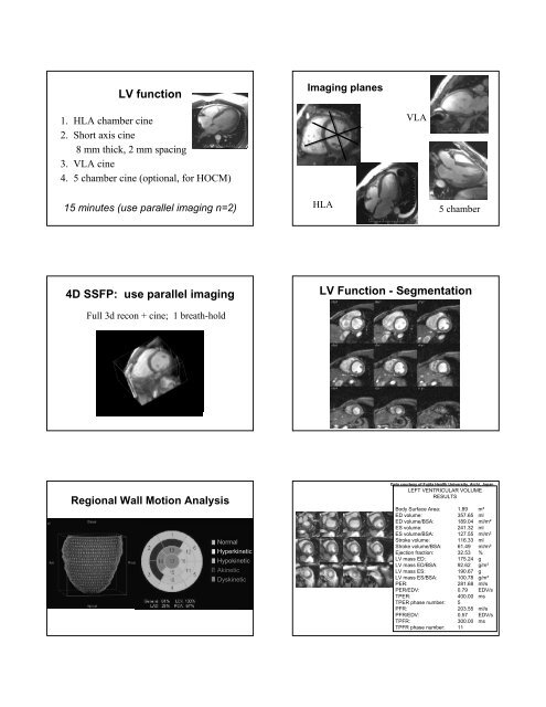
LV function<br />
<strong>Imaging</strong> planes<br />
1. HLA chamber cine<br />
2. Short axis cine<br />
8 mm thick, 2 mm spacing<br />
3. VLA cine<br />
4. 5 chamber cine (optional, for HOCM)<br />
VLA<br />
15 minutes (use parallel imaging n=2)<br />
HLA<br />
5 chamber<br />
4D SSFP: use parallel imaging<br />
LV Function - Segmentation<br />
Full 3d recon + cine; 1 breath-hold<br />
Regional Wall Motion Analysis<br />
Normal<br />
Hyperkinetic<br />
Hypokinetic<br />
Akinetic<br />
Dyskinetic<br />
Data courtesy of Fujita Health University, Aichi, Japan<br />
LEFT VENTRICULAR VOLUME<br />
RESULTS<br />
<strong>Body</strong> Surface Area: 1.89 m²<br />
ED volume: 357.65 ml<br />
ED volume/BSA: 189.04 ml/m²<br />
ES volume: 241.32 ml<br />
ES volume/BSA: 127.55 ml/m²<br />
Stroke volume: 116.33 ml<br />
Stroke volume/BSA: 61.49 ml/m²<br />
Ejection fraction: 32.53 %<br />
LV mass ED: 175.24 g<br />
LV mass ED/BSA: 92.62 g/m²<br />
LV mass ES: 190.67 g<br />
LV mass ES/BSA: 100.78 g/m²<br />
PER: 281.68 ml/s<br />
PER/EDV: 0.79 EDV/s<br />
TPER: 400.00 ms<br />
TPER phase number: 5<br />
PFR: 203.55 ml/s<br />
PFR/EDV: 0.57 EDV/s<br />
TPFR: 300.00 ms<br />
TPFR phase number: 11