Optimize Your Body MR Imaging Protocols - Johns Hopkins Radiology
Optimize Your Body MR Imaging Protocols - Johns Hopkins Radiology
Optimize Your Body MR Imaging Protocols - Johns Hopkins Radiology

You also want an ePaper? Increase the reach of your titles
YUMPU automatically turns print PDFs into web optimized ePapers that Google loves.
RC429: <strong>Optimize</strong> <strong>Your</strong> <strong>Body</strong> <strong>MR</strong> Practice:<br />
<strong>Optimize</strong> <strong>Your</strong> <strong>Body</strong> <strong>MR</strong><br />
<strong>Imaging</strong> <strong>Protocols</strong>:<br />
Cardiovascular<br />
David A. Bluemke, M.D., Ph.D.<br />
Associate Professor, Clinical Director, <strong>MR</strong>I<br />
Departments of <strong>Radiology</strong> and Medicine<br />
<strong>Johns</strong> <strong>Hopkins</strong> University School of Medicine<br />
Baltimore, Maryland<br />
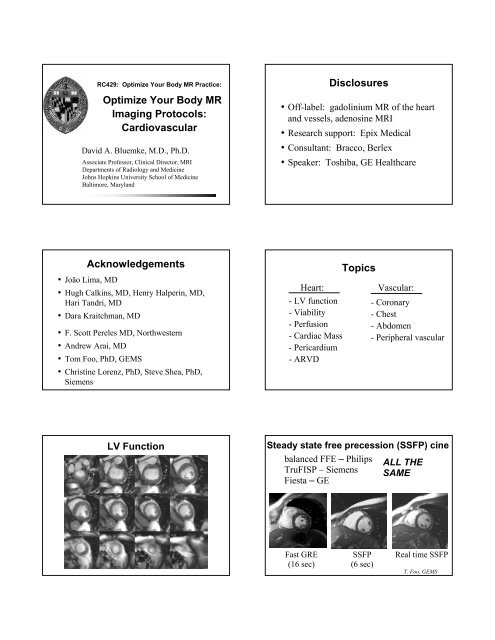
Disclosures<br />
• Off-label: gadolinium <strong>MR</strong> of the heart<br />
and vessels, adenosine <strong>MR</strong>I<br />
• Research support: Epix Medical<br />
• Consultant: Bracco, Berlex<br />
• Speaker: Toshiba, GE Healthcare<br />
Acknowledgements<br />
• João Lima, MD<br />
• Hugh Calkins, MD, Henry Halperin, MD,<br />
Hari Tandri, MD<br />
• Dara Kraitchman, , MD<br />
• F. Scott Pereles MD, Northwestern<br />
• Andrew Arai, MD<br />
• Tom Foo, , PhD, GEMS<br />
• Christine Lorenz, PhD, Steve Shea, , PhD,<br />
Siemens<br />
Heart:<br />
- LV function<br />
- Viability<br />
- Perfusion<br />
- Cardiac Mass<br />
- Pericardium<br />
-ARVD<br />
Topics<br />
Vascular:<br />
- Coronary<br />
- Chest<br />
- Abdomen<br />
- Peripheral vascular<br />
LV Function<br />
Steady state free precession (SSFP) cine<br />
balanced FFE – Philips<br />
TruFISP – Siemens<br />
Fiesta – GE<br />
ALL THE<br />
SAME<br />
Fast GRE<br />
(16 sec)<br />
SSFP<br />
(6 sec)<br />
Real time SSFP<br />
T. Foo, GEMS
LV function<br />
<strong>Imaging</strong> planes<br />
1. HLA chamber cine<br />
2. Short axis cine<br />
8 mm thick, 2 mm spacing<br />
3. VLA cine<br />
4. 5 chamber cine (optional, for HOCM)<br />
VLA<br />
15 minutes (use parallel imaging n=2)<br />
HLA<br />
5 chamber<br />
4D SSFP: use parallel imaging<br />
LV Function - Segmentation<br />
Full 3d recon + cine; 1 breath-hold<br />
Regional Wall Motion Analysis<br />
Normal<br />
Hyperkinetic<br />
Hypokinetic<br />
Akinetic<br />
Dyskinetic<br />
Data courtesy of Fujita Health University, Aichi, Japan<br />
LEFT VENTRICULAR VOLUME<br />
RESULTS<br />
<strong>Body</strong> Surface Area: 1.89 m²<br />
ED volume: 357.65 ml<br />
ED volume/BSA: 189.04 ml/m²<br />
ES volume: 241.32 ml<br />
ES volume/BSA: 127.55 ml/m²<br />
Stroke volume: 116.33 ml<br />
Stroke volume/BSA: 61.49 ml/m²<br />
Ejection fraction: 32.53 %<br />
LV mass ED: 175.24 g<br />
LV mass ED/BSA: 92.62 g/m²<br />
LV mass ES: 190.67 g<br />
LV mass ES/BSA: 100.78 g/m²<br />
PER: 281.68 ml/s<br />
PER/EDV: 0.79 EDV/s<br />
TPER: 400.00 ms<br />
TPER phase number: 5<br />
PFR: 203.55 ml/s<br />
PFR/EDV: 0.57 EDV/s<br />
TPFR: 300.00 ms<br />
TPFR phase number: 11
Cardiac <strong>Protocols</strong><br />
LV function<br />
Viability<br />
Perfusion<br />
Cardiac Mass<br />
Pericardium<br />
ARVD<br />
Viability Protocol<br />
• Purpose: evaluate delayed washout of<br />
gadolinium in infarction,<br />
inflammation, infiltrative disease<br />
Viability Protocol<br />
Time<br />
15 min 1. LV Function protocol<br />
Long, short axis cine images<br />
15<br />
2. Administer 0.15-0.2 mmol/kg<br />
gadolinium*, wait...<br />
3. TI scout<br />
{ 4. Delayed images, short and long<br />
axis, begin 10 min after gad was given<br />
Viability Protocol<br />
Alternative<br />
Time<br />
10 min 1. LV Function protocol<br />
Long axis cine images<br />
15<br />
2. Administer 0.15-0.2 mmol/kg<br />
gadolinium*,<br />
3. Short axis cines, then TI scout<br />
{ 4. Delayed images, short and long<br />
axis, begin 10 min after gad was given<br />
IR-prepared segmented fast GRE<br />
• segmentation factor: 24 OR single shot SSFP<br />
• TD: 300 ms (diastole)<br />
• TI: 200-250 ms (adjust)<br />
• 2 NEX<br />
• 8 mm thick/0 mm spacing.<br />
• acquire images ~10-20 min after 0.2 mmol/kg<br />
gadolinium, 12 hb/ slice<br />
Adjust the TI time for each patient<br />
• Optimal TI time depends on clearance of<br />
gadolinium from the normal myocardium<br />
• Typical range: 175-250 msec<br />
• Lower TI time when<br />
more gad is present:<br />
- decreased renal<br />
function<br />
-CHF
“TI Scout”<br />
“TI Scout”<br />
Single breath-hold, 50 phases,<br />
20 msec temporal resolution<br />
Images every 20<br />
msec<br />
Phase Sensitive Inversion Recovery<br />
Magnitude Reconstruction<br />
3D Viability Sequence<br />
Septal MI Antero- Septal MI<br />
TI 100 150 200 250<br />
Phase Sensitive Reconstruction<br />
TI 100 150 200 250<br />
Arai, AHA 2002<br />
Foo et al, <strong>Radiology</strong> 2004; 230:845<br />
Viability Protocol:<br />
Increasing Dyspnea<br />
14% EF<br />
EDV 210<br />
LV mass 232g<br />
RCA Infarct (old)<br />
14% EF<br />
EDV 210<br />
LV mass 232g
Hibernating<br />
Myocardium<br />
16%EF<br />
Viability Protocol: also for<br />
Nonischemic Cardiomyopathy<br />
• Hypertrophic cardiomyopathy<br />
• Myocarditis – inflammation<br />
• Amyloid<br />
• Sarcoid<br />
• Drug toxicity<br />
• Chagas disease (fibrosis)<br />
Hypertrophic Cardiomyopathy: Septum<br />
HOCM: Myocardial Fibrosis<br />
Cine<br />
Delayed contrast<br />
Pre Treatment<br />
HOCM, EtOH ablation<br />
2 mths Post Treatment<br />
Progressive RV<br />
failure<br />
Giant Cell<br />
Myocarditis<br />
Wu et al., <strong>Johns</strong> <strong>Hopkins</strong>
25 Myocarditis yo, acute chest with scar pain<br />
Cardiac <strong>Protocols</strong><br />
LV function<br />
Viability<br />
Perfusion<br />
Cardiac Mass<br />
Pericardium<br />
ARVD<br />
Adenosine Stress <strong>MR</strong>I - requirements<br />
1. Equipment<br />
Infusion pump<br />
2 IV’s (gadolinium and adenosine)<br />
2. Patient prep: withhold caffeine,<br />
methylxanthines<br />
3. Antidote (AV block, T1/2 = 2 min)<br />
(aminophylline 125 mg IV over 3 min)<br />
<strong>MR</strong>I perfusion<br />
• 0.05-0.1 mmol gad, 5<br />
ml/ sec<br />
• (Notched-interleaved)<br />
EPI-FGRE acquisition<br />
• 6-8 images / 2 R-R<br />
• 128x128 matrix<br />
• 8 mm thick, 2 mm gap<br />
• 40 phases<br />
Protocol – Stress Portion<br />
• Localize short axis:<br />
- 3 min adenosine @140<br />
ug/kg/min OR,<br />
- 2 min dipyridamole @0.56<br />
mg /kg over 4 min<br />
• 0.05 mmol/kg gadolinium bolus, 5 ml/sec<br />
• Short axis perfusion for 1 min<br />
Protocol – Rest Portion<br />
• Administer additional<br />
0.1 mmol/kg gadolinium<br />
• ~15 min delay:<br />
LV function protocol<br />
• Viability protocol<br />
• Optional: Repeat perfusion at rest, 0.1<br />
mmol/kg gadolinium @ 5 ml/sec<br />
(optional)
Saturation recovery SSFP<br />
Cardiac <strong>Protocols</strong><br />
Rest Stress<br />
LV function<br />
Viability<br />
Perfusion<br />
Cardiac Mass<br />
Pericardium<br />
ARVD<br />
}<br />
from Fenchel et al AJR 2005: 185<br />
Cardiac mass protocol<br />
1. Axial T1 images (find the mass!)<br />
2. Axial T2 images<br />
3. +/- fat suppressed T1 images<br />
4. Axial cine images<br />
5. Pre/ post gadolinium T1 images<br />
• fat sat double IR FSE (1x gadolinium) or<br />
“viability” T1 images with 2x dose<br />
gadolinium<br />
Primary benign tumors:<br />
1. Myxoma 41%<br />
2. Lipoma 14%<br />
3. Papillary fibroelastoma 13%<br />
4. Rhabdomyoma 11%<br />
(clot)<br />
Syncope, mass by echo:<br />
myxoma<br />
Emergency transfer for<br />
cardiac mass on echo<br />
Axial T1 T2<br />
Axial SSFP cine<br />
Axial T1<br />
Axial T1
Emergency transfer for cardiac<br />
mass on echo<br />
Pulmonary hypertension, RV<br />
dysfunction<br />
Axial STIR images<br />
(fat is dark, edema is bright)<br />
Axial T1 images with fat<br />
suppression<br />
cine SSFP<br />
viability image<br />
Malignant tumors:<br />
Secondary tumors 20x more common:<br />
Metastatic disease, lymphoma<br />
Primary:<br />
1. Angiosarcoma 31%<br />
2. Rhabodmyosarcoma 20%<br />
3. Other sarcoma 16%<br />
4. Mesothelioma 15%<br />
5. Primary Lymphoma 6%<br />
Leiomysarcoma metastatsis<br />
T2<br />
CHF, soft tissue mass by CT<br />
CHF, soft tissue mass by CT:<br />
angiosarcoma<br />
Axial T1<br />
Axial T2, fat sat
Pericardium - Protocol<br />
1. LV mass protocol<br />
2. Short axis cines for constriction<br />
quantitate LV/ RV function<br />
3. Axial tagging<br />
Constrictive pericarditis<br />
• ≥4mm pericardial<br />
thickness<br />
• Equalization of left/<br />
right heart pressures<br />
• Tubular right ventricle<br />
• Reduced diastolic<br />
filling<br />
• Enlarged right atrium,<br />
IVC<br />
3429369<br />
Pericardial line + mediastinal fat<br />
(chemical shift artifact)<br />
Cardiac <strong>Protocols</strong><br />
LV function<br />
Viability<br />
Perfusion<br />
Cardiac Mass<br />
Pericardium<br />
ARVD<br />
<strong>MR</strong>I tagging, axial images, stripe tags<br />
Arrhythmogenic RV Dysplasia<br />
• Fibrofatty infiltration of RV resulting in<br />
ventricular tachycardia<br />
• Palpitations, syncope, sudden death<br />
• Age 33 ± 14 yrs.<br />
• 30-50% cases are familial. <strong>MR</strong> screening<br />
of family members?<br />
RV dysplasia - Protocol<br />
1. Axial / short axis “T1” images, blood<br />
suppression (double IR FSE)<br />
- 5 mm slice thickness, ETL 24-32<br />
- Anterior coil, FOV 24-28<br />
2. same as (1), with fat suppression<br />
3. Cine: axial and short axis, HLA<br />
4. Delayed gadolinium images, form the<br />
viability protocol, axial and short axis
Black blood images<br />
• Axial “T1” images, blood/ ±fat suppression<br />
– TE min, ETL 24-32, 256x256, ZIP<br />
– 5x3 mm<br />
– Anterior coil, FOV 24-28<br />
Common protocol questions:<br />
1. What about prone imaging?<br />
• not necessary with breath-hold<br />
imaging.<br />
• difficult for patients to sustain for<br />
the duration of this protocol (45 +<br />
minutes).<br />
Common protocol questions:<br />
2. We have a double IR single shot<br />
sequence (ssfse, HASTE) that is much<br />
faster – should I use this?<br />
“Double IR” single shot (HASTE) FSE<br />
2 sec per image – do not use for heart <strong>MR</strong>I<br />
ARVD: morphology<br />
Right ventricle fat<br />
38 yo F athlete, ventricular tachycardia
RV and Pulmonary outflow<br />
tract enlarged, poor function<br />
Right ventricular aneurysm<br />
Typical ARVD<br />
Delayed Gadolinium Enhancement<br />
RV delayed enhancement<br />
• Delayed enhancement present in 8/13<br />
(61%) of ARVD patients.<br />
• 7 patients had biopsy, all showed fibrosis.<br />
• All of patients had other RV<br />
abnormalities (wall motion, morphology)<br />
Tandri, JACC 2005; 45<br />
*AICD, investigational<br />
RV delayed enhancement<br />
Topics<br />
Heart:<br />
- LV function<br />
- Viability<br />
- Perfusion<br />
- Cardiac Mass<br />
- Pericardium<br />
-ARVD<br />
•Vascular<br />
- Coronary<br />
- Chest<br />
- Abdomen<br />
- Peripheral vascular
Coronary <strong>MR</strong>A <strong>Protocols</strong><br />
VCATS: volume coronary angiography<br />
using targeted scans<br />
1. Targeted <strong>MR</strong>A (VCATS)<br />
- breath-hold 3d SSFP technique<br />
- double oblique images, oriented<br />
along the course of each coronary<br />
artery<br />
2. Whole heart coronary <strong>MR</strong>A<br />
Dirksen et al JC<strong>MR</strong> 2003 5: 365<br />
Breath-hold 3D SSFP of RCA<br />
• Advantages: quick, 20 sec, repeatable<br />
• Disadvantages: breath-hold time limits<br />
resolution, difficult at high heart rates,<br />
complex for technologist<br />
Multicenter Coronary <strong>MR</strong>A Study 1.5T:<br />
targeted <strong>MR</strong>A with navigator<br />
Aarhus Berlin Boston Leiden<br />
Köln<br />
Kim WY et al.: N Engl J Med;345(26):1863-1869 (2001).<br />
Texas Leeds Zürich<br />
Whole Heart 3d axial <strong>MR</strong>A:<br />
diaphragm tracking<br />
Whole Heart 3d <strong>MR</strong>A
Whole Heart 3d <strong>MR</strong>A<br />
Vascular <strong>Protocols</strong><br />
Coronary<br />
Chest<br />
<br />
}<br />
Abdomen<br />
Peripheral vascular<br />
Abdomen, Chest <strong>Protocols</strong><br />
Sequence Chest Abdomen<br />
3D <strong>MR</strong>A ≤ 3mm ≤ 2mm<br />
(fat suppression)<br />
Pre Ax, Sag T1 SSFSE<br />
(gated)<br />
(cysts, fluid)<br />
Post Post T1 VIBE, 3d T1 GRE<br />
(fat sat, gated) (liver, kidney, etc)<br />
<strong>MR</strong>I/A Chest: Contrast allergy<br />
History: septic emboli, cardiac failure<br />
• Black Blood:<br />
double IR<br />
breath-hold<br />
FSE<br />
• Option: single<br />
shot technique<br />
<strong>MR</strong>I/A Chest: combine with function<br />
Thrombosed Aortic Dissection<br />
Cardiac Fiesta cine<br />
3d Gad <strong>MR</strong>A<br />
“double IR” black blood FSE <strong>MR</strong>A
Aortic Dissection - intraluminal view<br />
Takayasu arteritis<br />
Takayasu arteritis<br />
Renal <strong>MR</strong>A (3T): with 3d T1<br />
T1 double fat sat, IR gad<br />
Vascular <strong>Protocols</strong><br />
Coronary<br />
Chest<br />
Abdomen<br />
Peripheral vascular<br />
Bolus<br />
Chase:<br />
Stepping<br />
Table <strong>MR</strong>A<br />
1<br />
2<br />
9 - 12 sec<br />
9 - 11 sec<br />
F. Scott Pereles MD<br />
3<br />
11-13 sec<br />
x 2 runs
Hybrid p<strong>MR</strong>A Approach<br />
3 stations BUT 2 Injections<br />
• Calf and foot station<br />
- 20 ml Gad and 2 or 3 acquisitions<br />
• Pelvis & Thigh stations with step table<br />
- 25 – 35 ml Gad bolus chase style<br />
• Improved resolution at all stations<br />
• Avoids venous contamination in the feet<br />
and calves.<br />
F. Scott Pereles MD<br />
Hybrid p<strong>MR</strong>A<br />
F. Scott Pereles MD<br />
Hybrid Approach<br />
3 stations BUT 2 Injections<br />
• 2 separate timing runs (pelvis & calves)<br />
- Axial timing run,<br />
proximal calf<br />
2 ml Gad @ 2 ml/sec (20ml<br />
saline flush @ 2ml/sec)<br />
- Axial timing run,<br />
aortic bifurcation<br />
2 ml Gad @ 2 ml/sec (20ml<br />
saline flush @ 2ml/sec)<br />
F. Scott Pereles MD<br />
Hybrid Technique<br />
Timing Run<br />
2 nd Acquisition, better<br />
visualization of foot<br />
vessels<br />
F. Scott Pereles MD<br />
Time Resolved <strong>MR</strong>A<br />
(TREAT, TRICKS)<br />
F. Scott Pereles MD<br />
Heart:<br />
- LV function<br />
- Viability<br />
- Perfusion<br />
- Cardiac Mass<br />
- Pericardium<br />
-ARVD<br />
Summary<br />
Thank you<br />
Vascular:<br />
- Coronary<br />
- Chest<br />
- Abdomen<br />
- Peripheral vascular