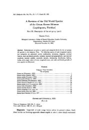
Pleistocene Gobiid Fishes of the Genus Rhinogobius
Pleistocene Gobiid Fishes of the Genus Rhinogobius
Pleistocene Gobiid Fishes of the Genus Rhinogobius

You also want an ePaper? Increase the reach of your titles
YUMPU automatically turns print PDFs into web optimized ePapers that Google loves.
Bull. Kitakyushu Mus. Nat. Hist., 7: 111-119. December 20, 1987<br />
<strong>Pleistocene</strong> <strong>Gobiid</strong> <strong>Fishes</strong> <strong>of</strong> <strong>the</strong> <strong>Genus</strong> <strong>Rhinogobius</strong><br />
from Kusu Basin, Oita Prefecture, Japan<br />
Yoshitaka Yabumoto<br />
Kitakyushu Museum <strong>of</strong> Natural History, Nishihonmachi,<br />
Kitakyushu, 805 Japan<br />
(Received August 21, 1987)<br />
Abstract Two species <strong>of</strong> <strong>Pleistocene</strong> gobiid fishes, <strong>Rhinogobius</strong> giurinus and R. brumous,<br />
were collected from <strong>the</strong> diatomite bed at Kusu Basin in Oita Prefecture, Japan. These<br />
species are identified by characters <strong>of</strong> <strong>the</strong> pelvic fin and scales. From Kusu Basin, a<br />
salmonid species, Oncorhyncus cf. 0. masou or 0. rhodums, and three cyprinid species<br />
Hemibarbus barbus, Zacco temminckii, and Acheilognathus lanceolata were reported by Uyeno et<br />
al. (1975). This is <strong>the</strong> first record <strong>of</strong> gobiid fishes which were found from this deposit.<br />
A number <strong>of</strong> fossil specimens belonging to <strong>the</strong> family <strong>Gobiid</strong>ae were collected from<br />
Nogami Member in Kusu Formation at Nogami in Kusu Basin, Oita Prefecture. This<br />
Formation and Member were first described by Shuto (1953) who made a geological<br />
survey <strong>of</strong> <strong>the</strong> upper area <strong>of</strong> Kusu River, and named it as Kusu Formation and recognized<br />
three Members: Hosenji, Nogami, and Hijiu Member in order from bottom to top. The<br />
age <strong>of</strong> Nogami Member is Middle <strong>Pleistocene</strong> (Iwauchi and Hase, 1987). Uyeno, et al.<br />
(1975) reported freshwater fish fossils from Nogami Member. These are a salmonid<br />
species, Oncorhyncus cf. 0. masou or 0. rhodums, and three cyprinid species, Hemibarbus<br />
barbus, Zacco temminckii, and Acheilognathus lanceolata.<br />
Two species <strong>of</strong> gobiid fossil fishes <strong>of</strong><br />
<strong>the</strong> genus <strong>Rhinogobius</strong> are reported here for <strong>the</strong> first time from Kusu Basin.<br />
Acknowledgments<br />
I am very grateful to Dr. Teruya Uyeno <strong>of</strong> <strong>the</strong> National Science Museum for his<br />
invaluable advice and critical reading <strong>of</strong> <strong>the</strong> manuscript. I would like to express my<br />
sincere gratitude to Mr. Akinori Takayama <strong>of</strong> <strong>the</strong> Hakusan Kogyo Corporation for <strong>the</strong><br />
donation <strong>of</strong> his specimens and his cooperation in collecting o<strong>the</strong>r specimens. I thank Mr.<br />
Tsugito Nagashima for <strong>the</strong> first information <strong>of</strong> <strong>the</strong> yielding <strong>of</strong> <strong>the</strong> specimens. I am<br />
grateful to Dr. Nobuaki Mizuno <strong>of</strong> Ehime University for <strong>the</strong> donation <strong>of</strong> some forms <strong>of</strong><br />
Recent specimens <strong>of</strong> R. brunneus, and to Mr. Muneyoshi Hayashi <strong>of</strong> <strong>the</strong> Yokosuka City<br />
Museum for information on <strong>the</strong> distribution <strong>of</strong> Recent species <strong>of</strong> <strong>the</strong> genus <strong>Rhinogobius</strong>. I<br />
am also grateful to Dr. Ryuzo Toriyama and Dr. Masamichi Ota <strong>of</strong> <strong>the</strong> Kitakyushu<br />
Museum <strong>of</strong> Natural History for <strong>the</strong>ir constant encouragement.
112 Yoshitaka Yabumoto<br />
Material:<br />
Class Osteichthyes<br />
Order Perciformes<br />
Family <strong>Gobiid</strong>ae<br />
<strong>Rhinogobius</strong> giurinus (Rutter)<br />
Specimens were collected by Akinori Takayama, Teruya Uyeno, and<br />
Yoshitaka Yabumoto between 1983-1986.<br />
KMNH (Kitakyushu Museum <strong>of</strong> Natural History) VP 100,117, almost complete<br />
specimen with dorsal side exposed, but first dorsal fin and anal fin missing. Head region<br />
well preserved. Standard length 19.8 mm and number <strong>of</strong> vertebrae 10+16=26. This<br />
specimen is identified by <strong>the</strong> form <strong>of</strong> <strong>the</strong> pelvic fin. KMNH VP 100,118, almost complete<br />
specimen with its left side exposed, but second dorsal fin missing. Scales <strong>of</strong> <strong>the</strong> posterior<br />
part <strong>of</strong> <strong>the</strong> body are well preserved. Standard length 24.8 mm and number <strong>of</strong> vertebrae<br />
10+16=26. This specimen is identified by <strong>the</strong> form <strong>of</strong> <strong>the</strong> scales. KMNH VP 100,119,<br />
almost complete specimen with ventral side exposed, but first dorsal fin, a part <strong>of</strong> 2nd<br />
dorsal fin and pelvic fin missing. Standard length 20.6 mm and number <strong>of</strong> vertebrae 10<br />
+ 16=26. This specimen is identified by <strong>the</strong> form <strong>of</strong> <strong>the</strong> scales. KMNH VP 100,120,<br />
almost complete specimen with its right side exposed, but lacking dorsal fin, a part <strong>of</strong> anal<br />
fin and some bones <strong>of</strong> head region. Standard length 26.1 mm and number <strong>of</strong> vertebrae<br />
10+16=26. This specimen is identified by <strong>the</strong> form <strong>of</strong> <strong>the</strong> pelvic fin. KMNH VP<br />
100,121, almost complete specimen with its right side exposed, but lacking pectoral fin,<br />
pelvic fin, anal fin and a part <strong>of</strong> head region. Scales are preserved on abdominal part<br />
and are detached from caudal part. Standard length 25.6 mm. This specimen is<br />
identified by <strong>the</strong> form <strong>of</strong> <strong>the</strong> scales. KMNH VP 100,122, almost complete specimen with<br />
its right side exposed. Scales are preserved. Standard length 26.0 mm and number <strong>of</strong><br />
vertebrae 10+16=26. This specimen is identified by <strong>the</strong> form <strong>of</strong> <strong>the</strong> scales. KMNH<br />
VP 100,123, anterior part <strong>of</strong> <strong>the</strong> body and caudal fin with its left side exposed, scales<br />
preserved on <strong>the</strong> part below second dorsal fin. Standard length 24.1 mm. This<br />
specimen is identified by <strong>the</strong> form <strong>of</strong> <strong>the</strong> scales. Takayama's specimen 1, almost<br />
complete specimen with its left side exposed, but lacking a part <strong>of</strong> head region and anal<br />
fin. Scales are preserved. Standard length 27.0 mm and number <strong>of</strong> vertebrae 10+16=<br />
26. This specimen is identified by <strong>the</strong> form <strong>of</strong> <strong>the</strong> scales. KMNH VP 100,124, some<br />
bones <strong>of</strong>head region, pectoral fin, dorsalspines, and scales are preserved. This specimen<br />
is identified by <strong>the</strong> form <strong>of</strong> <strong>the</strong> scales.<br />
Distinguishing characters: The branching point <strong>of</strong> <strong>the</strong> innermost pelvic fin ray is<br />
approximately in between <strong>the</strong> base and <strong>the</strong> branching point <strong>of</strong> <strong>the</strong> next ray. The<br />
posterior end <strong>of</strong> each scale is slightly pointed. The spines <strong>of</strong> each scale are short at <strong>the</strong><br />
posterior corner and gradually become longer toward <strong>the</strong> dorsal and ventral ends <strong>of</strong> <strong>the</strong><br />
posterior margin. Grooves extend close to <strong>the</strong> posterior margin <strong>of</strong> <strong>the</strong> scales.<br />
Description: Scales are ctenoid. The frontal bone is wide at <strong>the</strong> posterior part<br />
and rapidlynarrows at <strong>the</strong>anteriorpart. The sensory canalis presenton <strong>the</strong> lateral margin
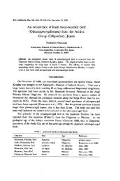
<strong>Pleistocene</strong> gobiid fishes from Oita 113<br />
preo<br />
max<br />
1mm<br />
Fig. 1. Dorsal view <strong>of</strong> head region <strong>of</strong> a <strong>Pleistocene</strong> <strong>Rhinogobius</strong> giurinus, KMNH VP 100,117.<br />
den, dentary; ecp, ectopterygoid; eth, ethmoid; exo, exoccipital; fro, frontal; max,<br />
maxillary;ope, opercle;pal, palatine; prem, premaxillary; preo, preopercle; pto, pterotic;<br />
qua, quadrate; supo, supraoccipital; sym, symplectic.<br />
<strong>of</strong> <strong>the</strong> frontal and opens at <strong>the</strong> lateral end. The melanin <strong>of</strong> <strong>the</strong> eye ball is preserved in<br />
KMNH VP 100,117 (Fig. 1). The dentary is narrow at <strong>the</strong> anterior part and deep at <strong>the</strong><br />
posterior part. Small unicuspid teeth are present on <strong>the</strong> premaxillary and <strong>the</strong> dentary<br />
(Fig. 1). The ceratohyal is narrow at <strong>the</strong> anterior part and wide at <strong>the</strong> posterior part.<br />
Four branchiostegals are attached on <strong>the</strong> ceratohyal. The epihyal is almost triangular in<br />
shape. The posterior margin <strong>of</strong> <strong>the</strong> pectoral fin is circular. O<strong>the</strong>r observable bones <strong>of</strong><br />
<strong>the</strong> head region are maxillary, quadrate, symplectic, palatine, ectopterygoid, opercle,<br />
preopercle, supraoccipital, pterotic and exoccipital (Fig. 1). The first and second and <strong>the</strong><br />
third and fourth hypurals are fused. The parhypural is short. The haemal spine <strong>of</strong> <strong>the</strong><br />
second preuralcentrum is thick and wide. O<strong>the</strong>r observable bones <strong>of</strong> <strong>the</strong> caudal region<br />
are a part <strong>of</strong> <strong>the</strong> 5th hypural and a part <strong>of</strong> <strong>the</strong> epural (Fig. 2).
114 Yoshitaka Yabumoto<br />
Fig. 2. Caudal region <strong>of</strong> a <strong>Pleistocene</strong> <strong>Rhinogobius</strong> giurinus, KMNH VP 100,122. epu, epural;<br />
hes, haemal spine <strong>of</strong> <strong>the</strong> second preuralcentrum; hyu, hypural; parh, parhypural.<br />
<strong>Rhinogobius</strong> brunneus (Temminck et Schlegel)<br />
Material: KMNH VP 100,125, almost complete specimen with its left side exposed.<br />
Standard length 26.0mm and number <strong>of</strong> vertebrae 10+16=26. This specimen is<br />
identified by <strong>the</strong> form <strong>of</strong> <strong>the</strong> pelvic fin. KMNH VP 100,126, almost complete specimen<br />
with its right side exposed. Standard length 24.3 mm and number <strong>of</strong>vertebrae 10+16=<br />
26. This specimen is identified by <strong>the</strong> form <strong>of</strong> <strong>the</strong> scales. KMNH VP 100,127, body<br />
twisted at anal origin, all elements <strong>of</strong> skeleton, fin rays and scales are detached. This<br />
specimen is identified by <strong>the</strong> form <strong>of</strong> <strong>the</strong> scales. KMNH VP 100,128, almost complete<br />
specimen with its left side exposed, but ribs, pectoral, pelvic and anal fins missing.<br />
Standard length 23.7 mm. This specimen is identified by <strong>the</strong> form <strong>of</strong> <strong>the</strong> scales.<br />
KMNH VP 100,129, almost complete specimen with its left side exposed, but pelvic fins<br />
missing. Standard length 35.8 mm and number <strong>of</strong> vertebrae 10+16=26. This speci<br />
men is identified by <strong>the</strong> form <strong>of</strong> <strong>the</strong> scales. KMNH VP 100,130, specimen with its left
<strong>Pleistocene</strong> gobiid fishes from Oita 115<br />
side exposed lacking first dorsal fin, caudal skeleton and caudal fin. This specimen is<br />
identified by <strong>the</strong> form <strong>of</strong> <strong>the</strong> scales. KMNH VP 100,131, specimen with ventral side<br />
exposed lacking caudal part. Some bones <strong>of</strong> head region preserved. This specimen is<br />
identified by <strong>the</strong> form <strong>of</strong> <strong>the</strong> scales. Takayama's specimen 2, almost complete specimen<br />
with its left side exposed, but first dorsal fin missing. Some bones <strong>of</strong> head region<br />
disarticulated. Standard length 37.5 mm and number <strong>of</strong> vertebrae 10+16=26. This<br />
specimen is identified by <strong>the</strong> form <strong>of</strong> <strong>the</strong> scales. KMNH VP 100,132, almost complete<br />
specimen with its right side exposed, but a part <strong>of</strong> <strong>the</strong> anal fin missing. Standard length<br />
37.4 mm and number <strong>of</strong> vertebrae 10+16=26. This specimen is identified by <strong>the</strong> form<br />
<strong>of</strong> <strong>the</strong> scales. KMNH VP 100,133, almost complete specimen with its left side exposed,<br />
and some bones <strong>of</strong> head region disarticulated. Standard length 27.0 mm and number <strong>of</strong><br />
vertebrae 10+16=26. This specimen is identified by <strong>the</strong> form <strong>of</strong> <strong>the</strong> scales. KMNH<br />
VP 100,134, almost complete specimen with its left side exposed, but lacking some bones<br />
<strong>of</strong> head region and a part <strong>of</strong> abdominal vertebrae. This specimen is identified by <strong>the</strong><br />
form <strong>of</strong> <strong>the</strong> scales. KMNH VP 100,135, caudal part <strong>of</strong> <strong>the</strong> body. This specimen is<br />
identified by <strong>the</strong> form <strong>of</strong> <strong>the</strong> scales. KMNH VP 100,136, anterior part <strong>of</strong> <strong>the</strong> body.<br />
This specimen is identified by <strong>the</strong> form <strong>of</strong> <strong>the</strong> scales. KMNH VP 100,137, caudal part <strong>of</strong><br />
<strong>the</strong> body, lacking upper part <strong>of</strong> <strong>the</strong> caudal skeleton.<br />
This specimen is identified by <strong>the</strong><br />
form <strong>of</strong> <strong>the</strong> scales.<br />
Distinguishing characters: The branching point <strong>of</strong> <strong>the</strong> innermost pelvic fin ray is<br />
beside <strong>the</strong> branching point <strong>of</strong> <strong>the</strong> next ray. The posterior margin <strong>of</strong> each scale is round<br />
in shape. The spines <strong>of</strong> each scale are almost equal in length and grooves extend two<br />
pro<br />
prem<br />
den<br />
max<br />
Fig.3. Ventral view <strong>of</strong> head region <strong>of</strong> a <strong>Pleistocene</strong> <strong>Rhinogobius</strong> brunneus, KMNH VP<br />
100,131. ang, angular; bra, branchiostegal; para, parashenoid; pro, prootic; o<strong>the</strong>r<br />
abbreviations see Fig. 1.
116 Yoshitaka Yabumoto<br />
thirds <strong>of</strong> <strong>the</strong> way from <strong>the</strong> anterior end.<br />
Description: Scales are ctenoid. In <strong>the</strong> fossil specimens, <strong>the</strong> observable bones <strong>of</strong><br />
<strong>the</strong> head region are maxillary, premaxillary, dentary, angular, palatine, ectopterygoid,<br />
quadrate, symplectic, frontal, parasphenoid, prootic, and branchiostegals. There are two<br />
condyles on <strong>the</strong> anterior end <strong>of</strong> <strong>the</strong> maxillary. The middle part <strong>of</strong> <strong>the</strong> maxillary is broad.<br />
Small unicuspid teeth are present on <strong>the</strong> premaxillary and <strong>the</strong> dentary. The palatine has<br />
two processes and one acetabulum at <strong>the</strong> anterior end. The posterior margin connects<br />
with <strong>the</strong> ectopterygoid which is pointed at <strong>the</strong> upper end. The lower end <strong>of</strong> <strong>the</strong><br />
ectopterygoid is wide and connects with <strong>the</strong> anterior margin <strong>of</strong> <strong>the</strong> quadrate which is<br />
approximately triangular with a long process ventrally. The antero-ventral corner <strong>of</strong> <strong>the</strong><br />
quadrate is a condyle for <strong>the</strong> angular (Fig. 3). A pair <strong>of</strong> foramina is present at <strong>the</strong> middle<br />
<strong>of</strong> <strong>the</strong> parasphenoid. The anterior quarter <strong>of</strong> <strong>the</strong> ventral surface <strong>of</strong> <strong>the</strong> parasphenoid is<br />
slightly concave for <strong>the</strong> attachment to <strong>the</strong> prevomer. The prootichas a large foramen for<br />
<strong>the</strong> facial nerve (Fig. 3). Forms <strong>of</strong> <strong>the</strong>se observable bones are similar to those <strong>of</strong> Recent<br />
R. brunneus.<br />
Remarks: In <strong>the</strong> Recent species <strong>of</strong> <strong>the</strong> genus <strong>Rhinogobius</strong>, <strong>the</strong> first pair <strong>of</strong> normal<br />
long ribs are attached to <strong>the</strong> third vertebra. The number <strong>of</strong> dorsal pterygiophores<br />
correspond to <strong>the</strong> number<strong>of</strong>dorsal fin rays. The space between <strong>the</strong> fifth and sixthspines<br />
<strong>of</strong> <strong>the</strong> first dorsal fin is much wider than spaces between o<strong>the</strong>r spines <strong>of</strong> <strong>the</strong> fin. The<br />
number <strong>of</strong> anal pterygiophores is one fewer than that <strong>of</strong>anal fin rays. The haemalspine<br />
<strong>of</strong> <strong>the</strong> first caudal vertebra is conspicuous (Fig.6). This information is useful in<br />
determining <strong>the</strong> numbers <strong>of</strong> abdominal and caudal vertebrae, and <strong>the</strong> numbers <strong>of</strong> dorsal<br />
and anal fin rays in <strong>Rhinogobius</strong> fossil material.<br />
The following characters indicate that <strong>the</strong> fossil specimens are members <strong>of</strong> <strong>the</strong><br />
teleostean fish family <strong>Gobiid</strong>ae <strong>of</strong> <strong>the</strong> order Perciformes: (1) <strong>the</strong> first and second dorsal<br />
fins are present and are notcontinuous; (2) both pelvic fins are close toge<strong>the</strong>r andsituated<br />
below <strong>the</strong> pectoral fin; (3) <strong>the</strong> body is covered with ctenoid scale; (4) <strong>the</strong> hypural bones<br />
arefused and simplified; (5) <strong>the</strong> dentary and premaxillary bear small conical teeth; (6) <strong>the</strong><br />
interorbital space is narrow. They have <strong>the</strong> following meristic characters: (1) <strong>the</strong><br />
number <strong>of</strong> abdominal vertebrae is 10 and <strong>the</strong> number <strong>of</strong> caudal vertebrae is 16; (2) <strong>the</strong><br />
first dorsal fin consists <strong>of</strong>6 spines and <strong>the</strong>second dorsal fin consists <strong>of</strong>onespine and 8 s<strong>of</strong>t<br />
rays; (3) <strong>the</strong> anal fin also consists <strong>of</strong>onespine and 8 s<strong>of</strong>t rays. According to Masuda et<br />
al. (1984), 35 species in 18 genera <strong>of</strong> gobiid fishes are found in freshwater in Japan.<br />
Among <strong>the</strong>m, <strong>the</strong> meristic characters <strong>of</strong> <strong>Rhinogobius</strong> giurinus and R. brunneus correspond to<br />
that <strong>of</strong> <strong>the</strong> fossil specimens. Threespecies <strong>of</strong><strong>the</strong> genus <strong>Rhinogobius</strong> have been reported in<br />
Japan. The fossil specimens are not <strong>Rhinogobius</strong> flumineus, because R. fiumineus has 11<br />
abdominal vertebrae. The differences between Recent R. giurinus and R. brunneus are<br />
recognized in <strong>the</strong>forms <strong>of</strong><strong>the</strong>sucking disk <strong>of</strong><strong>the</strong> pelvic fin andscales. Uyeno and Iwao<br />
(1966) described <strong>the</strong> differences in <strong>the</strong> shape <strong>of</strong> <strong>the</strong> sucking disk and <strong>the</strong> number <strong>of</strong><br />
branches <strong>of</strong> <strong>the</strong> innermost pelvic fin ray. But <strong>the</strong>se characters <strong>of</strong> <strong>the</strong> pelvic fin are not
<strong>Pleistocene</strong> gobiid fishes from Oita 117<br />
1mm<br />
CSs"^<br />
1mm<br />
Fig. 4. Pelvic fins <strong>of</strong> Recent and fossil specimens <strong>of</strong> <strong>the</strong> genus <strong>Rhinogobius</strong>. A, Recent R.<br />
giurinus; B, Recent R. brunneus; C, fossil R. giurinus, KMNH VP 100,118; D, fossil R.<br />
brunneus, KMNH VP 100,125. The lines indicate <strong>the</strong> branching points <strong>of</strong> fin rays.<br />
1mm<br />
Fig. 5. Scales <strong>of</strong> Recent and fossil specimens <strong>of</strong> <strong>the</strong> genus <strong>Rhinogobius</strong>. A and B, Recent R.<br />
giurinus; C and D, Recent R. brunneus; E, fossil R. giurinus, KMNH VP 100,118; F,<br />
fossil R. brunneus, KMNH VP 100,132.
<strong>Pleistocene</strong> gobiid fishes from Oita 119<br />
observable in <strong>the</strong> fossil specimens from Kusu Basin, because <strong>the</strong> posterior part <strong>of</strong> <strong>the</strong><br />
pelvic fin is not preserved. In Recent R. giurinus and R. brunneus, <strong>the</strong>re is a difference in<br />
<strong>the</strong> position <strong>of</strong> <strong>the</strong> branching point <strong>of</strong> <strong>the</strong> innermost ray <strong>of</strong> <strong>the</strong> pelvic fin. The branching<br />
point <strong>of</strong> <strong>the</strong> innermost ray is approximately in between <strong>the</strong> base and <strong>the</strong> branching point<br />
<strong>of</strong> <strong>the</strong> next ray in R. giurinus (Fig. 4A). The branching point <strong>of</strong> <strong>the</strong> innermost ray is<br />
beside to <strong>the</strong> branching point <strong>of</strong> <strong>the</strong> next ray in R. brunneus (Fig. 4B). This character is<br />
observable in <strong>the</strong> fossil specimens (Fig. 4C and D).<br />
In R.giurinus, <strong>the</strong> posterior end <strong>of</strong> <strong>the</strong> scale is slightly pointed and <strong>the</strong> spines are short<br />
on <strong>the</strong> posterior corner and gradually become longer toward <strong>the</strong> dorsal and ventral ends <strong>of</strong><br />
<strong>the</strong> posteriormargin. Grooves extendclose to <strong>the</strong> posteriormargin <strong>of</strong> <strong>the</strong> scale (Fig.5B).<br />
In R. brunneus, <strong>the</strong> posterior margin <strong>of</strong> <strong>the</strong> scale is round in shape and <strong>the</strong> spines are<br />
almostequal in length. Grooves extend two thirds<strong>of</strong> <strong>the</strong> way from <strong>the</strong> anterior end (Fig.<br />
5C). In R. giurinus, scales similar to those <strong>of</strong> R. brunneus occur in a ratio <strong>of</strong> approximately<br />
one to twenty (Fig. 5A). In R. brunneus, scales similar to those <strong>of</strong> R. giurinus occur in a<br />
ratio <strong>of</strong> one to five or six (Fig. 5D). Two types <strong>of</strong> scales are recognized in <strong>the</strong> fossil<br />
specimens (Fig. 5E and F). All <strong>of</strong> <strong>the</strong> observablescales in a fossil specimen are examined<br />
for identification <strong>of</strong> species. Two species, R. giurinus and R. brunneus, are recognized<br />
among fossil specimens on <strong>the</strong> bases <strong>of</strong> <strong>the</strong> characters <strong>of</strong> <strong>the</strong> pelvic fin and scales (Figs. 4C<br />
and D, 5E and F).<br />
Literature Cited<br />
Iwauchi, A. and Y. Hase. 1987. Late Cenozoic vegetation and paleoenvironment <strong>of</strong> nor<strong>the</strong>rn and<br />
central Kyushu, Japan. Part 3. Sou<strong>the</strong>rn part <strong>of</strong> Kusu Basin (Lower and Middle <strong>Pleistocene</strong>).<br />
Jour. Geol. Soc. Japan, 93 (7): 469-489, figs. 1-13, tabs. 1-2, pis. 1-2.<br />
Masuda, H., K. Amaoka, C. Araoa, T. Uyeno, and T. Yoshino (ed.). 1984. The fishes <strong>of</strong> <strong>the</strong><br />
Japanese Archipelago. Text. Tokai Univ. Press, xxii+437 pp., 245 figs.<br />
Shuto, T. 1953. Younger Cenozoic history <strong>of</strong> Oita District, Kyushu (I). Jour. Geol. Soc. Japan, 59<br />
(693): 225-240, figs. 1-8, tabs. 1-6.<br />
Uyeno, T., S. Kimura, and Y. Hasegawa. 1975. Freshwater fishes from Late Cenozoic deposits in<br />
Kusu Basin, Oita Prefecture, Japan. Mem. Natn. Sci. Mus. (8): 57-66, fig. 1, tabs. 1-2, pis. 6-9.<br />
Uyeno, T. and Y. Iwao. 1975. Late Cenozoic gobiid fish from Togo Formation in Kagoshima<br />
Prefecture, Japan. Bull. Natn. Sci. Mus., Ser. C (Geol.), 1 (2): 55-60, figs. 1-2, pis. 1-2.
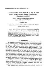
<strong>Pleistocene</strong> <strong>Gobiid</strong> <strong>Fishes</strong> <strong>of</strong> <strong>the</strong> <strong>Genus</strong> <strong>Rhinogobius</strong><br />
from Kusu Basin, Oita Prefecture, Japan.<br />
Yoshitaka Yabumoto<br />
Plates 1-2
Explanation <strong>of</strong> Plate 1.<br />
Photographs <strong>of</strong> <strong>the</strong> fossils <strong>of</strong> <strong>Rhinogobius</strong> giurinus (Rutter) from Kusu<br />
Basin, Oita Prefecture.<br />
A. lateral view <strong>of</strong> <strong>the</strong> left side (KMNH VP 100,118).<br />
B. ventral view (KMNH VP 100,119).<br />
C dorsal view (KMNH VP 100,117).<br />
Scales indicate 10 mm.
Yabumoto, Y. <strong>Pleistocene</strong> gobiid fishes from Oita Plate 1<br />
wtmm...<br />
B<br />
C
Plate2<br />
I ;•••; !
Explanation <strong>of</strong> Plate 2.<br />
Photographs <strong>of</strong> <strong>the</strong> fossils <strong>of</strong> <strong>Rhinogobius</strong> brunneus (Temminck et Schlegel)<br />
from Kusu Basin, Oita Prefecture.<br />
A. lateral view <strong>of</strong> <strong>the</strong> left side (KMNH VP 100,125).<br />
B. lateral view <strong>of</strong> <strong>the</strong> left side (KMNH VP 100,129).<br />
C lateral view <strong>of</strong> <strong>the</strong> right side (KMNH VP 100,132).<br />
Scales indicate 10 mm.
Yabumoto, Y. <strong>Pleistocene</strong> gobiid fishes from Oita Plate 2<br />
B<br />
irp