A revision of the genus Deltote RL and its allied genera from Japan ...
A revision of the genus Deltote RL and its allied genera from Japan ...
A revision of the genus Deltote RL and its allied genera from Japan ...

Create successful ePaper yourself
Turn your PDF publications into a flip-book with our unique Google optimized e-Paper software.
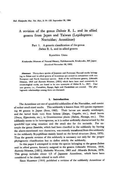
Bull. Kitakyushu Mus. Nat. Hut., 5: 91-133. September20, 1984<br />
A <strong>revision</strong> <strong>of</strong> <strong>the</strong> <strong>genus</strong> <strong>Deltote</strong> R. L. <strong>and</strong> <strong>its</strong> <strong>allied</strong><br />
<strong>genera</strong> <strong>from</strong> <strong>Japan</strong> <strong>and</strong> Taiwan (Lepidoptera:<br />
Noctuidae; Acontiinae)<br />
Part 1. A generic classification <strong>of</strong> <strong>the</strong> <strong>genus</strong><br />
<strong>Deltote</strong> R. L. <strong>and</strong> <strong>its</strong> <strong>allied</strong> <strong>genera</strong><br />
Kyoichiro Ueda<br />
Kitakyushu Museum <strong>of</strong> Natural History, Nishihonmachi, Kitakyushu, 805 <strong>Japan</strong><br />
(Received November 30, 1983)<br />
Abstract Thirty-three species <strong>of</strong><strong>Japan</strong>ese <strong>and</strong> Formosan Noctuid moths belong<br />
ing to <strong>Deltote</strong> <strong>and</strong> <strong>its</strong> <strong>allied</strong> <strong>genera</strong> <strong>of</strong> Acontiinae are revised in comparison with two<br />
European <strong>and</strong> North American species. Both <strong>of</strong> <strong>the</strong> well-known <strong>genera</strong>, Lithacodia<br />
Hubner, 1818 <strong>and</strong> Eustrotia Hubner, [1821] which have been used extensively in<br />
entomological works, arc found to be new synonyms <strong>of</strong> <strong>Deltote</strong> R. L., 1817. Four<br />
new <strong>genera</strong>, i.e., Protodeltote, Koyaga, Sugia <strong>and</strong> Pseiidodeltote are erected. The phylogenetic<br />
relationships among <strong>the</strong>m arc discussed.<br />
1. Introduction<br />
The Acontiinae are one<strong>of</strong> quadrifid subfamilies <strong>of</strong> <strong>the</strong> Noctuidae, <strong>and</strong> consist<br />
<strong>of</strong> ra<strong>the</strong>r small-sized moths. This subfamily is known <strong>from</strong> 105species represent<br />
ing 44 <strong>genera</strong> in <strong>Japan</strong> (Suci, 1982). Their larvae are usually semilooper,<br />
<strong>and</strong> <strong>the</strong> larval foods vary <strong>from</strong> lichens (Enispa, Corgatha, etc.), dead leaves<br />
(Oruza, Hyperstrotia, etc.), to Graminaceous plants (<strong>Deltote</strong>, Naranga, etc.). This<br />
subfamily seems to be heterogenous, as it is ra<strong>the</strong>r artficially characterized by <strong>the</strong><br />
quadrifid type wing venation <strong>and</strong> <strong>the</strong> small size for <strong>the</strong> noctuids. For ex<br />
ample <strong>the</strong> <strong>genus</strong> Stenoloba, which had been classified in <strong>the</strong> subfamily by having<br />
<strong>the</strong> above-mentioned two characters, wasrecently transferred <strong>from</strong> thissubfamily<br />
to <strong>the</strong> subfamily Bryophilinae mainly based on <strong>the</strong> larval structure (Sugi, 1970).<br />
Thus <strong>the</strong> generic <strong>revision</strong> <strong>of</strong> this subfamily is necessary not only for <strong>the</strong> proper<br />
phylogenetic classification but to define strictly <strong>the</strong> subfamily.<br />
In this paper I attempted to revise <strong>the</strong> species belonging to <strong>the</strong> <strong>genus</strong> <strong>Deltote</strong><br />
<strong>and</strong> <strong>its</strong> <strong>allied</strong> <strong>genera</strong>, formerly assigned to <strong>the</strong> <strong>genera</strong> Lithacodia Hubner, 1818,<br />
Eustrotia Hubner, [1821], Maliattha Walker, 1863 <strong>and</strong> Micardia Butler, 1878.<br />
This group includes about 1/3 <strong>of</strong> <strong>Japan</strong>ese Acontiinae, which have been<br />
considered to be closely related to each o<strong>the</strong>r.<br />
Since Hampson (1910) published a <strong>revision</strong> <strong>of</strong> <strong>the</strong> subfamily Acontiinae <strong>of</strong>
92 Kyoichiro Ueda<br />
Ozarba Amyna Lithacodia {Maliattha)<br />
Eustrotia (Micardia)<br />
Eulocastra<br />
Naranga<br />
Fig. 1.<br />
Phylogeny <strong>of</strong>Acontiinae by Hampson (1910) (part).<br />
<strong>the</strong> world, <strong>the</strong> <strong>genera</strong> Lithacodia, Eustrotia, Maliattha <strong>and</strong> Micardia have been consid<br />
ered to be closely related to each o<strong>the</strong>r (Fig. 1). Although Hampson (1910) treated<br />
<strong>the</strong> <strong>genus</strong> Maliattha as one <strong>of</strong>three sections <strong>of</strong>Lithacodia <strong>and</strong> <strong>the</strong> <strong>genus</strong> Micardia<br />
as one <strong>of</strong> <strong>the</strong> synonyms <strong>of</strong> Eustrotia, Warren (1912) regarded <strong>the</strong>se two <strong>genera</strong><br />
distinct <strong>from</strong> Lithacodia <strong>and</strong> Eustrotia, respectively. The Warren's system has been<br />
adopted by <strong>Japan</strong>ese authors (Inoue <strong>and</strong> Sugi, 1958 <strong>and</strong> o<strong>the</strong>rs). Recently<br />
Sugi (1982) used <strong>the</strong> name <strong>Deltote</strong> bankiana (Fabricius) for <strong>the</strong> species hi<strong>the</strong>rto<br />
known as Eustrotia olivana (Fabricius) following Nye (1975), <strong>and</strong> Pseudeustrotia<br />
Warren, 1913 for <strong>the</strong> c<strong>and</strong>idula group following Warren (1913), <strong>and</strong> erected two<br />
new <strong>genera</strong>, Neustrotia for <strong>the</strong> japonica group, <strong>and</strong> Inabaia for Eustrotia culta.<br />
<strong>Japan</strong>ese species <strong>of</strong><strong>the</strong>se <strong>genera</strong> have been described orrecorded by Butler<br />
(1878, 1881, 1885), Leech (1889, 1900), Hampson (1910), Wileman (1911),<br />
Warren (1912, 1913) <strong>and</strong> Sugi (1958, 1959, 1982). Thus <strong>the</strong>se 8'<strong>genera</strong> are<br />
known by 30 species in<strong>Japan</strong> at present.<br />
These species are typical small Noctuid moths. Most <strong>of</strong> <strong>the</strong>m are common<br />
in <strong>Japan</strong> <strong>and</strong> distributed <strong>from</strong> Hokkaido to Amami Is. inhabiting plaines or hills<br />
covered with various Graminaceous plants. Some <strong>of</strong> <strong>the</strong>m were recorded as<br />
rice-pests (Kawada et al., 1959). Most <strong>of</strong> <strong>the</strong>m are bivoltine judging <strong>from</strong><br />
<strong>the</strong>ir records <strong>of</strong>collection, but <strong>the</strong>ir early stages remain unknown except for some<br />
species (Kawada et al., 1959; Nakatomi, 1959; Yamamoto, 1977, etc.).<br />
In this paper, I tried to clarify <strong>the</strong> intra- <strong>and</strong> intergeneric relationships <strong>of</strong>37<br />
known <strong>and</strong> 3new species <strong>of</strong> <strong>the</strong> <strong>Deltote</strong> group <strong>from</strong> <strong>Japan</strong> <strong>and</strong> Taiwan based mainly<br />
on <strong>the</strong> comparative morphology <strong>of</strong><strong>the</strong> male <strong>and</strong> female genitalia.<br />
In <strong>the</strong> course <strong>of</strong> this study, Nearctic Lithacodia bellicula Hubner (<strong>the</strong> typespecies<br />
<strong>of</strong> Lithacodia), Palaearctic Eustrotia uncula (Clerk) (<strong>the</strong> type-species <strong>of</strong><br />
Eustrotia) <strong>and</strong> <strong>Deltote</strong> bankiana (Fabricius) (<strong>the</strong> type-species <strong>of</strong> <strong>Deltote</strong>) are con-
Revision <strong>of</strong> <strong>the</strong> <strong>genus</strong> <strong>Deltote</strong> R. L. 93<br />
sidered to belong to <strong>the</strong> same monophyletic <strong>genus</strong>, toge<strong>the</strong>r with <strong>the</strong> Palaearctic<br />
Lithacodia deceptoria (Scopoli) <strong>and</strong> Lithacodia nemorum (Oberthur). Therefore,<br />
<strong>Deltote</strong>, <strong>the</strong> oldest available name, should be used for <strong>the</strong> group, <strong>and</strong> consequently,<br />
<strong>the</strong> well-known Lithacodia <strong>and</strong> Eustrotia are sunkas newsynonyms <strong>of</strong> <strong>Deltote</strong>.<br />
The inclusion <strong>of</strong><strong>the</strong> type-species <strong>of</strong>Lithacodia <strong>and</strong> Eustrotia in <strong>Deltote</strong> necessi<br />
tated <strong>the</strong> generic classification <strong>of</strong> many species hi<strong>the</strong>rto assigned to Lithacodia <strong>and</strong><br />
Eustrotia to be revised, so I erected <strong>the</strong> new <strong>genera</strong>, Protodeltote, Koyaga, Sugia <strong>and</strong><br />
Pseudodeltote for some <strong>of</strong> <strong>the</strong>m.<br />
According to <strong>the</strong> Hennig's principles <strong>of</strong>reconstruction <strong>of</strong>phylogeny (1966),<br />
I attempted to establish <strong>the</strong> phylogenetic relationships <strong>of</strong> 35 species under 8<br />
<strong>genera</strong> based on <strong>the</strong> morphological characters.<br />
2. Acknowledgements<br />
1 express my cordial thanks to Pr<strong>of</strong>essor Yoshihiro Hirashima <strong>and</strong> Associate<br />
Pr<strong>of</strong>essor Katsura Morimoto <strong>of</strong> Kyushu University for <strong>the</strong>ir valuable criticism<br />
<strong>and</strong> constant encouragement during this study. My cordial thanks are also<br />
due to Pr<strong>of</strong>essor Ryuzo Toriyama, Dr. Masamichi Ota <strong>and</strong> Emeritus Pr<strong>of</strong>essor<br />
Takashi Shirozu <strong>of</strong> Kitakyushu Museum <strong>of</strong> Natural History, Pr<strong>of</strong>essors Toyohei<br />
Saigusa <strong>and</strong> Akinori Nakanishi <strong>of</strong> Kyushu University, Pr<strong>of</strong>essor Hiroshi Inoue<br />
<strong>of</strong> Otsuma Woman's University, Mr. Shigero Sugi, Tokyo, <strong>and</strong> Pr<strong>of</strong>essor Kqji<br />
Yano <strong>of</strong> Yamaguchi University for <strong>the</strong>ir useful suggestions <strong>and</strong> lending me<br />
valuable materials <strong>and</strong> literatures. Pr<strong>of</strong>essor T. Saigusa kindly read this manu<br />
script throughly <strong>and</strong> gave me critical comments.<br />
I also express my hearty thanks to <strong>the</strong> following entomologists for <strong>the</strong>ir kind<br />
help <strong>and</strong> <strong>of</strong>fering valuable materials: Drs. Yoshihiko Kurosawa <strong>and</strong> Mamoru<br />
Owada (National Science Museum, Tokyo), Dr. Isoko Hattori (National Insti<br />
tute <strong>of</strong>Agricultural Science, Ibaraki), Dr. Yutaka Yoshiyasu (Kyoto Prefectural<br />
University), Pr<strong>of</strong>. Hiroshi Shima, Messrs. Osamu Yata, Masaru Yamamoto,<br />
Kenji Ohara, Kenzou Yamagishi, Nobutoyo Koda <strong>and</strong> Tadao Goto (Kyushu<br />
University), Dr. Furumi Komai, Mr. Kyoichi Tanaka <strong>and</strong> Dr. Satoshi<br />
Hashimoto (University <strong>of</strong> Osaka Prefecture), Pr<strong>of</strong>. Yutaka Arita (Meijo Uni<br />
versity), Mr. Jun Emoto (Nanzan University), Mr. Ban Tanaka (Nagoya), Mr.<br />
Izumi Ochiai (Tochigi), Mr. Hideaki Fuse (Fujioka), Dr. Rikio Sato <strong>and</strong> Mr.<br />
Akio Seino (Niigata), Mr. Hiroshi Endo (Gifu), Mr. Kqji Setoya (Beppu), Mr.<br />
Tadashi Kawamura (Kitakyushu) <strong>and</strong> Mr. Naomi Gyotoku (Fukuoka).<br />
My thanks are extended to Mr. Hisakatsu Minei <strong>and</strong> Pr<strong>of</strong>essor Takeshi<br />
Kawarabata <strong>of</strong>Kyushu University for <strong>the</strong>irkind advices for taking photographs.<br />
Finally but not least I wish to express my hearty thanks to Dr. D. F.<br />
Hardwick <strong>and</strong> Mr. E. Rockburne (Biosystematics Research Institute, Agriculture<br />
Canada, Ottawa), Dr. E. S. Nielsen (Division <strong>of</strong> Entomology, CSIRO,
94 Kyoichiro Ueda<br />
Canberra), Dr. W. Hogenes (Institute voor Taxonomische Zoologie, Amsterdam),<br />
Drs. D. R. Davis <strong>and</strong> J. B. Heppner (National Museum <strong>of</strong> Natural History,<br />
Smithsonian Institution Washington, D. C), Dr. G. S. Robinson <strong>and</strong> Miss. R<br />
Gilbert (British Museum <strong>of</strong> Natural History) <strong>and</strong> Dr. Kauri Mikkola (Zoological<br />
Museum <strong>of</strong> <strong>the</strong> Helsinki University, Helsinki) for <strong>the</strong>ir kindness in giving me <strong>the</strong><br />
opportunities to examine <strong>the</strong>ir valuable specimens <strong>and</strong> literatures.<br />
3. Character states <strong>and</strong> phylogenetic considerations<br />
In <strong>the</strong> classification <strong>of</strong> Acontiinae, <strong>the</strong> characters used in <strong>the</strong> grouping <strong>of</strong><br />
<strong>genera</strong> or species have been restricted to venation, wing patterns, <strong>the</strong> number <strong>of</strong><br />
dorsal crests <strong>and</strong> shape <strong>of</strong>labial palpus (Hampson, 1910). Infact, <strong>the</strong>se characters<br />
are very useful ones, because we could observe <strong>the</strong>se character states without<br />
dissection. However, <strong>the</strong>se characters have ra<strong>the</strong>r limited phylogenetic significance,<br />
as we could easily find many examples <strong>of</strong> individual variations <strong>and</strong> parallelism<br />
on <strong>the</strong>se characters, i.e., <strong>the</strong> individual variation on <strong>the</strong> venation <strong>from</strong> <strong>the</strong> areole<br />
on fore wing (Fig. 2), <strong>the</strong> same number <strong>of</strong> dorsal crests among many <strong>genera</strong>,<br />
which are apparently not related to each o<strong>the</strong>r, <strong>and</strong> so on. Consequently, each<br />
<strong>genus</strong> <strong>of</strong>Acontiinae will be <strong>of</strong>ten found to embrace heterogeneous elements in<strong>the</strong>m.<br />
Therefore I reexamined <strong>the</strong>se character states according to <strong>the</strong> criteria men<br />
tioned below to make tentative judgement <strong>of</strong> each character state-plesiomorphic<br />
or apomorphic. In addition to characters used in Hampson's classification I<br />
examined <strong>the</strong> structure <strong>of</strong> male external genitalia, <strong>the</strong>ir musculature <strong>and</strong> <strong>the</strong><br />
structure <strong>of</strong>female genitalia, because <strong>the</strong>se structures have been used in systematics<br />
<strong>of</strong>Lepidoptera <strong>and</strong> <strong>the</strong>ir validity has been found in many works.<br />
Fig. 2.<br />
Individual variations on <strong>the</strong> venations arising <strong>from</strong> <strong>the</strong> areole <strong>of</strong>fore wing<br />
{Koyaga falsa (Butler)).
Revision <strong>of</strong> <strong>the</strong> <strong>genus</strong> <strong>Deltote</strong> R. L. 95<br />
In <strong>the</strong> course <strong>of</strong> this examination, I used <strong>the</strong> following criteria for judgement<br />
<strong>of</strong> character state, if it is apomorphic or plesiomorphic (Saigusa, 1980).<br />
d. Criteria based on <strong>the</strong> distribution patterns <strong>of</strong> each character state in<br />
recent species.<br />
Criteria based on <strong>the</strong> conditions <strong>of</strong> distribution <strong>of</strong> character states within<br />
<strong>the</strong> group in question.<br />
(1) If a character state <strong>of</strong> a morphocline is more frequently distributed in<br />
"primitive" subgroups than in "derived" subgroups, this character state is primi<br />
tive. The "primitive" subgroups are those which have more character states<br />
presumed to be primitive based on o<strong>the</strong>r criteria than o<strong>the</strong>r subgroups (<strong>and</strong><br />
moreover are "phylogenetically" isolated), or those less differentiated in sexual<br />
dimorphism.<br />
Criteria based on <strong>the</strong> distribution <strong>of</strong> character states in "related groups".<br />
(4) It is primitive extreme, when a character state is more frequently distributed<br />
in "primitive" subgroups than "derived" subgroups within <strong>the</strong> "related group".<br />
(5) It is primitive extreme, when a character state ismore frequently distributed<br />
in "primitive related groups".<br />
(6) It is primitive extreme, when a character state is distributed in various or<br />
many related groups.<br />
f. Criteria peculiar to <strong>the</strong> comparative morphology<br />
(3) Criterion based on symmetry<br />
In <strong>the</strong> structure where symmetry is definitive, asymmetrization <strong>of</strong> <strong>the</strong> bilateral<br />
character, distortion by twisting <strong>of</strong> plane <strong>of</strong> symmetry, sinuation or spiralling<br />
caused by elongations in internal organs, etc. are considered to be deviation <strong>from</strong><br />
symmetry, <strong>and</strong> <strong>the</strong>y are inferred as apomorphic character states.<br />
3-1-1. Hairs <strong>of</strong> antenna.<br />
In <strong>genera</strong>l, <strong>the</strong> male antenna <strong>of</strong> Acontiinae, which were examined in this<br />
paper, has minute hairs on each segment <strong>of</strong> flagellum. The hairs <strong>of</strong> Koyaga are<br />
distinctly longer than those <strong>of</strong> any o<strong>the</strong>r <strong>genera</strong> <strong>and</strong> this condition apparently<br />
shows an apomorphic state <strong>of</strong> this character. The male antenna <strong>of</strong> <strong>Deltote</strong> is<br />
plesiomorphic condition as well as o<strong>the</strong>r <strong>genera</strong>. The female antenna <strong>of</strong> Koyaga<br />
has short hairs as in those <strong>of</strong> o<strong>the</strong>r <strong>genera</strong>, which are usually sparser <strong>and</strong> shorter<br />
than those <strong>of</strong> male antenna.<br />
3-1-2. Wing patterns.<br />
Many systems have been proposed to explain <strong>the</strong> wing patterns <strong>of</strong> <strong>the</strong> Noc<br />
tuidae mainly concerning <strong>the</strong> fore wing (Herrich-Schafter, 1843-1856,<br />
Forbes, 1954, Heath, 1976, Sergent, 1976 etc.). In this paper, <strong>the</strong> system <strong>of</strong><br />
Heath (1976) is adopted for description, because his system is <strong>the</strong> most compre<br />
hensive <strong>and</strong> useful one. I made <strong>the</strong> following minor changes in <strong>the</strong> system, i.e.,
96<br />
Kyoichiro Ueda<br />
Costa) spots<br />
Cilia<br />
Orbicular<br />
stigma<br />
1 Terminal<br />
line<br />
Antemedial<br />
line Clavifonn Postmedial<br />
stigma line<br />
Fig. 3-A. Terminology <strong>of</strong> wing pattern used in this paper (Protodeltote distinguenda<br />
(Staudinger)).<br />
fascia <strong>and</strong> basal spot arc changed to line <strong>and</strong> basal line respectively, <strong>and</strong> median<br />
shade <strong>and</strong> costal spot were newly added (Fig. 3-A). The elements <strong>of</strong> markings<br />
in <strong>the</strong> system are as follows:<br />
Basal line: A very short patch at wing base, usually absent in <strong>the</strong> species <strong>of</strong><br />
<strong>Japan</strong>ese Acontiinae.<br />
Subbasal line: Ashort <strong>and</strong> more or less sinous line <strong>from</strong> costa to inner margin<br />
near subbasal portion <strong>of</strong> wing.<br />
Antemedial line: A sinuous, usually double linesrunning <strong>from</strong> costa to inner<br />
margin a little distad to subbasal line; sometimes this line interruped or absent.<br />
Orbicular stigma: A small annulus or lunulc at <strong>the</strong> middle <strong>of</strong> cell.<br />
Claviform stigma: A small lunulc below vein 2.<br />
Medial line: Usually absent; if this line is present, it is represented by an<br />
excurved weak line <strong>from</strong> lower angle <strong>of</strong> cell to inner margin.<br />
Reniform stigma: Large annulus surrounding discocellular <strong>and</strong> usually<br />
constircted at <strong>the</strong> middle.<br />
Postmedial line: A distinct line, usually represented by double dark lines<br />
<strong>and</strong> filled with lighter (<strong>of</strong>ten white) interspace; this line running <strong>from</strong> <strong>the</strong> middle<br />
<strong>of</strong> costa, angled outwards at vein 7, minutely waved to vein 4, <strong>the</strong>n incurved to<br />
inner margin.<br />
Costal spots: Small light spots on costa between postmedial line <strong>and</strong> subterminal<br />
line.<br />
Subterminal line:<br />
sometimes excurved below vein 7 or vein 4.<br />
A sinuous line <strong>from</strong> costa to inner margin near termen <strong>and</strong>
Revision <strong>of</strong> <strong>the</strong> <strong>genus</strong> <strong>Deltote</strong> R. L. 97<br />
10 1112<br />
Fig. 3-B.<br />
Inferred basic plan <strong>of</strong> <strong>the</strong> wing pattern in Noctuidae.<br />
loilr-4? 10 » 12<br />
12 Fig. 4. Fore wing patterns <strong>of</strong> Notodontidac<br />
<strong>and</strong> Noctuidae, which are tentatively<br />
analysed compared with <strong>the</strong> basic plan<br />
(Fig. 3-B). A: Neocerura tattakmui<br />
(Matsumura). B: Centra vinula felina<br />
Butler. C: Trichosea champa (Moore).<br />
D: Polia goliath Oberthur. E: Amphipoea<br />
ussuriensis (Pertersen). Large arrows<br />
indicate <strong>the</strong> 2nd cells where two<br />
separate striae arc still present.
98 Kyoichiro Ueda<br />
11 12<br />
8 9<br />
Fig. 5. Fore wing patterns <strong>of</strong> Acontiinae. which arc tentatively analysed compared with<br />
basic plan (Fig. 3-B). A: Sugia idiostygia (Sugi). B: Koyaga falsa (Butler). C:<br />
Maliattha signifera (Walker). D: Maliattha arefacta (Butler). E: Protodeltote wiscotti<br />
(Staudinoer). F: <strong>Deltote</strong> uncula (Clerk). G: Erastroidesfenloni (Butler).
Revision <strong>of</strong> <strong>the</strong> <strong>genus</strong> <strong>Deltote</strong> R. L. 99<br />
Terminal line: Usually represented by a series <strong>of</strong>striae on <strong>the</strong> termen.<br />
The basic plan <strong>of</strong>fore wing pattern <strong>of</strong>Noctuidae is tentatively inferred as<br />
Fig. 3-B. This system consists <strong>of</strong> twelve lines. Reniform stigma <strong>and</strong> terminal<br />
series <strong>of</strong> striae are treated as "line" (7th <strong>and</strong> 12th line, respectively) for <strong>the</strong><br />
present. This system is based on <strong>the</strong> wing patterns <strong>of</strong> Neocerura tattakana (Fig.<br />
4-A), Centra vinulafelina (Fig. 4-B), Trichosea champa (Fig. 4-C), Polia goliath (Fig.<br />
4-D), <strong>and</strong> so on. These wing patterns are primitive ones, because two separate<br />
striae are still present on <strong>the</strong> terminal portion <strong>of</strong>2nd cell (arrows in Fig. 4). In<br />
<strong>genera</strong>l, <strong>the</strong> wing patterns <strong>of</strong> 2nd cell tend to be fused with each o<strong>the</strong>r <strong>and</strong> to be<br />
complete line orpatch after <strong>the</strong> loss <strong>of</strong> vein Ic (Cup). Moreover, most <strong>of</strong><strong>the</strong> lines<br />
(4th-6th, 8th-l 1th) could be well traced <strong>from</strong> costa to inner margin. Therefore<br />
<strong>the</strong>se wing patterns are considered as primitive ones <strong>and</strong> <strong>the</strong> basic plan is<br />
inferred as Fig. 3-B.<br />
Thefore wing patterns <strong>of</strong>Fig. 5 (A-G) are <strong>the</strong> results applying this system to<br />
some <strong>of</strong>species in this study. The most primitive condition seems to be preserved<br />
in Protodeltote distinguenda (Fig. 3-A) <strong>and</strong> Sugia idiostygia (Fig. 5-A), because each<br />
line in basic plan could be traced except for 10th <strong>and</strong> 11th lines. Median shade<br />
is considered to be <strong>the</strong> enlargement <strong>of</strong> <strong>the</strong> 6th line in <strong>the</strong> discoidal cell. But,<br />
<strong>the</strong>se two species are not closely related to each o<strong>the</strong>r judging <strong>from</strong> <strong>the</strong>ir male<br />
external <strong>and</strong> female genital structures. Then, this apparent resemblance on fore<br />
wingpatterns is due to symplesiomorphy.<br />
In Koyaga faba (Fig. 5-B), <strong>the</strong> wing patterns are almost same as in <strong>the</strong> above<br />
two species. But <strong>the</strong> so-called medial line is distinct <strong>and</strong> broad. This medial line<br />
seems to be composed <strong>of</strong><strong>the</strong> union <strong>of</strong>a part <strong>of</strong>6th line below vein 3 <strong>and</strong> a part <strong>of</strong><br />
8th line below vein 3, <strong>and</strong> is strongly curved inwards <strong>and</strong> shifted basally <strong>from</strong> <strong>its</strong><br />
normal position.<br />
In Maliattha, 6th line tends tobedivided into two lines (longitudinal subdivi<br />
sion) (Fig. 5-C, D). In M. arefacta 5th <strong>and</strong> 6th lines run inwards to inner<br />
margin in <strong>the</strong> contrast with <strong>the</strong>ir primitive conditions (Fig. 5-F).<br />
In <strong>the</strong> wing patterns <strong>of</strong> Protodeltote wiscotti <strong>and</strong> <strong>Deltote</strong> uncula (Fig. 5-E, F),<br />
8th line below vein 6 (in <strong>the</strong> latter below vein 7) is distinctive; this line is almost<br />
straight to inner margin. This condition is also observed in Micardia argentata<br />
(Fig. 29-D). However, <strong>the</strong>se species are not closely related to each o<strong>the</strong>r judging<br />
<strong>from</strong> <strong>the</strong>ir genital structures, <strong>and</strong> <strong>the</strong>se resemblances will be due to <strong>the</strong> parallelism.<br />
In Erastroides fentoni* <strong>the</strong> simplification <strong>of</strong> wing pattern is characteristic.<br />
Most <strong>of</strong> <strong>the</strong> patterns are substituted by orange scales.<br />
As shortly discussed above, <strong>the</strong> considerable modifications could be observed<br />
* As Suoi (1982) suggested, <strong>the</strong>systematic postition <strong>of</strong> this species is indistinct, <strong>and</strong> here I ten<br />
tatively assigned it to <strong>the</strong> <strong>genus</strong> Erastroides Hampson because <strong>of</strong> <strong>the</strong> lack <strong>of</strong> <strong>the</strong> areole on <strong>the</strong><br />
fore wing.
100 Kyoichiro Ueda<br />
in <strong>the</strong> wing patterns <strong>of</strong> <strong>Deltote</strong> <strong>and</strong> <strong>its</strong> <strong>allied</strong> <strong>genera</strong>, <strong>and</strong> more examinations are<br />
needed to trace <strong>the</strong> transformation series <strong>of</strong>each wing pattern <strong>and</strong> to discuss <strong>the</strong><br />
phylogenetic relationships <strong>of</strong> each species based on <strong>the</strong> wing patterns. There<br />
fore I used Heath's system in descriptions <strong>of</strong>each species in <strong>the</strong> present work.<br />
3-1-3. Male external genital structure.<br />
The terms <strong>of</strong>each structure <strong>of</strong><strong>the</strong> male external genitalia are mainly based<br />
on <strong>the</strong> work <strong>of</strong>Sibatani etal. (1954-1957). This system has been used frequently<br />
by many authors <strong>and</strong> is most useful to describe a small but important structure.<br />
I added to this system <strong>the</strong>process <strong>of</strong>uncus in this paper.<br />
Fig. 6. Diagrams showing musculature <strong>of</strong> male external genitalia. A: Lateral view (left<br />
valva removed). B: Frontal view. A; anus, A. p.; anal plate, Co.; costa, De.;<br />
ductus ejaculatorius, Fe.; fenestrula, Hrp.; harpe, Jx.; juxta, Ph.; phallus,<br />
Scl.; sacculus, Tg.; tegumen, Un.; uncus, Vin.; vinculum.<br />
The tegumen is usually broad <strong>and</strong> strongly <strong>and</strong> uniformly sclerotized. But,<br />
in Pseudodeltote, <strong>the</strong> tegumen is separated into anterior <strong>and</strong> posterior parts by a<br />
deep groove on <strong>its</strong> lateral portion. The presence <strong>of</strong>this groove is only observed<br />
in Pseudodeltote, <strong>and</strong> considered to be apomorphic condition. In Protodeltote,<br />
<strong>Deltote</strong> <strong>and</strong> some species <strong>of</strong> Koyaga, <strong>the</strong> tegumen is separated into strongly scle<br />
rotized anterior portion <strong>and</strong> weakly sclerotized posterior portion respectively. In<br />
Sugia <strong>and</strong> Maliattha, <strong>the</strong> tegumen is very long <strong>and</strong> slender, <strong>and</strong> such a separation<br />
is not observed. These character states are also observed among <strong>the</strong> <strong>genera</strong> <strong>of</strong><br />
<strong>Japan</strong>ese Acontiinae. In Neustrotia <strong>and</strong> Pseudoeustrotia, <strong>the</strong> dorsum <strong>of</strong>tegumen is<br />
innerted V-shaped <strong>from</strong> <strong>the</strong> dorsal view, being swollen at <strong>the</strong> middle <strong>and</strong><br />
narrowing posteriorly. This state is widely observed in <strong>the</strong> <strong>genera</strong> <strong>of</strong><strong>Japan</strong>ese<br />
Acontiinae <strong>and</strong> Hypeninae, <strong>the</strong>n it is considered to be plesiomorphic. In <strong>the</strong>
Revision <strong>of</strong><strong>the</strong><strong>genus</strong> <strong>Deltote</strong> R. L. 101<br />
Noctuidae, when <strong>the</strong> ventral margin <strong>of</strong> tegumen is bulged, this portion is called<br />
peniculus. The peniculus is well developed especially in Koyaga <strong>and</strong> <strong>Deltote</strong>. In<br />
<strong>the</strong> latter it is developed into a large broad flap with evenly curved ventral margin.<br />
The posterior part <strong>of</strong> <strong>the</strong> tegumen is sometimes separated into a pair <strong>of</strong> weakly<br />
sclerotized portion by dorsomedian sclerite or concaved area, fenestrula, which is<br />
a characteristic structure <strong>of</strong> <strong>the</strong> Protodeltote <strong>and</strong> <strong>Deltote</strong>. But <strong>the</strong> presence <strong>of</strong><br />
fenestrula seems to be plesiomorphic state, because this structure tends to be<br />
narrowed in Pseudodeltote, Koyaga <strong>and</strong> finally unrecognized at all in Sugia,<br />
Maliattha Neustrotia <strong>and</strong> Pseudoeustrotia.<br />
The gnathos is usually absent in <strong>the</strong> Noctuidae except for some species <strong>of</strong> <strong>the</strong><br />
subfamily Westermaniinae, consequently no element <strong>of</strong> gnathos is found in <strong>the</strong><br />
Acontiinae.<br />
The basic pattern <strong>of</strong>uncus is a simple <strong>and</strong> long falciform process with short<br />
hairs, <strong>and</strong> it is completely united with <strong>the</strong> posterior margin <strong>of</strong> tegumen or<br />
fenestrula. This condition is widely observed in Noctuidae. The uncus isra<strong>the</strong>r<br />
uniform inshape, but it <strong>of</strong>ten bears small acute process on <strong>the</strong> apical portion.<br />
In Sugia <strong>the</strong> uncus is short, ra<strong>the</strong>r straight <strong>and</strong> weakly sclerotized. In <strong>Deltote</strong><br />
uncus is more or less twisted in caudal view. A small <strong>and</strong> flat apodemal process<br />
extending fowards <strong>from</strong> <strong>the</strong> base <strong>of</strong> uncus is observed exclusively in Maliattha <strong>and</strong><br />
it provides <strong>the</strong> muscle M.l with <strong>the</strong> attachment (Fig. 7, B). These character<br />
states <strong>of</strong> uncus mentioned above are considered to be apomorphic.<br />
The vinculum is moderately long<strong>and</strong> broad. The small triangular process,<br />
which is connected with valva by tough membrane, is developed in some species<br />
<strong>of</strong>Sugia. The saccus is usually short <strong>and</strong> broad.<br />
The valva is almost uniformly sclerotized on <strong>its</strong> outer wall which is <strong>of</strong>ten<br />
streng<strong>the</strong>ned proximally by leiste. In <strong>Deltote</strong>, Koyaga <strong>and</strong> Sugia, <strong>the</strong> valva is<br />
elongated <strong>and</strong> atleast four times as long as wide. The shape <strong>of</strong>inner wall <strong>of</strong>valva<br />
is complicated. The base <strong>of</strong>costa is strongly produced dorsally at a right angle to<br />
<strong>the</strong> dorsal margin <strong>of</strong>costa. This condition is observed in Protodeltote, <strong>Deltote</strong> <strong>and</strong><br />
Koyaga. Ino<strong>the</strong>r <strong>genera</strong>, <strong>the</strong> base <strong>of</strong>costa is moderately swollen. The ampulla<br />
is usually not developed except for some species <strong>of</strong> Maliattha <strong>and</strong> <strong>Deltote</strong>. The<br />
anellifer is broad. The small plate or process is present on median region <strong>of</strong><br />
anellifer in Neustrotia <strong>and</strong> this peculiar condition seems to be apomorphic. The<br />
sacculus is large with <strong>its</strong> dorsal margin sometimes bearing alarge process orsmall<br />
serrations. In Protodeltote this process is broad <strong>and</strong> well developed, <strong>and</strong> shows <strong>its</strong><br />
apomorphic state. Upper portion <strong>of</strong>posterior margin <strong>of</strong>sacculus extends poste<br />
riorly <strong>and</strong>forms a long acute process in some species <strong>of</strong><strong>Deltote</strong>. In Maliattha, <strong>the</strong><br />
base <strong>of</strong> <strong>the</strong> dorsal margin <strong>of</strong> sacculus isprovided witha slender club-like process.<br />
The harpe is usually slender <strong>and</strong> has a small process on <strong>its</strong> dorsal margin. The<br />
muscle M. 5 <strong>from</strong> sacculus attaches to <strong>the</strong> inner ridge <strong>of</strong> <strong>the</strong> harpe (Fig. 7, C).<br />
Thecucullus tends tobeelongated <strong>and</strong> ends in an acute process or rounded distal
102 Kyoichiro Ueda<br />
margin. The distal margin <strong>of</strong> cucullus has acute processes in Maliattha. The<br />
inner wall <strong>of</strong> <strong>the</strong> cucullus+harpe <strong>of</strong> Koyaga has many special hairs. In Micardia,<br />
<strong>the</strong> inner wall <strong>of</strong> <strong>the</strong> cucullus is distinctly concaved. The dorsal margin <strong>of</strong><br />
cucullus-f-harpe is irregularly rugged in Pseudodeltote. The valvula is designated<br />
as semimembranous area or process ventrad <strong>of</strong> <strong>the</strong> cucullus (Sibatani et al., 1954),<br />
but <strong>the</strong> distinction among harpe, cucullus <strong>and</strong> valvula is not definitely maintained<br />
in Acontiinae, because <strong>of</strong> <strong>the</strong>ir fusion in various way. Ventral margin <strong>of</strong> valva is<br />
strongly curved upwards in Protodeltote, Pseudodeltote <strong>and</strong> Micardia. Considerable<br />
asymmetrization between right <strong>and</strong> left valva is observed in Pseudodeltote subcoenia<br />
<strong>and</strong> Maliattha pulverosa.<br />
The diaphragma is broad <strong>and</strong> has a pair <strong>of</strong>lateral plates (transtilla?) on each<br />
side <strong>of</strong><strong>the</strong> manica. The lateral plate is fused with costa basally <strong>and</strong> extends to<br />
manica apically.<br />
The shape <strong>of</strong>juxta is various <strong>and</strong> is normally present except for Neustrotia, in<br />
which <strong>the</strong> juxta is obliterated <strong>and</strong> it is functionally substituted by <strong>the</strong> fused bases<br />
<strong>of</strong>sacculi.<br />
The phallus is long <strong>and</strong> strongly curved ventrally beyond zone in most <strong>genera</strong><br />
except for Maliattha <strong>and</strong> Neustrotia. This condition ischaracteristic in Acontiinae.<br />
The subzonal sheath forms a small coecum penis in Maliattha <strong>and</strong> Neustrotia<br />
<strong>and</strong>it isapomorphic condition. Theventral portion <strong>of</strong>subzonal sheath sometimes<br />
has a concaved area, on <strong>the</strong> edge <strong>of</strong> which <strong>the</strong> muscle M. 7or M. 7b is inserted<br />
(Fig. 7, G). The suprazonal sheath <strong>of</strong> phallus shows two types: (1) <strong>the</strong> suprazonal<br />
sheath <strong>of</strong>phallus is uniformly sclerotized <strong>and</strong> cylindrical in Neustrotia, <strong>and</strong><br />
(2) <strong>the</strong> dorsal half <strong>of</strong>suprazonal sheath is entirely membranous in o<strong>the</strong>r <strong>genera</strong>.<br />
Judging <strong>from</strong> <strong>the</strong> fact that primitive ditrysian Lepidoptera like Cossidae <strong>and</strong><br />
many species <strong>of</strong> <strong>the</strong> Noctuidae has uniformly sclerotized cylindrical suprazonal<br />
sheath, this state is plesiomorphic. The sclerotized portion <strong>of</strong>suprazonal sheath<br />
is characteristically narrow inSugia. The vesica is very large in Neustrotia. The<br />
vesica is usually provided with cornuti, which are furnished with various spines,<br />
plates <strong>and</strong> processes. In Koyaga, a plate with long <strong>and</strong> acute processes is present<br />
among cornuti. In Neustrotia, a large <strong>and</strong> acute process is present in vesica.<br />
3-1-4. Musculature <strong>of</strong> male external genitalia.<br />
The musculature <strong>of</strong> male external genitalia <strong>of</strong> <strong>the</strong> Acontiinae has hi<strong>the</strong>rto<br />
not been investigated at all. These has been many discussions on <strong>the</strong> reliability <strong>of</strong><br />
<strong>the</strong> muscles as Stekol'nikov stated that "<strong>the</strong> mobility <strong>of</strong>many muscles detracts<br />
considerable <strong>from</strong> <strong>the</strong>ir value inestablishing homology <strong>and</strong> phylogenetic relations.<br />
The criterion <strong>of</strong>"constancy in <strong>the</strong> insertion <strong>of</strong>muscles" in <strong>the</strong> homologization <strong>of</strong><br />
sclerites has already been criticized by a number <strong>of</strong> authors (Du Porte, 1946;<br />
Gerasimov, 1952; Medvedev, 1960). In <strong>the</strong> course <strong>of</strong> organ modification <strong>the</strong><br />
muscles may transfer <strong>from</strong> one sclerite to ano<strong>the</strong>r (Stekol'nikov, 1967), may
Revision <strong>of</strong> <strong>the</strong> <strong>genus</strong> <strong>Deltote</strong> R. L 103<br />
Fig. 7.<br />
Male genital musculature <strong>of</strong> Acontiinae.<br />
A: Dorsum <strong>of</strong> Protodeltote pygarga in dorsal view, showing M. 1. M. 2 <strong>and</strong> T. I. B: Dorsum <strong>of</strong><br />
Maliattha arefacta in dorsal view, showing M. 1. M. 2. T. 1 <strong>and</strong> process <strong>of</strong> uncus (arrow). C:<br />
Inner view <strong>of</strong> left valva <strong>of</strong> Al. arefacta showing M. 5 <strong>and</strong> M. (i. D: Male genitalia <strong>of</strong> P.<br />
pygarga showing M. 3, M. I. M. (i. M. 7a <strong>and</strong> M. 7b. E: Male genitalia <strong>of</strong> Neustrotia c<strong>and</strong>idula<br />
showing M. 4, M. (i. M. 7 <strong>and</strong> M. 8. 1'": Male genitalia <strong>of</strong> M. arefacta showing M. 3.<br />
M. 4. M. 6. M. 7 <strong>and</strong> T. 2. G: Ventral view <strong>of</strong> phallus <strong>of</strong> /'. pygarga showing M. 7bl <strong>and</strong><br />
M. 7b2. H: Juxta <strong>of</strong> Micardia pulchra showing M. 8. 1: Juxta <strong>of</strong> Sugia idiostygia showing<br />
M. 8a <strong>and</strong> M. 8b.<br />
pass across o<strong>the</strong>r muscles (Medvedev, 1960), <strong>and</strong> may also alter <strong>the</strong>ir function."<br />
(Stekol'nikov, 1967: 406). But he also stated "Never<strong>the</strong>less, this reliability <strong>of</strong> <strong>the</strong><br />
muscles does not exclude <strong>the</strong> possibility <strong>of</strong> homologization on <strong>its</strong> basis. Detailed<br />
investigation <strong>of</strong> <strong>the</strong> muscles within any group makes it possible to establish <strong>the</strong><br />
range <strong>of</strong> variability <strong>of</strong> insertion <strong>and</strong> functioning <strong>of</strong> <strong>the</strong> muscles, <strong>and</strong> also <strong>genera</strong>l<br />
trends in <strong>the</strong> evolution <strong>of</strong> organ function. This gives rise to <strong>the</strong> possibility <strong>of</strong>
104 Kyoichiro Ueda<br />
evaluating phylogenetic relations within <strong>the</strong> groups under investigation <strong>and</strong> <strong>of</strong><br />
clarifying many disputed aspects <strong>of</strong> <strong>the</strong> homologization <strong>of</strong> sclerites." (ibid.: 406-<br />
407). In this section, I also attempt to clarify <strong>the</strong> musculature <strong>of</strong> male external<br />
genitalia <strong>of</strong> this group <strong>and</strong> evaluate some <strong>of</strong> <strong>the</strong> character states for phylogenetic<br />
analysis. Forbes's system (1939) is adopted for muscle terminology.<br />
M.1 (adductor <strong>of</strong> uncus) is strong, flat <strong>and</strong> triangular, <strong>and</strong> originates <strong>from</strong><br />
<strong>the</strong> anterior edge, or sometimes <strong>the</strong> ventrolateralwall, <strong>of</strong> tegumen, <strong>and</strong> is inserted<br />
to <strong>the</strong> base <strong>of</strong> uncus. But, in Maliattha, it appears to be a large dorsal transverse<br />
muscle as a whole, <strong>and</strong>, as already staed, it is inserted on <strong>the</strong> apodemal process <strong>of</strong><br />
uncus (Fig. 7, B). This state is observed only in this <strong>genus</strong> among <strong>Japan</strong>ese<br />
Acontiinae.<br />
M.2 (retractor <strong>of</strong> anal plate) is long <strong>and</strong> slender, originates <strong>from</strong> <strong>the</strong> anterodorsal<br />
edge <strong>of</strong> <strong>the</strong> tegumen, extends caudally under M. 1, <strong>and</strong> is inserted on <strong>the</strong><br />
posterior part <strong>of</strong> anal plate. The M.2 in Neustrotia c<strong>and</strong>idula <strong>and</strong> Erastroidesfentoni<br />
originates more ventrad, <strong>and</strong> in <strong>the</strong> former, it is inserted on <strong>the</strong> basal edge <strong>of</strong> anal<br />
plate.<br />
M.3 (adductor <strong>of</strong> valva) originates <strong>from</strong> <strong>the</strong> edge <strong>of</strong> tegumen usually ventrad<br />
to M.2, <strong>and</strong> is inserted on <strong>the</strong> costa <strong>of</strong> valva (Fig. 7, D, F).<br />
M.4 (adductor <strong>of</strong> valva) is short <strong>and</strong> broad, usually originates <strong>from</strong> <strong>the</strong><br />
dorsal end <strong>of</strong>vinculum, <strong>and</strong> is inserted on <strong>the</strong> costa <strong>of</strong>valva. This muscle is more<br />
or less twisted. In Maliattha, M.4 is broad <strong>and</strong> originates broadly <strong>from</strong> <strong>the</strong> dorsal<br />
half <strong>of</strong> vinculum (Fig. 7, F).<br />
M.5 (flexor <strong>of</strong> harpe) is broad <strong>and</strong> triangular, originates <strong>from</strong> <strong>the</strong> ventroproximal<br />
edge <strong>of</strong> sacculus <strong>and</strong> is inserted on <strong>the</strong> edge <strong>of</strong> <strong>the</strong> narrow swellingin <strong>the</strong><br />
middle <strong>of</strong> valva, which is <strong>the</strong> harpe. Sometimes, as already stated, <strong>the</strong> border <strong>of</strong><br />
harpe becomes untraceable integumentally by <strong>the</strong> fusion <strong>of</strong> cucullus with valvula.<br />
In such cases, <strong>the</strong> inserted point <strong>of</strong> this muscle is useful for one <strong>of</strong> <strong>the</strong> criteria to<br />
homologize <strong>the</strong> harpe (Fig. 7, C).<br />
M.6 (protractor <strong>of</strong> phallus) is a massive muscle <strong>and</strong> connects between <strong>the</strong><br />
dorsal margin <strong>of</strong> <strong>the</strong> vinculum <strong>and</strong> <strong>the</strong> lateral wall <strong>of</strong> anterior extremity (usually<br />
coecum penis) <strong>of</strong> subzonal sheath <strong>of</strong> <strong>the</strong> phallus. Sometimes M.6 connects <strong>the</strong><br />
valva <strong>and</strong> <strong>the</strong> phallus (Fig. 7, C, E). But <strong>the</strong> same condition is also observed in<br />
many <strong>genera</strong> <strong>of</strong> Noctuidae.<br />
M.7 (retractor <strong>of</strong> phallus) is usually a single muscle <strong>from</strong> <strong>the</strong> saccus to <strong>the</strong><br />
anteroventrad extremity <strong>of</strong> <strong>the</strong> subzonal sheath, but in <strong>Japan</strong>ese Acontiinae, this<br />
muscle tends to be divided into two parts (M.7a <strong>and</strong> M.7b). M.7a originates<br />
<strong>from</strong> <strong>the</strong> saccus <strong>and</strong> is inserted on <strong>the</strong> distal portion <strong>of</strong> subzonal sheath <strong>of</strong> phallus,<br />
<strong>and</strong> M.7b is <strong>the</strong> same as <strong>the</strong> normal M.7 in <strong>its</strong> origin <strong>and</strong> inserted portion. In<br />
Erastroides fentoni M.7b is inserted on <strong>the</strong> lateral wall near zone. In Protodeltote<br />
M.7b is again divided into two parts (M.7bl <strong>and</strong> M.7b2) (Fig. 7, D, G).<br />
M.8 (pronator <strong>of</strong>juxta) is large <strong>and</strong> flat, usually originates <strong>from</strong> <strong>the</strong> anterior
Revision <strong>of</strong> <strong>the</strong> <strong>genus</strong> <strong>Deltote</strong> R. L. 105<br />
edge <strong>of</strong> vinculum <strong>and</strong> is inserted on <strong>the</strong> anterior portion <strong>of</strong> juxta. But, in Jap<br />
anese Acontiinae, this muscle tends to originate <strong>from</strong> <strong>the</strong> posterior portion <strong>of</strong><br />
sacculus. Only in Sugia, this muscle is divided into two parts, M.8a <strong>and</strong> M.8b,<br />
which originates <strong>from</strong> <strong>the</strong> sacculus <strong>and</strong> <strong>the</strong> saccus, respectively (Fig. 7, H, I).<br />
3-1-5. Female terminalia <strong>and</strong> genitalia.<br />
The female genital structure is almost uniform in <strong>Japan</strong>ese Acontiinae, <strong>and</strong><br />
<strong>the</strong> differences in <strong>its</strong> shape <strong>and</strong> size among species or <strong>genera</strong> are ra<strong>the</strong>r smaller<br />
than those <strong>of</strong> male genitalia. The terms <strong>of</strong> female genitalia is mainly based on<br />
Shirozu <strong>and</strong> Yamamoto (1956) <strong>and</strong> Callahan (1960).<br />
A small membranous invagination is sometimes present on <strong>the</strong> membranous<br />
area between 7th abdominal tergum <strong>and</strong> sternum. This invagination has many<br />
special hairs <strong>and</strong> may be a gl<strong>and</strong>ular organ, but <strong>its</strong> real function is unknown.<br />
The dorsum <strong>of</strong> <strong>the</strong> 8th abdominal segment is well sclerotized. But it is usu<br />
allyseparated into two lateral tergites by <strong>the</strong>dorsomedial membranization. Each<br />
lateral tergite in <strong>Deltote</strong> has a narrow membranous area at <strong>the</strong> middlejust behind<br />
<strong>the</strong> base <strong>of</strong> <strong>the</strong> apophysis anterioris. In Koyaga, this membranous area is well<br />
developed. The apophysis anterioris is slender <strong>and</strong> extends anteriorly <strong>from</strong> <strong>the</strong><br />
ventrodistal margin <strong>of</strong> <strong>the</strong> 8th abdominal tergum.<br />
The genital plate is composed <strong>of</strong> lamella antevaginalis <strong>and</strong> lamella postvaginalis.<br />
These two plates are <strong>of</strong>ten fused into a complicated skeleton circumfusing<br />
<strong>the</strong> ostium-copulatory cavity, so that <strong>the</strong> lamella postvaginalis is not found<br />
separately in most species. The shape <strong>and</strong> size <strong>of</strong> lamella antevaginalis is very<br />
various. It may be a single quadrate plate or transverse b<strong>and</strong>.<br />
The ostium bursae, i.e., <strong>the</strong> copulatory cavity is deep <strong>and</strong> trapezoidal in<br />
shape. No spines or hairs are found in <strong>the</strong> ostium bursae.<br />
The ductus bursae is long, membranous, sometimes sclerotized <strong>and</strong> usually<br />
has many minute spines on <strong>its</strong> inner wall. Some authors (Olsen <strong>and</strong> Nielsen,<br />
1977,etc.) designate <strong>the</strong> part <strong>of</strong> ductus bursae as antrum, ventral margin <strong>of</strong> antrum<br />
<strong>and</strong> colliculum, etc., according to <strong>the</strong>ir shapes <strong>and</strong> degrees <strong>of</strong> sclerotization.<br />
But, in <strong>the</strong> present analysis, <strong>the</strong>y are included into ductus bursae as a whole.<br />
The rounded diverticulum may be found on <strong>the</strong> ventral area <strong>of</strong> ductus<br />
bursae near <strong>the</strong> ostium.<br />
The ductus seminalis is a slender membranous tube connecting <strong>the</strong> bursa<br />
copulatrix <strong>and</strong> <strong>the</strong> vagina. It is <strong>of</strong>ten greatly bulged near <strong>the</strong> vagina.<br />
The sperma<strong>the</strong>ca (receptaculum seminalis) consists <strong>of</strong> a tubular sperma<strong>the</strong>cal<br />
gl<strong>and</strong>, a large lobe (utriculus) <strong>and</strong> a small lobe (lagena) (Weidner, 1935). The<br />
surface <strong>of</strong> <strong>the</strong> utriculus is convoluted in some species. The ductus receptaculi is<br />
long, twisted twice or three times <strong>and</strong> consists <strong>of</strong> a membranous duct <strong>and</strong> a weakly<br />
sclerotized b<strong>and</strong> surrounding <strong>the</strong> duct.<br />
The bursa copulatrix is very large <strong>and</strong> <strong>its</strong> anterior margin almost reaches <strong>the</strong>
106 Kyoichiro Ueda<br />
2nd abdominal segmentin mostspecies. The shape <strong>of</strong> bursa copulatrix is various<br />
<strong>and</strong> may be rectangular, or bifurcated, or egg-like. The inner wall <strong>of</strong> bursa<br />
copulatrix <strong>of</strong>ten bears one or more signa usually in <strong>the</strong> form <strong>of</strong> spines or as<br />
dentatepatches. When<strong>the</strong> signa are absent, <strong>the</strong>re are many minute spines at<br />
r<strong>and</strong>om on <strong>the</strong> inner wall <strong>of</strong> bursa copulatrix.<br />
The papilla analis is a weakly sclerotized lobe <strong>and</strong> bears many setae on <strong>its</strong><br />
outer surface <strong>and</strong> apophysis posterioris at <strong>the</strong> middle <strong>of</strong> <strong>its</strong> anterior margin. In<br />
Neustrotia a large weakly sclerotized triangular plate bearing minute spines is<br />
present on <strong>the</strong> ventral membranous area between <strong>the</strong> papillae anales.<br />
3-1-6. Character states<br />
The states <strong>of</strong> each character mentioned above are summarized as follows<br />
(p: plesiomorphic, a: apomorphic). The number <strong>of</strong> each character state also<br />
corresponds to <strong>the</strong> number indicated in Fig. 8.<br />
1. Tip <strong>of</strong> phallus strongly curved downwards (a) or straight (p).<br />
2. Dorsal half portion <strong>of</strong> suprazonal sheath entirely membranous (a) or<br />
uniformly sclerotized (p).<br />
3. Muscle 7 divided into two parts (a) or single (p).<br />
4. Process <strong>of</strong> uncus present (a) or absent (p).<br />
5. Muscle 1 inserted on <strong>the</strong> process <strong>of</strong> uncus (a) or in base <strong>of</strong> uncus (p).<br />
6. Muscle 4 originated <strong>from</strong> <strong>the</strong> middle <strong>of</strong> vinculum (a) or <strong>from</strong> dorsal<br />
end <strong>of</strong> vinculum (p).<br />
7. Slender club-like process present (a) or absent (p) on base <strong>of</strong> dorsal<br />
margin <strong>of</strong> sacculus.<br />
8. Juxta absent (a) or present (p).<br />
9. Bases <strong>of</strong> both sacculi fused (a) or separated (p).<br />
10. Small plate or process present (a) or absent (p) on middle region <strong>of</strong><br />
anellifer.<br />
11. Vesica very large (a) or small (p).<br />
12. A large acute process present (a) or absent (p) in cornuti.<br />
13. Triangular plate present (a) or absent (p) between papillae anales.<br />
14. Ventral margin <strong>of</strong> valva strongly curved upwards (a) or faintly curved<br />
upwards (p).<br />
15. Valva at least four times as long as wide (a) or short (p).<br />
16. A deep groove present (a) or absent (p) on lateral wall <strong>of</strong> tegumen.<br />
17. Dorsal margin <strong>of</strong> cucullus irregularly rugged (a) or smooth (p).<br />
18. Inner wall <strong>of</strong> cucullus concaved (a) or flat (p).<br />
19. Reniform <strong>and</strong> claviform stigmata absent (a) or present (p).<br />
20. A long <strong>and</strong> broad process present (a) or absent (p) on middle <strong>of</strong> dorsal<br />
margin <strong>of</strong> sacculus.<br />
21. Peniculus bulged (a) or slender (p).
Revision <strong>of</strong> <strong>the</strong> <strong>genus</strong> <strong>Deltote</strong> R. L. 107<br />
22. Hairs <strong>of</strong> male antenna long (a) or minute (p).<br />
23. A plate with long acute processes (cornuti) present (a) or absent (p).<br />
24. Uncus more or less twisted (a) or straight (p) in caudal aspect.<br />
25. Membranous region present (a) or absent (p) on lateral wall <strong>of</strong><br />
female 8th abdominal tergum.<br />
26. Muscle 8 divided into two parts (a) or single (p).<br />
27. Fensetrula separated <strong>from</strong> adjacent sclerites (a) or fused with <strong>the</strong>m (p).<br />
28. Sclerotized portion <strong>of</strong>suprazonal sheath very narrow (a) or broad (p).<br />
29. Uncus weakly sclerotized <strong>and</strong> short (a) or well sclerotized <strong>and</strong> long (p).<br />
•8<br />
•8<br />
§><br />
17 18,19<br />
22,23 24,25<br />
20 16<br />
8,9,10,<br />
11,12,13<br />
1,2,3<br />
Fig. 8. Inferred phylogenetic relationship among <strong>the</strong> <strong>genus</strong> <strong>Deltote</strong> <strong>and</strong> <strong>its</strong> <strong>allied</strong> <strong>genera</strong><br />
mainly occurring in <strong>Japan</strong> <strong>and</strong> Taiwan..<br />
3-2. Phylogenetic relationships <strong>of</strong> each <strong>genus</strong>.<br />
The phylogenetic relationships among <strong>the</strong><strong>genera</strong><strong>of</strong> <strong>Japan</strong>ese Acontiinae have<br />
not been fully studied. In this section <strong>the</strong> inferred phylogenetic relationships <strong>of</strong><br />
each <strong>genus</strong> are shortly discussed based on <strong>the</strong> morphological data stated above.<br />
3-2-1. The systematic position <strong>of</strong> Neustrotia Sugi.<br />
This <strong>genus</strong> is here for four species, costimacula noloides, <strong>and</strong> japonica, which
108 Kyoichiro Ueda<br />
have been included in <strong>the</strong> <strong>genus</strong> Eustrotia, <strong>and</strong> N. sp. A<strong>from</strong> Taiwan. Comparing<br />
this <strong>genus</strong> with o<strong>the</strong>r <strong>genera</strong> <strong>of</strong><strong>Japan</strong>ese Acontiinae, many peculiar character<br />
states could be pointed outas follows. On <strong>the</strong> middle portion <strong>of</strong>anellifer, a small<br />
plate or process with minute setae on <strong>its</strong> apical portion is present. This thin<br />
rectangular plate is plesiomorphic in costimacula <strong>and</strong> noloides, in which it is faintly<br />
attached basally to <strong>the</strong> border region between costa <strong>and</strong> cucullus by <strong>the</strong> weakly<br />
sclerotized portion. Injaponica <strong>its</strong> surface is slightly swollen <strong>and</strong> separated <strong>from</strong><br />
that region by narrow membranous area. This plate <strong>of</strong> sp. A is peculiarly<br />
modified. It is a slender <strong>and</strong> long process produced dorsally <strong>and</strong> completely<br />
separated <strong>from</strong> that region.<br />
The juxta is absent in this <strong>genus</strong> <strong>and</strong> substituted for <strong>the</strong> strongly sclerotized<br />
ridge which is resulted <strong>from</strong> <strong>the</strong> fusion <strong>of</strong>basal portions <strong>of</strong>sacculi.<br />
The suprazonal sheath is entirely sclerotized <strong>and</strong> cylindrical through <strong>its</strong><br />
length. But, this character state may be plesiomorphic.<br />
The vesica is very large <strong>and</strong> <strong>the</strong> cornuti consist <strong>of</strong>many small spines, plates<br />
<strong>and</strong> an acute process. This process is 1/4 as long as subzonal sheath in noloides.<br />
In sp. A, it is ra<strong>the</strong>r long <strong>and</strong> 1/3 as long as subzonal sheath. In japonica <strong>and</strong><br />
costimacula, it is greatly developed <strong>and</strong> as long as subzonal sheath.<br />
A scent organ (brush organ) is present between <strong>the</strong> vinculum <strong>and</strong> 8th ab<br />
dominal sternum in sp. A.<br />
In female, between <strong>the</strong> papillae anales, Neustrotia has a more or less scle<br />
rotized triangular plate, which has many minute spines on<strong>its</strong> apical portion.<br />
Judging <strong>from</strong> <strong>the</strong> above-mentioned character states, which have never found<br />
in o<strong>the</strong>r <strong>genera</strong> <strong>of</strong><strong>Japan</strong>ese Acontiinae, this <strong>genus</strong> may be inferred to occupy a<br />
peculiar position in this subfamily. As <strong>its</strong> sister group has not been found, I<br />
exclude it <strong>from</strong> <strong>Deltote</strong> <strong>and</strong> <strong>its</strong> <strong>allied</strong> <strong>genera</strong> as Fig. 8 at <strong>the</strong> present.<br />
3-2-2. The systematic position <strong>of</strong> Maliattha Walker<br />
Since Hampson's work (1910), <strong>the</strong> <strong>genus</strong> Maliattha has been considered to be<br />
closely related to <strong>the</strong> <strong>genus</strong> Lithacodia. But, this <strong>genus</strong> also has many peculiar<br />
character states as <strong>the</strong> preceeding <strong>genus</strong> Neustrotia. The process <strong>of</strong> uncus seems<br />
to be an apomorphic character only common to <strong>the</strong> species <strong>of</strong>this <strong>genus</strong>, even<br />
though it is various in shape <strong>from</strong> flat triangular plate to slender process.<br />
Muscle 1 originates <strong>from</strong> all over <strong>the</strong>lateral wall <strong>of</strong> tegumen <strong>and</strong> inserts on this<br />
process. It appears to be transverse muscle in tegumen.<br />
The muscle 4, which originates <strong>from</strong> <strong>the</strong> dorsal half <strong>of</strong> vinculum, is very<br />
broad <strong>and</strong> triangular in <strong>its</strong> shape.<br />
Aslender club-like process bearing sparse setae <strong>and</strong> dentations ispresent on<br />
<strong>the</strong>base <strong>of</strong><strong>the</strong>dorsal margin <strong>of</strong>sacculus except for bella. The base <strong>of</strong>this process<br />
isvery weakly sclerotized in culta. In bella more broad <strong>and</strong>short process ispresent<br />
in this region. The cucullus seems to be fused with <strong>the</strong> sclerotized valvula or <strong>the</strong>
Revision <strong>of</strong> <strong>the</strong> <strong>genus</strong> <strong>Deltote</strong> R. L. 109<br />
harpe <strong>and</strong> is curved dorsally. Their separation is distinctive inrosacea <strong>and</strong> arefacta,<br />
butinsignifera, vialis <strong>and</strong> bella, it is indistinct. The distal margin <strong>of</strong>cucullus has a<br />
series <strong>of</strong>acute processes. In culta, <strong>the</strong> valva is very different <strong>from</strong> o<strong>the</strong>r species <strong>of</strong><br />
this <strong>genus</strong>; costa with a large process on <strong>the</strong> base, <strong>the</strong> dorsal margin <strong>of</strong>cucullus<br />
strongly swollen at <strong>the</strong> middle, <strong>the</strong> harpe large triangular with a large process<br />
<strong>and</strong> two small processes on <strong>the</strong> apical portion.<br />
As well as <strong>the</strong> <strong>genus</strong> Neustrotia this <strong>genus</strong> has many peculiar character states<br />
mentioned above. But, <strong>the</strong> sister group <strong>of</strong> <strong>the</strong> <strong>genus</strong> Maliattha also has not been<br />
found in <strong>Japan</strong>ese Acontiinae. Therefore I treat <strong>the</strong> <strong>genus</strong> Maliattha <strong>and</strong> <strong>the</strong><br />
<strong>genus</strong> Neustrotia as different <strong>genera</strong> <strong>from</strong> <strong>the</strong> <strong>genus</strong> <strong>Deltote</strong> <strong>and</strong> <strong>its</strong> <strong>allied</strong> <strong>genera</strong><br />
at present (Fig. 8).<br />
3-2-3. The phylogenetic relationship <strong>of</strong> <strong>the</strong> <strong>genus</strong> <strong>Deltote</strong> R. L. <strong>and</strong> <strong>its</strong> <strong>allied</strong><br />
<strong>genera</strong>.<br />
This group consists <strong>of</strong>six <strong>genera</strong>, i.e., Protodeltote, Pseudodeltote, Micardia Butler,<br />
Koyaga, <strong>Deltote</strong> R. L. <strong>and</strong> Sugia. The monophyly <strong>of</strong> this group is shown by <strong>the</strong><br />
following three apomorphic character states: 1) tip to pahllus strongly curved<br />
ventrally, 2) upper half <strong>of</strong> suprazonal sheath entirely membranous, <strong>and</strong> 3) <strong>the</strong><br />
muscle 7 divided into two parts. Fur<strong>the</strong>rmore this group is divided into two<br />
major monophyletic groups. One<strong>of</strong><strong>the</strong>m consists <strong>of</strong>Protodeltote, Pseudodeltote <strong>and</strong><br />
Micardia <strong>and</strong> <strong>the</strong> o<strong>the</strong>r consists <strong>of</strong> Koyaga, <strong>Deltote</strong> <strong>and</strong> Sugia. The former group<br />
seems to be more primitive than <strong>the</strong> latter. The monophyly <strong>of</strong> <strong>the</strong> former group<br />
is shown by <strong>the</strong> valva with strongly curved ventral margin <strong>and</strong> that <strong>of</strong> <strong>the</strong> latter<br />
is shown by long valva (valva at least 4 times as long as wide).<br />
The <strong>genus</strong> Protodeltote represents <strong>the</strong> most primitive position in <strong>the</strong> phylogeny<br />
<strong>of</strong> thisgroup. This <strong>genus</strong> iserected hereforfour species, i.e.,pygarga, distinguenda,<br />
which have been included into <strong>the</strong> <strong>genus</strong> Lithacodia, wiscotti, which has been in<br />
cluded into <strong>the</strong> <strong>genus</strong> Micardia or Lithacodia, <strong>and</strong> sp B. The overall resemblance<br />
<strong>of</strong> <strong>the</strong>se four species, however, is apparently due to symplesiomorphy on <strong>the</strong> most<br />
characters. But a long <strong>and</strong> broad process on <strong>the</strong> middle <strong>of</strong> <strong>the</strong> dorsal margin<br />
<strong>of</strong> sacculus is an apomorphic condition only common to this <strong>genus</strong>. The dense<br />
<strong>and</strong> small serrations on <strong>the</strong> apical portion <strong>of</strong> suprazonal sheath, <strong>and</strong> <strong>the</strong> cornuti<br />
which consist <strong>of</strong> a slender oblong plate <strong>and</strong> two congregations <strong>of</strong> small spines may<br />
by apomorphic conditions respectively. But <strong>the</strong> judgement <strong>of</strong> <strong>the</strong>se character<br />
states needs more careful study.<br />
The <strong>genus</strong> Pseudodeltote is erected here for four species, i.e., brunnea,formosana,<br />
coenia <strong>and</strong> subcoenia. They have been included into <strong>the</strong> <strong>genus</strong> Lithacodia. These<br />
species also have some plesiomorphies as those <strong>of</strong> Protodeltote. But <strong>the</strong>y have<br />
peculiar apomorphies respectively, in comparison with <strong>the</strong> species <strong>of</strong> Protodeltote.<br />
For example, <strong>the</strong> dorsal margin <strong>of</strong> <strong>the</strong> uncus <strong>of</strong> brunnea is evenly curved upwards<br />
<strong>and</strong> a large slit is present on it, <strong>the</strong> valvae <strong>of</strong> subcoenia are drastically asymmetrical
HO<br />
KyoichiroUeda<br />
each o<strong>the</strong>r, <strong>the</strong> muscle 3 is absent informosana <strong>and</strong> so on.<br />
Such character states<br />
are only common to each species. They are included into a monophyletic group<br />
for <strong>the</strong> present based on an apomorphic character state, i.e., <strong>the</strong> dorsal margin<br />
<strong>of</strong> cuculus+harpe irregularly rugged. Considering peculiar character states<br />
mentioned above, <strong>the</strong>y may be divided into some monophyletic groups with o<strong>the</strong>r<br />
unexamined species <strong>of</strong> o<strong>the</strong>r regions.<br />
The systematic position <strong>of</strong> <strong>the</strong> <strong>genus</strong> Micardia has been <strong>of</strong>ten changed.<br />
Hampson (1910) treated this <strong>genus</strong> is one <strong>of</strong>synonyms <strong>of</strong><strong>the</strong> <strong>genus</strong> Eustrotia because<br />
<strong>of</strong> <strong>the</strong> abdomen with dorsal crest at base only. Warren (1912) separated this<br />
<strong>genus</strong> <strong>from</strong> <strong>the</strong> <strong>genus</strong> Eustrotia mainly based on <strong>the</strong> shape <strong>of</strong>fore wing, even though<br />
he overlooked <strong>the</strong> dorsal crest <strong>of</strong>abdomen in <strong>the</strong>se two <strong>genera</strong>. In <strong>the</strong> present<br />
analysis <strong>the</strong> <strong>genus</strong> Micardia may be inferred a sister group <strong>of</strong><strong>the</strong> <strong>genus</strong> Pseudodeltote<br />
based on <strong>the</strong> presence <strong>of</strong> a deep groove on <strong>the</strong> lateral wall <strong>of</strong> tegumen. But<br />
Micardia has o<strong>the</strong>r peculiar character states. Especially, <strong>the</strong> shape <strong>of</strong>egg is very<br />
different <strong>from</strong> that <strong>of</strong>o<strong>the</strong>r <strong>genera</strong> <strong>of</strong>Acontiinae. The egg <strong>of</strong>Micardia is trape<br />
zoidal in lateral view.<br />
The <strong>genus</strong> Koyaga is also erected here for five species, i.e., falsa, numisma,<br />
senex, virescens <strong>and</strong> viriditincta, which have been included into <strong>the</strong> <strong>genus</strong> Lithacodia.<br />
The <strong>genus</strong> Koyaga seems to be related to <strong>the</strong> <strong>genus</strong> <strong>Deltote</strong>, because <strong>the</strong>y share well<br />
developed peniculus in common. In o<strong>the</strong>r characters <strong>the</strong>y differ much <strong>from</strong> each<br />
o<strong>the</strong>r. In Koyaga <strong>the</strong> cornuti are present, but <strong>the</strong>y are absent in <strong>Deltote</strong>. The<br />
cornuti <strong>of</strong> Koyaga consist <strong>of</strong>many small spines on a thin plate normally. How<br />
ever this character state is not easily arranged into a transformation series. For<br />
example, in <strong>the</strong> cornuti <strong>of</strong> viriditincta, this thin plate is absent <strong>and</strong> very slender<br />
plateispresent on <strong>the</strong>lateral region <strong>of</strong>congregation <strong>of</strong>spines. On <strong>the</strong>o<strong>the</strong>rh<strong>and</strong>,<br />
in virescens, <strong>the</strong> cornuti are highly specialized. The cornuti <strong>of</strong> this species consist<br />
<strong>of</strong>a wedge-shaped plate which is swollen dorsally, many small spines on this plate<br />
<strong>and</strong> a well sclerotized rod extending posteriorly beneath this plate. These two<br />
specialization seem to be developed along different lineages. In addition to<br />
<strong>the</strong>se facts, judging <strong>from</strong> <strong>the</strong> apomorphic antennal character state <strong>and</strong> ra<strong>the</strong>r<br />
plesiomorphic wing patterns, <strong>the</strong> <strong>genus</strong> Koyaga seems to take so-called "interme<br />
diate" position between Protodeltote <strong>and</strong> Sugia. It is noteworthy that <strong>the</strong> base <strong>of</strong><br />
costa is strongly curved upwards in all species <strong>of</strong> this <strong>genus</strong>.<br />
The <strong>genus</strong> <strong>Deltote</strong> consists <strong>of</strong> five species, i.e., bellicula, deceptoria <strong>and</strong> nemorum<br />
which have been included into <strong>the</strong> <strong>genus</strong> Lithacodia, uncula which has been included<br />
into <strong>the</strong> <strong>genus</strong> Eustrotia <strong>and</strong> bankiana. The twisted uncus <strong>and</strong> <strong>the</strong> membranous<br />
region on <strong>the</strong> lateral wall <strong>of</strong> female 8th abdominal tergum are apomorphic<br />
characterstates which are present only in this <strong>genus</strong>. Their wing patterns are<br />
various <strong>and</strong> it is very difficult to arrange <strong>the</strong>se characters into a transformation<br />
series at <strong>the</strong> present. As mentioned previously, <strong>the</strong>se wide differences <strong>of</strong> wing<br />
patterns may be ascribed to <strong>the</strong> result <strong>of</strong> parallelism or peculiar specialization.
Revision <strong>of</strong> <strong>the</strong> <strong>genus</strong> <strong>Deltote</strong> R. L.<br />
Ill<br />
Therefore I place <strong>the</strong> more reliance on <strong>the</strong> conformation <strong>of</strong> structure <strong>of</strong> male<br />
external genitalia than that <strong>of</strong> wing patterns in <strong>the</strong> present analysis.<br />
The <strong>genus</strong> Sugia is erected here for five species, i.e., stygia, stygiodes, idiostygia<br />
which have been included into <strong>the</strong> <strong>genus</strong> Lithacodia <strong>and</strong> elaeostygia Sugi <strong>and</strong> sp C.<br />
In this group <strong>the</strong> <strong>genus</strong> Sugia is <strong>the</strong> most specialized on <strong>the</strong> male external<br />
genital structure. Those are summarized as follows: 1) <strong>the</strong> elongation <strong>of</strong><br />
tegumen is remarkable <strong>and</strong> <strong>its</strong> most advanced form is observed in idiostygia,<br />
in which <strong>the</strong> tegumen is about two times as long as vinculum, 2) <strong>the</strong> dorsum <strong>of</strong><br />
tegumen is evenlysclerotized <strong>and</strong> <strong>the</strong> fenestrula is not recognizable at all in every<br />
species, 3) <strong>the</strong> uncus is short, weakly sclerotized <strong>and</strong> almost straight except for<br />
idiostygia in which <strong>the</strong> uncus gently curved upwards, 4) <strong>the</strong> valva is very long, at<br />
least four times as long as wide, 5) <strong>the</strong> sclerotized portion <strong>of</strong> suprazonal sheath is<br />
very narrow, <strong>and</strong> 6) <strong>the</strong> muscle 8 is divided into two parts. On <strong>the</strong> contrary,<br />
<strong>the</strong> wing patterns <strong>of</strong> this <strong>genus</strong> are basically <strong>the</strong> same as those<strong>of</strong> Protodeltote. This<br />
resemblance may be ascribed to <strong>the</strong> symplesiomorphy.<br />
The <strong>genus</strong> Pseudeustrotia waserected by Warren (1913) <strong>and</strong> <strong>its</strong> type-species is<br />
Noctua c<strong>and</strong>idula Denis <strong>and</strong> Schiffermuller, which had been assigned to <strong>the</strong><br />
<strong>genus</strong> Eustrotia or Lithacodia. Sugi (1982) used this <strong>genus</strong> for c<strong>and</strong>idula <strong>and</strong><br />
Eustrotia bipartita Wileman was also included in this <strong>genus</strong> by him. As I could<br />
not get enough materials <strong>of</strong> bipartita <strong>and</strong> o<strong>the</strong>r species in <strong>the</strong> <strong>genus</strong> Pseudeustrotia<br />
(most <strong>of</strong> <strong>the</strong>m distributed in Australia), <strong>the</strong> systematic position <strong>of</strong> this <strong>genus</strong> is<br />
unknown to me. However, P. c<strong>and</strong>idula seems to be sister species <strong>of</strong> Eustrotia<br />
carneola (Guenee) <strong>of</strong> North America, judging <strong>from</strong> <strong>the</strong>ir wing patterns <strong>and</strong> male<br />
external genital structure.<br />
The inferred phylogenetic relationship <strong>of</strong> <strong>the</strong> <strong>genus</strong> <strong>Deltote</strong> <strong>and</strong> <strong>its</strong> <strong>allied</strong><br />
<strong>genera</strong> occurring in <strong>Japan</strong> <strong>and</strong> Taiwan is shown in Fig. 8.<br />
4. Key to <strong>the</strong> <strong>genera</strong> (mainly based on <strong>the</strong> male external genital structure)<br />
1. Process <strong>of</strong> uncus absent; phallus with well-developed coecum 2<br />
— Process <strong>of</strong> uncus present; phallus with small coecum...MaliatthaWalker<br />
2. Accessory cell <strong>of</strong> fore wing moderate in size 3<br />
— Accessorycell <strong>of</strong> fore wing very minute or absent Erastroides Hampson<br />
3. Juxta present <strong>and</strong> bases <strong>of</strong> both sacculi not fused; small plate or<br />
process absent on median region <strong>of</strong> anellifer; tip <strong>of</strong> phallus curved<br />
downwards strongly; upper half <strong>of</strong> suprazonal sheath membranous 4<br />
— Juxta absent <strong>and</strong> bases <strong>of</strong> both sacculi fused; small plate or process<br />
present on median region <strong>of</strong> anellifer; tip <strong>of</strong> phallus straight;<br />
suprazonal sheath cylindrical <strong>and</strong> entirely sclerotized Neustrotia Sugi<br />
4. Tegumen without lateral groove 5<br />
— Tegumen with a deep groove laterally 8
112 Kyoichiro Ueda<br />
5. Tegumen moderately large; uncus long, slender falciform <strong>and</strong><br />
strongly sclerotized; fenestrula more or less distinguished <strong>from</strong><br />
adjoiningsclerites by distinctive suture or distinctly concaved when<br />
this suture is absent; ventral portion <strong>of</strong> tegumen enlarged; base <strong>of</strong><br />
costa strongly produced dorsally 6<br />
— Tegumen very long <strong>and</strong> slender; uncus short <strong>and</strong> thin; fenestrula<br />
not recognizable; ventral portion <strong>of</strong> tegumen not enlarged; base <strong>of</strong><br />
costa notproduced dorsally<br />
Sugia gen. nov.<br />
6. Cucullus4-harpe long; process <strong>of</strong> sacculus small or absent, <strong>and</strong> if<br />
it is present, this process is situated on distal corner <strong>of</strong> sacculus with<br />
irregular dentations on <strong>its</strong> tip 7<br />
— Cucullus+harpe short; process <strong>of</strong>sacculus verylarge<strong>and</strong> broad, <strong>and</strong><br />
present on <strong>the</strong> middle portion <strong>of</strong> <strong>the</strong> dorsal margin <strong>of</strong> sacculus<br />
Protodeltote gen. nov.<br />
7. Hairs <strong>of</strong> male antenna long; sacculus very large <strong>and</strong> swollen at<br />
middle; phallus with cornuti<br />
Koyaga gen. nov.<br />
— Antenna <strong>of</strong> male minutely ciliated; sacculus moderately large <strong>and</strong><br />
not swollen at middle; phallus without cornuti <strong>Deltote</strong> R. L.<br />
8. Dorsal margin <strong>of</strong> cucullus4-harpe smooth; inner margin <strong>of</strong><br />
cucullus4-harpe concaved; reniform <strong>and</strong> claviform stigmata<br />
entirely absent<br />
Micardia Butler<br />
— Dorsal margin <strong>of</strong> cucullus+harpe irregularly rugged; inner wall <strong>of</strong><br />
cucullus+harpe not concaved; reniform, orbicular <strong>and</strong> claviform<br />
stigmata present<br />
Pseudodeltote gen. nov.<br />
5. Descriptions <strong>of</strong><strong>the</strong> <strong>genera</strong><br />
Type-species:<br />
5-1. Genus Protodeltote gen. nov.<br />
(Figs. 9, 10)<br />
Phalaena pygarga Hufnaoel, 1766 (Berl. Mag. 3: 408, by present designation.)<br />
Frons smooth without prominences; labial palpus upturned, <strong>its</strong> tip reaching<br />
to vertex <strong>and</strong> <strong>the</strong> 2nd fringed with scales in front; antenna ventrally minutely<br />
ciliated in male, <strong>and</strong> more sparsely <strong>and</strong> shorter ciliated in female. The<br />
number<strong>of</strong>dorsal crests <strong>of</strong>abdomen varied <strong>from</strong> <strong>the</strong> basal one to four amongeach<br />
species. Fore wing with rounded apex; termen excurved at vein 4; 9 <strong>and</strong> 10<br />
anastomosing with 8 formingan areole. Hind wing with veins3 <strong>and</strong> 4 not stalked<br />
<strong>and</strong> <strong>from</strong> lower angle <strong>of</strong> cell; 5 well developed <strong>and</strong> <strong>from</strong> below middle <strong>of</strong> discocellulars.<br />
Wing pattern various <strong>from</strong> typical noctudis form to much modified<br />
one.
Revision <strong>of</strong> <strong>the</strong> <strong>genus</strong> <strong>Deltote</strong> R. L<br />
113<br />
mm<br />
Fig. 9.<br />
Male external genitalia <strong>of</strong> Protodeltote pygarga (Hufnagel).<br />
A: Dorsum in dorsal view. B: Ring. C: Right valva, inner view. D: Juxta. E<br />
in lateral view (left). F: Phallus in ventral view. G: Caudal portion <strong>of</strong> phallus<br />
view. H: Cornuti. Scale 1 mm on A-F. 0."> mm on (i-H.<br />
: Phallus<br />
in dorsal<br />
Male external genitalia: Tegumen broad <strong>and</strong> separated into strongly<br />
sclerotized anterior portion <strong>and</strong> weakly sclerotized posterior portion; fenestrula<br />
usually strongly sclerotized <strong>and</strong> deeply concaved; peniculus moderately developed;<br />
vinculum about 1.3 times as deep as tegumen <strong>and</strong> broad; saccus very short.<br />
Uncus long falciform <strong>and</strong> bearing short hairs sparsely. Valva moderately short;<br />
costa strongly swollen dorsally at an angle <strong>of</strong> 45° <strong>from</strong> <strong>its</strong> middle portion:<br />
cucullus-i- harpe short, about 2/3 as long as sacculus <strong>and</strong> <strong>the</strong> ventral margin<br />
<strong>of</strong> it irregularly rugged or produced; <strong>the</strong> base <strong>of</strong> harpe narrow <strong>and</strong> bearing<br />
some small denticles; sacculus large with a large process on <strong>its</strong> dorsal margin.<br />
Juxta strongly sclerotized <strong>and</strong> spade-shaped. Phallus long; dorsal half <strong>of</strong> supra<br />
zonal sheath entirely membranous; coecum long; a deep concaved area present<br />
vcntrallv near zone on <strong>the</strong> subzonal sheath; dense <strong>and</strong> small serrations on <strong>the</strong><br />
apical portion <strong>of</strong> suprazonal sheath; cornuti composed <strong>of</strong> a slender rectangular
114 Kyoichiro Ueda<br />
Fig. 10.<br />
Female genitalia <strong>and</strong> terminalia <strong>of</strong> Protodeltote pygarga (Hufnagel).<br />
A: Whole genitalia in lateral view (left). B: Ostium in ventral view. C: Female terminalia<br />
in lateral view (left). D: 7th abdominal sternum in ventral view. Scales 1 mm.<br />
plate <strong>and</strong> two congregations <strong>of</strong> small spines.<br />
Female terminalia: The ventrolateral portion <strong>of</strong> 7th abdominal tergum with<br />
a large concaved area. The region <strong>of</strong>special hairs broad on <strong>the</strong> posterior portion<br />
<strong>of</strong> 7th abdominal sternum.<br />
Female genitalia: The lateral wall <strong>of</strong> 8<strong>the</strong> abdominal tergum broad <strong>and</strong><br />
strongly sclerotized; copulatory cavity deep <strong>and</strong> triangular; ventral sclerotized<br />
portion <strong>of</strong> copulatory cavity strongly sclerotized <strong>and</strong> abruptly dilating to evenly<br />
curved apex; lamella postvaginalis absent; a small membranous pouch present<br />
at <strong>the</strong> end <strong>of</strong> copulatory cavity. Ductus bursae membranous <strong>and</strong> shorter than<br />
bursa copulatrix. Bursa copulatrix membranous <strong>and</strong> composed <strong>of</strong> curved<br />
elongated corpus bursae <strong>and</strong> a broad, shallow cervix bursae. Signa absent.<br />
Ductus seminalis slender <strong>and</strong> bulged beyond <strong>the</strong> middle. Papilla analis broad,<br />
strongly sclerotized <strong>and</strong> gently concaved at <strong>the</strong> middle portion <strong>of</strong> <strong>its</strong> distal margin;<br />
apophysis posterioris about 2 times as long as apophysis anterioris.<br />
Type-species:<br />
present designation.)<br />
5-2. Genus Pseudodeltote gen. nov.<br />
(Fig.s 11, 12)<br />
Erastria brunnea Leech, 1889, (Proc. Zool. Soc. Loud.. 1889: 527, pi. 54, fig. 2, by
Revision <strong>of</strong> <strong>the</strong> <strong>genus</strong> <strong>Deltote</strong> R. L. 115<br />
Fig. 11.<br />
Male external gen<br />
italia <strong>of</strong> Pseudodeltote<br />
brunnea (Leech).<br />
A: Dorsum in dorsal view.<br />
B: Ring in lateral view.<br />
C: Uncus in caudal view.<br />
D: Right valva, inner view.<br />
E: Phallus in lateral view<br />
(left).<br />
F: Phallus in ventral view.<br />
Scale 1mm.<br />
Head: Frons smooth without prominences; labial palpus upturned, <strong>the</strong><br />
2nd joint reaching to vertex <strong>of</strong> head <strong>and</strong> <strong>the</strong> 3rd short; antenna <strong>of</strong> male with<br />
short hairs; antenna <strong>of</strong> female sparsely ciliated. Dorsal crests <strong>of</strong> abdomen<br />
2nd-4th or 3rd-5th. Fore wing with veins 3-5 <strong>from</strong> lower angle <strong>of</strong> cell; 6<br />
<strong>from</strong> upper angle; 9 <strong>from</strong> 10 anastomosing with 8 to <strong>from</strong> areole; termen evenly<br />
curved. Hind wing with <strong>the</strong> veins 3-4 <strong>from</strong> <strong>the</strong> lower angle <strong>of</strong> cell; 5 <strong>from</strong>
116 Kyoichiro Ueda<br />
Fig. 12. Female genitalia <strong>and</strong> terminalia <strong>of</strong> Pseudodeltote brunnea (Leech).<br />
A: Whole genitalia in lateral view (left). B: Ostium in ventral view. C: Female terminalia<br />
in lateral view (left). D: 7th abdominal sternum in ventral view. Scales 1 mm.<br />
below middle <strong>of</strong> discocellulars; 6, 7 <strong>from</strong> <strong>the</strong> upper angle. Wing pattern:<br />
Basic patterns almost traceable except for some species, which are with<br />
modifications on antemedial <strong>and</strong> medial areas.<br />
Male external genitalia: Teguemn moderately large, separated into anterior<br />
<strong>and</strong> posterior portions bydeepgroove or a line; in dorsal view, <strong>the</strong> lateral portion<br />
<strong>of</strong> tegumen produced posteriorly beyond <strong>the</strong> base <strong>of</strong> uncus; fenestrula narrow<br />
<strong>and</strong> well sclerotized; peniculus not developed; vinculum slender, as deep as<br />
tegumen; a keel-like inner ridge running through at <strong>the</strong> middle <strong>of</strong> vinculum;<br />
saccus small. Uncus slender, falciform <strong>and</strong> densely long-haired beyond <strong>the</strong><br />
middle. Valva short, 2.5-4.0 times as long as wide; in some species <strong>the</strong> valvae<br />
asymmetrical; costa narrow <strong>and</strong> moderately curved dorsally at <strong>the</strong> base,<br />
cucullus4-harpe short, <strong>and</strong> <strong>its</strong> dorsal margin irregularly swollen dorsally;<br />
dorsal margin <strong>of</strong> <strong>the</strong> base <strong>of</strong> harpe slightly swollen; sacculus deep <strong>and</strong> large,<br />
<strong>and</strong> <strong>its</strong> dorsal margin with a process. Juxta small, tapering towards distal<br />
margin. Phallus moderately large; in lateral view phallus curved ventrally<br />
near <strong>its</strong> apical portion; ventral sclerotized portion <strong>of</strong> suprazonal sheath broad;<br />
coecum well developed, more than 1/2 as long as <strong>the</strong> subzonal sheath; cornuti<br />
absent or present.
Revision <strong>of</strong> <strong>the</strong> <strong>genus</strong> <strong>Deltote</strong> R. L. 117<br />
Female genitalia: Eighth abdominal tergum well sclerotized <strong>and</strong> broad;<br />
copulatory cavity broad, almost triangular <strong>and</strong> <strong>its</strong> ventral portion well scle<br />
rotized. Ductus bursae short 1/3-1/7 as long as bursa copulatrix. Ductus<br />
seminalis slender <strong>and</strong> bulged near <strong>the</strong> vagina. Bursa copulatrix large; cervix<br />
bursae short. Signa absent. Papilla analis large <strong>and</strong> densely long-haired.<br />
5-3. Genus Micardia Butler, 1878<br />
(Figs. 13-14)<br />
Micardia BUTLER, 1878. Aim. Mag. uat. Hist. (5) 1: 81.<br />
Type-species: Micardia argenlata BuTLBR, 1878. (ibidem (5) 1:81. by original designation.)<br />
Head:<br />
Frons smooth without prominences; labial palpus upturned, <strong>the</strong><br />
2nd joint reaching to vertex <strong>and</strong> <strong>the</strong> 3rd short; antenna <strong>of</strong> male with dense <strong>and</strong><br />
short hairs, which are minutely ciliated. Thorax with distinct crests. Dorsal<br />
crest <strong>of</strong> abdomen on lst-2nd. Fore wing with veins 3-5 <strong>from</strong> lower angle <strong>of</strong><br />
Fig. 13. Male external genitalia <strong>of</strong> Micardia argenlata BlJTLER.<br />
A: Dorsum in dorsal view. B: Ring in lateral view. C: Right valva, inner view. D: Juxta.<br />
E: Phallus in lateral view (left). F: Phallus in dorsal view. Scale 1 mm.
118 Kyoichiro Ueda<br />
Fig. 14. Female genitalia <strong>and</strong> terminalia <strong>of</strong>Micardia argenlata Butler.<br />
A: Whole genitalia in lateral view (left). B: Ostium in ventral view. C: Female terminalia<br />
in lateral view (left). D: 7th abdominal sternum in ventral view. E: 7th abdominal tergum<br />
in dorsal view. Scales 1 mm.<br />
cell; 6 <strong>from</strong> upper angle, 9 <strong>from</strong> 10, anastomosing with 8 to form <strong>the</strong> areole;<br />
termen excurved at <strong>the</strong> middle. Wing patterns very much specialized; subbasal<br />
<strong>and</strong> antemedial lines <strong>and</strong> claviform absent; orbicular represented by large annulus;<br />
postmedial line acutely angled outwards below costa to subterminal line <strong>and</strong><br />
incurved to inner margin; on postmedial area a broad line running almost<br />
parallel to termen below vein 7, <strong>and</strong> extending to inner margin.<br />
Male external genitalia: Tegumen large, separated into anterior <strong>and</strong> posterior<br />
portions by deep lateral groove; in dorsal view <strong>the</strong> lateral portion <strong>of</strong> tegumen<br />
produced posteriorly beyond <strong>the</strong> base <strong>of</strong> uncus; fenestrula well sclerotized narrow<br />
groove; peniculus well developed; vinculum slender <strong>and</strong> almost as deep<br />
as tegumen; saccus small. Uncus broad, densely long-haired beyond <strong>the</strong> middle<br />
<strong>and</strong> dilating towards blunt tip, which bears an acute spine. Valva long,
Revision <strong>of</strong> <strong>the</strong> <strong>genus</strong> <strong>Deltote</strong> R. L. 119<br />
5 times as long as wide; costa broad; cucullus4-harpe narrow <strong>and</strong> curved dorsally<br />
beyond <strong>the</strong> middle; inner wall <strong>of</strong>cucullus4-harpe concaved at <strong>the</strong> apical portion;<br />
sacculus broad. Juxta large, oblong <strong>and</strong> swollen at <strong>the</strong> ventromedial portion.<br />
Phallus moderate; in lateral view apical portion <strong>of</strong> suprazonal sheath strongly<br />
curved ventrally; ventral sclerotized portion <strong>of</strong>suprazonal sheath broad; coecum<br />
large, almost 1/2 as long as subzonal sheath; cornuti composed <strong>of</strong> some series<br />
<strong>of</strong> minute spines.<br />
Female genitalia: Eighth abdominal tergum moderate; apophysis anterioris<br />
short <strong>and</strong> less than 1/2 <strong>of</strong> width <strong>of</strong> 8th abdominal tergum; copulatory cavity<br />
broad <strong>and</strong> deep, <strong>its</strong> ventral portion well sclerotized. Ductus bursae moderate,<br />
1/3 as long as bursa copualtrix. Ductus seminalis slender <strong>and</strong> bulged beyond<br />
<strong>the</strong> middle. Bursa copulatrix large; cervix bursae short <strong>and</strong> 1/5 as long as<br />
bursa copulatrix. Signa absent. Papillaanalis densely long-haired; apophysis<br />
posterioris2.0-4.0 times as long as apophysisanterioris.<br />
Type-species:<br />
5-4. Genus Koyaga gen. nov.<br />
(Figs. 15, 16)<br />
Miana falsa Butler, 1885, {Cist. Ent. 3: 132, by present designation.)<br />
Head: Frons smooth without prominences; labial palpus upturned,<br />
2nd joint reaching to vertex <strong>of</strong> head <strong>and</strong> <strong>the</strong> 3rd moderate; antenna <strong>of</strong> male<br />
ciliated with long <strong>and</strong> dense hairs. Dorsal crests <strong>of</strong> abdomen on 3rd-5th or 3rd-<br />
4th. Fore wing with veins 3-5 <strong>from</strong> lower angle <strong>of</strong> cell; 6 <strong>from</strong> upper angle;<br />
9 <strong>from</strong> 10 anastomosing with 8 to form areole; middle portion <strong>of</strong> termen<br />
excurved. Hind wing with veins 3-4 <strong>from</strong> <strong>the</strong> angle <strong>of</strong> cell; 5 <strong>from</strong> below<br />
middle <strong>of</strong> discocellulars; 6, 7 <strong>from</strong> <strong>the</strong> upper angle. Wing pattern: The basic<br />
patterns almost traceable with some modifications; reniform open at <strong>the</strong> below.<br />
Male external genitalia: Tegumen moderately large; dorsum well sclerotized<br />
<strong>and</strong> fenestrula represented by concaved portion or indistinct; peniculus moderately<br />
developed; vinculum broad; saccus large <strong>and</strong> produced dorsally. Uncus long,<br />
falciform <strong>and</strong> sparsely long-haired. Valva long, 6-9 times as long as wide;<br />
base <strong>of</strong> costa strongly swollen dorsally or moderately produced; base <strong>of</strong><br />
harpe with some processes dorsally; cucullus+harpe well sclerotized; sacculus<br />
large <strong>and</strong> swollen dorsally. Juxta oblong or triangular <strong>and</strong> pointed posteriorly.<br />
Phallus long; ventral sclerotized portion <strong>of</strong> suprazonal sheath narrow <strong>and</strong><br />
strongly curved ventrally near <strong>the</strong> tip; coecum well developed, almost 1/2 <strong>of</strong> subzonal<br />
sheath; cornuti present <strong>and</strong> typically composed <strong>of</strong> a plate with many long<br />
spines.<br />
Female genitalia: Eighth abdominal tergum weakly sclerotized <strong>and</strong> narrow;<br />
apophysis anterioris slender <strong>and</strong> long; ventral wall <strong>of</strong> copulatory cavity weakly<br />
sclerotized; lamella antevaginalis present or absent. Ductus bursae longer than
120 Kyoichiro Ueda<br />
bursa copulatrix <strong>and</strong> sometimes produced dorsally. Bursa copulatrix simple;<br />
cervix bursae short. Papilla analis quadrate <strong>and</strong> long-haired; apophysis pos<br />
terioris as long as apophysis anterioris.<br />
Fig. 15.<br />
Male external genitalia <strong>of</strong> Koyaga falsa (Butler).<br />
A: Dorsum in dorsal view. B: Ring in lateral view. C: Right valva, inner view. D: Juxta.<br />
E: Phallus in lateral view (left). F: Phallus in ventral view. G: Cornuti. Scales 1 mm.<br />
F
Revision <strong>of</strong> <strong>the</strong> <strong>genus</strong> <strong>Deltote</strong> R. L. 121<br />
Fig. 16. Female genitalia <strong>and</strong> terminalia <strong>of</strong> Koyaga falsa (Butler).<br />
A: Whole genitalia in lateral view (left). B: Ostium in ventral view showing lamella post<br />
vaginalis. C: Female terminalia in lateral view (left) (arrow indicates <strong>the</strong> 7th abdominal<br />
spiracle). D: 7th abdominal sternum in ventral view. Scales 1 mm.<br />
5-5. Genus <strong>Deltote</strong> R. L., 1817<br />
(Figs. 17-21)<br />
<strong>Deltote</strong> R.L., 1817, Jenaische Allg. Lit. -Zlg. 1: 288.<br />
Type-species: Phalaena argentula Huber. [1787], Beitr. Gesch. Schmett. 1 (2): 9, pi. 2, fig. F.<br />
(Designation by Nye, 1975, in <strong>the</strong> Generic Names <strong>of</strong> Mollis <strong>of</strong> <strong>the</strong> World. 1: 152)*<br />
Lithacodia Hubner 1818, Zulrdge Samml. exot. Schmett. 1: 18. Syn. nov.<br />
Type-species: Lithacodia bellicula Hubner, 1818, ibidem 1: 18, figs. 85, 86, by monotypy.<br />
Eustrotia Hubner, [1821] 1816, Verz. bekannter Schmett.: 253. Syn. nov.<br />
Type-species: Phalaena uncula Clerck, 1759, Icones Insect, rariorum 1: pi. 3. fig. 7, by monotypy.<br />
Head: Frons smooth without prominences; labial palpus upturned,<br />
with 2nd segment reaching about to vertex <strong>of</strong> head, <strong>the</strong> 3rd short; antenna<br />
minutely ciliated. Number <strong>of</strong> dorsal crest <strong>of</strong> abdomen varies <strong>from</strong> 0 to 4 among<br />
"...As a result <strong>of</strong> Opin. Decl. int. Commn zool. Norn. (1958) 19 Opinion 516, <strong>the</strong> Fabrician name<br />
has priority <strong>and</strong> thus <strong>the</strong> valid name <strong>of</strong> <strong>the</strong> type-species is <strong>Deltote</strong> bankiana (Fabricius, 1775),<br />
Syst. Ent.: 645." (Nye, 1975: 152).
122 Kyoichiro Ueda<br />
Fig. 17. Male external genitalia <strong>of</strong> <strong>Deltote</strong> bankiana (FABRICIUS).<br />
A: Dorsum in dorsal view. B: Ring in lateral view. C: Right valva. innerview. D: Uncus<br />
in caudal view. E: Juxta. F: Phallus in lateral view (left). G: Phallus in ventral view.<br />
Scale 1 mm.<br />
species. Fore wing with moderately produced apex; 9 <strong>and</strong> 10 anastomosing with<br />
8 forming arelolc, <strong>and</strong> <strong>its</strong> size somewhat vairablc; termen evenly curved.<br />
Hind wing with veins 3 <strong>and</strong> 4 short stalked or <strong>from</strong> lower angle <strong>of</strong> cell; 5 well<br />
developed, <strong>from</strong> below middle <strong>of</strong> discocellulars.<br />
Male external genitalia: Tegumen broad, separated into strongly sclerotized<br />
anterior portion <strong>and</strong> weakly sclerotized posterior portion; fenestrula reduced;<br />
peniculus well developed; vinculum slender <strong>and</strong> as deep as tegumen; saccus<br />
moderately large; uncus long, more or less slightly twisted <strong>and</strong> falciform. Valva<br />
long, about 5 times as long as wide; costa narrow; base <strong>of</strong> costa strongly projected<br />
dorsally at a right angle to <strong>the</strong> dorsal margin <strong>of</strong> valva; cucullus + harpe simple<br />
<strong>and</strong> long; sacculus with a large or small process at <strong>the</strong> postcrodorsal edge. Juxta<br />
large <strong>and</strong> strongly narrowed posteriorly. Phallus moderate; sclerotized portion<br />
<strong>of</strong> suprazonal sheath moderate; coecum well developed; cornuti absent.<br />
Female terminalia: A small depression present on <strong>the</strong> lateral membranous<br />
portion <strong>of</strong> 7th abdominal segment.
Revision <strong>of</strong> <strong>the</strong> <strong>genus</strong> <strong>Deltote</strong> R. L.<br />
12.'!<br />
Fig. 18. Male external genitalia <strong>of</strong> <strong>Deltote</strong> bellicula (Hubner).<br />
A: Dorsum in dorsal view. B: Ring. C: Right valva, inner view. D: Uncus in caudal<br />
view. E: Juxta. F: Phallus inlateral view (left). G: Phallus in ventral view. Scale 1 mm.<br />
Fig. 19. Male external genitalia <strong>of</strong> <strong>Deltote</strong> uncula (Clerk).<br />
A: Dorsum in dorsal view. B: Ring. C: Right valva, inner view. D: Uncus in caudal<br />
view. E: Juxta. F: Phallus in lateral view (left). G: Phallus in ventral view. Scale 1 mm.
124 Kyoichiro Ueda<br />
Fig. 20.<br />
Female genitalia <strong>and</strong> ostium.<br />
A-C: Whole genitalia in lateral view (left).<br />
D-F:<br />
Ostium in ventral view.<br />
A, D: <strong>Deltote</strong> bankiana (¥.). B, E: <strong>Deltote</strong> bellicula (H.).<br />
C, F: <strong>Deltote</strong> uncula (C.). Scales 1 mm.<br />
Female genitalia: Eighth abdominal tergum with a membranous incision at<br />
anteroventral portion; copulatory cavity small; a small triangular depression<br />
on ventromedial portion <strong>of</strong> ventral sclerotized area <strong>of</strong> copulatory cavity; ductus<br />
bursae moderate, 1/2 as long as bursa copulatrix; bursa copulatrix long; cervix<br />
bursae well developed <strong>and</strong> long. Signa absent. Papilla analis large.<br />
Remarks: McDunnough (1937) had also recognized <strong>the</strong> resemblance <strong>of</strong><br />
genitalia between Lithacodia bellicula (H.) <strong>and</strong> Eustrotia nucula (C.) (in his paper<br />
Eraslria uncana (C.)).
Revision <strong>of</strong> <strong>the</strong> <strong>genus</strong> <strong>Deltote</strong> R. L.<br />
125<br />
Fig. 21. Female terminalia <strong>and</strong> 7th abdominal sternum.<br />
A-C: Female terminalia in lateral view (left).<br />
D-F: 7th abdominal sternum in ventral view.<br />
A, D: <strong>Deltote</strong> bankiana (F.). B, E: <strong>Deltote</strong> bellicula (H.). C, F: <strong>Deltote</strong> uncula (C.).<br />
Type-species:<br />
designation.)<br />
5-6. Genus Sugia gen. nov.<br />
(Figs. 22, 23)<br />
faspidia idiostygia Sugi, 1958, {Tinea 4(1): 244, figs. 3,4 <strong>and</strong> 10, by<br />
present<br />
Head: Frons smooth without prominences; labial palpus upturned, 2nd<br />
joint reaching to vertex <strong>of</strong> head <strong>and</strong> <strong>the</strong> 3rd moderate; antenna <strong>of</strong> male ciliated<br />
with short hairs. Dorsal crests <strong>of</strong> abdomen on 2nd-4th or 3rd-5th. Fore wing<br />
with veins 3-5 <strong>from</strong> lower angle <strong>of</strong> cell; 6 <strong>from</strong> upper angle; 9 <strong>and</strong> 10 anastom<br />
osing with 8 forming areole; termen evenly curved. Hind wing with veins 3-4<br />
<strong>from</strong> lower angle <strong>of</strong> cell; 5 <strong>from</strong> below middle <strong>of</strong> discocellulars. Wing pattern:<br />
The basic pattern completely traceable.<br />
Male external genitalia: Tegumen broad, well sclerotized <strong>and</strong> much flattened;<br />
fenestrula untraceable; peniculus undeveloped; vinculum slender, 1/2 as deep<br />
as tegumen; saccus large <strong>and</strong> truncated. Uncus short, weakly sclerotized <strong>and</strong><br />
densely long-haired. Valva very long, 6-10 times as long as wide; costa narrow;<br />
base <strong>of</strong> costa moderately curved dorsally; cucullus4-harpe well sclerotized, slender,<br />
curved dorsally beyond <strong>the</strong> middle <strong>and</strong> ended in a pointed tip; sacculus large.<br />
Juxta large with an excavated distal margin. Phallus very long; ventral<br />
sclerotized portion <strong>of</strong> suprazonal sheath very narrow <strong>and</strong> curved ventrally near<br />
<strong>the</strong> tip; coecum well developed, 1/2 as long as subzonal sheath.
126 Kyoichiro Ueda<br />
Fig. 22. Male external genitalia <strong>of</strong> Sugia idiostygia (Sugi).<br />
A: Dorsum indorsal view. B: Ring in lateral view. C: Right valva, innerview. D: Juxta<br />
inventralview. E: Juxta in lateral view. F: Phallus in lateral view (left). G: Phallus in<br />
ventral view. Scales 1 mm.<br />
Female genitalia: Eighth abdominal tergum weakly sclerotized; a narrow<br />
membranous incision on <strong>its</strong> anteroventral portion; copulatory cavity broad <strong>and</strong><br />
deep; ventral sclerotized portion <strong>of</strong> copulatory cavity narrow. Ductus bursae<br />
1/2 as long as bursa copulatrix <strong>and</strong> curved dorsally. Ductus seminalis verylong,<br />
more than 2 times aslong as ductus bursae. Bursa copulatrix large; cervix<br />
bursae short. Signa absent. Papilla analis weakly sclerotized <strong>and</strong> densely shorthaired.
Revision <strong>of</strong> <strong>the</strong> <strong>genus</strong> <strong>Deltote</strong> R. L. 127<br />
Fig. 23. Female genitalia <strong>and</strong> terminalia <strong>of</strong> Sugia idiostygia<br />
(Suoi).<br />
A: Whole genitalia in lateral view (left). B: Ostium in<br />
ventral view. C: Female terminalia in lateral view (left).<br />
D: 7th abdominal sternum in ventral view. Scales 1 mm.<br />
5-7. Genus Neustrotia Sugi, 1982<br />
(Figs. 24, 25)<br />
Neustrotia Sugi, Noctuidae, In Inoue, H. et al., Moths <strong>of</strong><strong>Japan</strong> 1: 819.<br />
Type-species: Eustrotia japonica Warren, 1912, (in Seitz, Gross-Schmett. Erde 3: 281, t. 52f, by<br />
original designation.)<br />
Head: Frons smooth without prominences; labial palpus upturned, 2nd<br />
joint reaching to vertex <strong>of</strong> head, <strong>the</strong> 3rd very short; antenna <strong>of</strong> male ciliated<br />
with short hairs; antenna <strong>of</strong> female sparsely ciliated. Dorsal crests absent or<br />
on 1st only. Fore wing with 9-11 stalked, or 9 <strong>from</strong> 8 <strong>and</strong> anastomosing with<br />
10 to form areole; termen evenly curved. Hind wing with veins 3-4 stalked;<br />
5 fully developed <strong>from</strong> below middle <strong>of</strong> discocellulars; 6, 7 <strong>from</strong> <strong>the</strong> upper<br />
angle. Wing pattern: Large costal patches on antemedial <strong>and</strong> medial areas; a<br />
large suffusion on <strong>the</strong> submedian fold; reniform stigma modified or untraceable;<br />
<strong>the</strong> basic patterns almost traceable on postmedial, subterminal <strong>and</strong> terminal<br />
areas.<br />
Male external genitalia: Tegumen moderately large; in dorsal view tegumen<br />
tapering towards posterior margin <strong>and</strong> swollen dorsally at <strong>the</strong> dorsomedial
128 Kvoirhiro Ueda<br />
Fig. 24. Male external genitalia <strong>of</strong> Neustrotia japonica (Warren).<br />
A: Ring in dorsal view. B: Ring in lateral view. C: Right valva, inner view. D: Phallus<br />
in lateral view (left). E: Phallus in ventral view. F: Everted ductus ejaculatorius showing<br />
cornuti. G: Cornutus indicated by arrow in F. Scales lmm.<br />
portion; fenestrula untraceable; peniculus undeveloped: vinculum slender<br />
<strong>and</strong> very short; saccus large <strong>and</strong> deep. Uncus ra<strong>the</strong>r short, slender, pointed<br />
at <strong>the</strong> tip <strong>and</strong> densely long-haired. Valva 2.5-5.0 times aslong as wide; cucullus<br />
distinctly separated <strong>from</strong> harpe by membranous incision on anellifer <strong>and</strong> well<br />
developed respectively; a small process on basal portion <strong>of</strong> cucullus <strong>and</strong> in<br />
some species this process separated <strong>from</strong> base <strong>of</strong> cucullus; sacculi moderatelylarge,<br />
fused with each o<strong>the</strong>r on basal 1/2 <strong>of</strong> <strong>the</strong>ir dorsal margins or not. Juxta<br />
absent or present. Phallus very large; <strong>the</strong> suprazonal sheath completely
Revision <strong>of</strong> <strong>the</strong> <strong>genus</strong> <strong>Deltote</strong> R. L. 129<br />
Fig. 25. Female genitalia <strong>and</strong> terminalia <strong>of</strong> Neustrotia japonica (Warren).<br />
A: Whole genitalia in lateral view (left). B: Whole genitalia in ventral view. C: Female<br />
terminalia in lateral view (left). D: 7th abdominal sternum in ventral view. E: Sperma<strong>the</strong>ca<br />
<strong>and</strong> <strong>its</strong> gl<strong>and</strong>. Scale 1 mm.<br />
sclerotized <strong>and</strong> cylindrical or membranous dorsally; coecum very short, 1/4 as<br />
long assubzonal sheath <strong>and</strong> rounded; cornuti composed <strong>of</strong> a large <strong>and</strong> acute<br />
apical process <strong>and</strong> minute spines on vesica.<br />
Female genitalia: Eighth abdominal tergum well sclerotized; <strong>its</strong> ventral<br />
portion projecting ventrally <strong>and</strong> in some species ventral portions fused with each<br />
o<strong>the</strong>r on<strong>the</strong> ventromedial area; copulatory cavity moderate <strong>and</strong> <strong>its</strong> ventral portion<br />
well sclerotized. Ductus bursae well sclerotized <strong>and</strong> short. Ductus seminalis<br />
slender, long, almost 3 times as long as ductus bursae <strong>and</strong> bulged beyond <strong>the</strong><br />
middle. Bursa copulatrix small; cervix bursae long or short. Signa represented<br />
by small triangular plate or short spines. Papilla analis quadrate; a triangular<br />
process with minute spines on <strong>its</strong> apical portion present on ventromedial portion<br />
between papillae anales.<br />
5-8. Genus Maliattha Walker, 1863<br />
(Figs. 26, 27)<br />
Maliattha Walker, 1863, List Specimens Lepid. Insects Colin Br. Mus. 27: 86.<br />
Type-species: Maliattha separata Walker, 1863, ibidem 27: 86. by monotypy.
130 Kyoichiro Ueda<br />
Head: Frons smooth without prominences; labial palpus upturned, 2nd<br />
joint reaching to vertex <strong>of</strong> head; 3rd moderate, antenna <strong>of</strong> male ciliated with<br />
short hairs; antenna <strong>of</strong> female sparsely ciliated. Dorsal crests variable in<br />
number, but normally on 1st—4th. Fore wing with veins 3-5 <strong>from</strong> lower angle<br />
<strong>of</strong> cell; 6 form upper angle; 9 <strong>from</strong> 10 anastomosing with 8 to form areole;<br />
termen evenly curved. Hind wing with veins 3-4 stalked; 5 fully developed<br />
<strong>from</strong> below middle <strong>of</strong> discocellulars; 6, 7 <strong>from</strong> <strong>the</strong> upper angle. Wing patterns<br />
variable in many respects except for rosacea, in which <strong>the</strong> basic patterns are<br />
well preserved; antemedial line represented by oblique line, which is curved<br />
inwards; medial area suffused; orbicular <strong>and</strong> claviform absent; reniform oblique,<br />
or absent; postmedial, subtcrminal <strong>and</strong> terminal areas almost <strong>of</strong> basic patterns.<br />
Male external genitalia: Tegumen broad <strong>and</strong> slender; fenestrula untraceable;<br />
peniculus undeveloped: vinculumshort: saccus moderate <strong>and</strong> projecting dorsally.<br />
Fig. 26. Male external genitalia <strong>of</strong> Maliattha signifera (Walker).<br />
A: Dorsum in dorsal view. B: Ring in lateral view. C: Right valva, inner view. D: Left<br />
valva, inner view. E: Juxta. F: Phallus in lateral view (left). G: Phallus in ventral view.<br />
Scale 1 mm.
Revision <strong>of</strong> <strong>the</strong> <strong>genus</strong> <strong>Deltote</strong> R. L. 131<br />
Fig. 27.<br />
Female genitalia <strong>and</strong> terminalia <strong>of</strong> Maliattha signifera (Walker).<br />
A: Whole genitalia in lateral view (left). B: Ostium in ventral view, showing large mem<br />
branous pouch <strong>of</strong> ductus bursae. C: Female terminalia in lateral view (left). D: 7th<br />
abdominal sternum in dorsal view, showing <strong>the</strong> invaginations <strong>from</strong> lateral membranous regions<br />
between tergum <strong>and</strong> sternum. E: Spermatophore. Scales 1 mm.<br />
Uncus broad, long <strong>and</strong> almost straight; basal portion <strong>of</strong> uncus weakly sclerotized;<br />
process <strong>of</strong> uncus projecting <strong>from</strong> base <strong>of</strong> uncus anteriorly <strong>and</strong> forming a<br />
muscle attachment <strong>of</strong> M. 1. Valva large <strong>and</strong> in some species valvae asymmet<br />
rical; costa narrow; cucullus 4-harpe well developed, broad <strong>and</strong> curved dorsally;<br />
distal margin <strong>of</strong> cucullus4-harpe with acute spines; ampulla developed in some<br />
species; sacculus large; in some species <strong>the</strong> border between sacculus <strong>and</strong><br />
cucullus4-harpe is distinct; a slender <strong>and</strong> long process at base <strong>of</strong> dorsal margin<br />
<strong>of</strong> sacculus. Juxta small. Phallus small; in lateral view <strong>the</strong> sclerotized portion<br />
<strong>of</strong> suprazonal sheath curved ventrally near <strong>its</strong> apical portion; coecum very small,<br />
1/4 as long as subzonal sheath; cornuti absent.<br />
Female genitalia: Eighth abdominal segment well sclerotized; copulatory<br />
cavity small; <strong>its</strong> ventral portion well sclerotized or not. Ductus bursae almost<br />
as long as bursa copulatrix; membranous pouch on <strong>its</strong> posterior portion in some<br />
species. Ductus seminalis long, 1.5-2.0 times as long as ductus bursae <strong>and</strong> bulged<br />
near vagina. Bursa copulatrix moderate; cervix bursae very short. Signa,<br />
if <strong>the</strong>y are present, represented by small plate or minute spines. Papilla analis<br />
small, weakly sclerotized <strong>and</strong> sparsely short-haired.<br />
(to be continued)
132 Kyoichiro Ueda<br />
Fig. 28.<br />
A: <strong>Deltote</strong> bankiana (Fahricius); male; <strong>Japan</strong>. B: <strong>Deltote</strong> bellicula (Hubner); male; Canada.<br />
C: <strong>Deltote</strong> uncula (Clerk); male; Europe. D: Protodeltote pygarga (Hufnagel) ; male; <strong>Japan</strong>.<br />
E: Protodeltote pygarga (Hufnagel); male; Europe. F: Koyaga falsa (Butler); male: <strong>Japan</strong>.<br />
G: Koyagafalsa (Butler); female;<strong>Japan</strong>. H: Sugia idiostygia (Sugi); female;<strong>Japan</strong>.
Revision <strong>of</strong> <strong>the</strong> <strong>genus</strong> <strong>Deltote</strong> R. L.<br />
133<br />
Fig. 29.<br />
A: Pseudodeltote brunnea (Leech); male;<strong>Japan</strong>. B: Neustrotia japonica (Warren); male:<strong>Japan</strong>.<br />
C: Maliattha signifera (Walker); male; <strong>Japan</strong>. D: Micardia argenlata Butler; male; <strong>Japan</strong>.