

PhD thesis
PhD thesis
PhD thesis

Create successful ePaper yourself
Turn your PDF publications into a flip-book with our unique Google optimized e-Paper software.
24 Results and discussion<br />
Growth patterns of Terebratalia transversa<br />
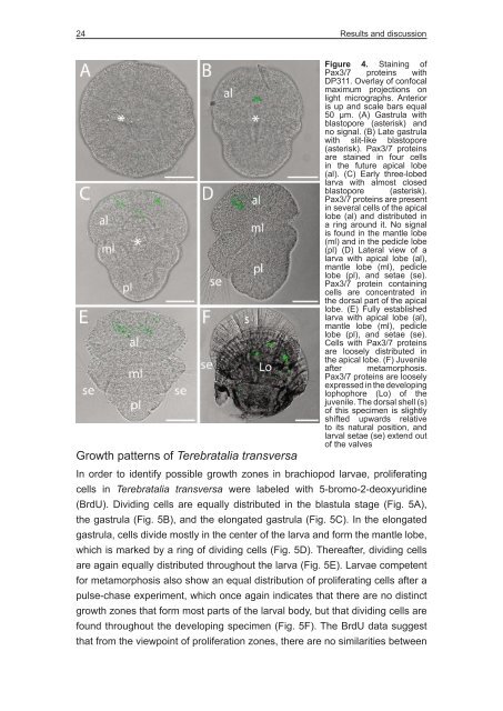
Figure 4. Staining of<br />
Pax3/7 proteins with<br />
DP311. Overlay of confocal<br />
maximum projections on<br />
light micrographs. Anterior<br />
is up and scale bars equal<br />
50 µm. (A) Gastrula with<br />
blastopore (asterisk) and<br />
no signal. (B) Late gastrula<br />
with slit-like blastopore<br />
(asterisk). Pax3/7 proteins<br />
are stained in four cells<br />
in the future apical lobe<br />
(al). (C) Early three-lobed<br />
larva with almost closed<br />
blastopore (asterisk).<br />
Pax3/7 proteins are present<br />
in several cells of the apical<br />
lobe (al) and distributed in<br />
a ring around it. No signal<br />
is found in the mantle lobe<br />
(ml) and in the pedicle lobe<br />
(pl) (D) Lateral view of a<br />
larva with apical lobe (al),<br />
mantle lobe (ml), pedicle<br />
lobe (pl), and setae (se).<br />
Pax3/7 protein containing<br />
cells are concentrated in<br />
the dorsal part of the apical<br />
lobe. (E) Fully established<br />
larva with apical lobe (al),<br />
mantle lobe (ml), pedicle<br />
lobe (pl), and setae (se).<br />
Cells with Pax3/7 proteins<br />
are loosely distributed in<br />
the apical lobe. (F) Juvenile<br />
after metamorphosis.<br />
Pax3/7 proteins are loosely<br />
expressed in the developing<br />
lophophore (Lo) of the<br />
juvenile. The dorsal shell (s)<br />
of this specimen is slightly<br />
shifted upwards relative<br />
to its natural position, and<br />
larval setae (se) extend out<br />
of the valves<br />
In order to identify possible growth zones in brachiopod larvae, proliferating<br />
cells in Terebratalia transversa were labeled with 5-bromo-2-deoxyuridine<br />
(BrdU). Dividing cells are equally distributed in the blastula stage (Fig. 5A),<br />
the gastrula (Fig. 5B), and the elongated gastrula (Fig. 5C). In the elongated<br />
gastrula, cells divide mostly in the center of the larva and form the mantle lobe,<br />
which is marked by a ring of dividing cells (Fig. 5D). Thereafter, dividing cells<br />
are again equally distributed throughout the larva (Fig. 5E). Larvae competent<br />
for metamorphosis also show an equal distribution of proliferating cells after a<br />
pulse-chase experiment, which once again indicates that there are no distinct<br />
growth zones that form most parts of the larval body, but that dividing cells are<br />
found throughout the developing specimen (Fig. 5F). The BrdU data suggest<br />
that from the viewpoint of proliferation zones, there are no similarities between


