Download - forensic medicine
Download - forensic medicine
Download - forensic medicine

You also want an ePaper? Increase the reach of your titles
YUMPU automatically turns print PDFs into web optimized ePapers that Google loves.
JIAFM, 2007 - 29(4); ISSN: 0971-0973<br />
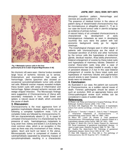
Fig. 4 Metastatic tumour cells in the liver<br />
parenchyma [H & E stain Original Magnification X 40]<br />
No chorionic villi were seen. Uterine fundus revealed<br />
large focus of ischemic necrosis up to serosa.<br />
Endometrium and myometrium had areas of<br />
hemorrhage. Uterine specimen also showed an<br />
occasional syncytiotrophoblastic tumor cells amidst<br />
red cells. Bilateral ovarian masses revealed multiple<br />
theca leutein cysts with areas of inflammation and<br />
hemorrhage. Spleen showed ischemic necrosis with<br />
wedge shaped infarction. The Microscopic findings<br />
were suggestive of Choriocarcinoma of Uterus with<br />
secondary metastasis in liver, small bowel and<br />
spleen, a natural cause of death, which unraveled<br />
the cause of death.<br />
3. Discussion:<br />
Choriocarcinoma is the most aggressive form of<br />
gestation trophoblastic disease, which mostly occurs<br />
following a complete hydatiform mole, 1-2% of<br />
complete moles are followed by choriocarcinoma [1].<br />
Villi are characteristically absent [1, 2]. In cases of<br />
untreated Choriocarcinoma it is charecterised by the<br />
presence of early haematogenous metastasis to<br />
lung ,brain liver, kidney and bowel [1,3,4,5] being the<br />
most common sites, which is an exception in this<br />
case as there was only involvement of liver, kidney,<br />
spleen, ileum and burnt out lesion in the uterus.<br />
Microscopically tumor is composed of clusters of<br />
cytotrophoblasts separated by streaming masses of<br />
syncytiotrophoblasts resulting in a characterstic<br />
dimorphic plexiform pattern. Hemorrhage and<br />
necrosis are usually present [1, 6].<br />
The presence of residual tumour in the uterus of<br />
patient dying of disseminated choriocarcinoma may<br />
be inconspicuous or altogether absent [1, 7] as in<br />
our case we found tumour cells in uterine scrapings<br />
as evidence of primary tumour.<br />
A natural history of an untreated choriocarcinoma is<br />
characterized by development of early<br />
hematogenous metastasis as seen in developing<br />
countries like ours were the patient with low<br />
economic background rarely go for regular<br />
checkups.<br />
The morphological changes seen in other organs in<br />
patients with choriocarcinoma are the result of<br />
increased secretion of β-hCG and other hormones<br />
by the tumour cells like hyperplasia of endocrinal<br />
glands, decidual reaction, Arias-stella phenomenon,<br />
bilateral enlargement of ovaries by theca luteal cysts<br />
and hyperplasia of mammary lobules. Detection of<br />
ovarian theca-lutein cysts long after a case of<br />
choriocarcinoma has been treated is usually a sign<br />
of persistent disease [1,7] but In this case bilateral<br />
enlargement of ovaries by theca luteal cysts and<br />
hyperplasia of mammary lobules and pigmentation<br />
around areola is seen however increased β- h CG<br />
levels were not detected.<br />
4. Conclusion:<br />
As illustrated in our case, the patient can succumb<br />
of Choriocarcinoma, as a sudden natural cause of<br />
death. Forensic pathologists should be aware of<br />
such an evolution. With an appropriate history and<br />
Clinico-pathological review such entities can be<br />
considered in cases of sudden death.<br />
References:<br />
[1] Rosai J, ed. Rosai and Ackerman's Surgical Pathology. 9th ed.<br />
Edinburgh, Scotland: Elsevier ;2004: 1744 –1746.<br />
[2] Elston CW, Bagshawe KD. The diagnosis of trophoblastic<br />
tumors from uterine currettings. J Clinical Pathol 1972; 25: 111 –<br />
118.<br />
[3] Ishizuka T, Tomoda Y, Kaseki S , Goto S, Hara T, Kobayashi<br />
T. Intracranial metastases of choriocarcinoma - A<br />
clinicopathologic study.Cancer 1983; 52: 1896 – 1903.<br />
[4] Mazur MT, Lurain JR, Brewer JI. Fetal gestational<br />
choriocarcinoma. Clinicopathologic study of patients treated at a<br />
trophoblastic disease centre.Cancer 1982; 50: 1833 – 1846.<br />
[5] Sober JT, Mutch DG, Chin N, Clarke, Pearson DL, Hammond<br />
CB. Renal metastases of gestational trophoblastic disease - A<br />
report of eight cases. Obstetric Gynaecol 1988; 72: 796 – 798.<br />
[6] Redline Rw, Abdul-Karim FW. Pathology of gestational<br />
trophobastic disease. Semin Oncol 1955; 22: 96 – 108.<br />
[7] Ober WB, Edgcoms JH, Price GB Jr. The pathology of<br />
choriocarcinoma. Ann NY Acad Sci 1971; 172:299-321.<br />
61

![syllabus in forensic medicine for m.b.b.s. students in india [pdf]](https://img.yumpu.com/48405011/1/190x245/syllabus-in-forensic-medicine-for-mbbs-students-in-india-pdf.jpg?quality=85)
![SPOTTING IN FORENSIC MEDICINE [pdf]](https://img.yumpu.com/45856557/1/190x245/spotting-in-forensic-medicine-pdf.jpg?quality=85)

![JAFM-33-2, April-June, 2011 [PDF] - forensic medicine](https://img.yumpu.com/43461356/1/190x245/jafm-33-2-april-june-2011-pdf-forensic-medicine.jpg?quality=85)

![JIAFM-33-4, October-December, 2011 [PDF] - forensic medicine](https://img.yumpu.com/31013278/1/190x245/jiafm-33-4-october-december-2011-pdf-forensic-medicine.jpg?quality=85)