Download - forensic medicine
Download - forensic medicine
Download - forensic medicine

You also want an ePaper? Increase the reach of your titles
YUMPU automatically turns print PDFs into web optimized ePapers that Google loves.
JIAFM, 2007 - 29(4); ISSN: 0971-0973<br />
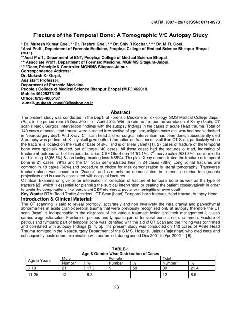
Fracture of the Temporal Bone: A Tomographic V/S Autopsy Study<br />
* Dr. Mukesh Kumar Goel, ** Dr. Rashmi Goel, *** Dr. Shiv R Kochar, **** Dr. M. R. Goel,<br />
*Asst Proff , Department of Forensic Medicine, People,s College of Medical Science Bhanpur Bhopal<br />
(M.P.).<br />
**Asst Proff , Department of ENT, People,s College of Medical Science Bhopal.<br />
***Associate Proff , Department of Forensic Medicine, MGNIMS Sitapura-Jaipur.<br />
****Dean, Principle & Controller MGNIMS Sitapura-Jaipur.<br />
Correspondence Address:<br />
Dr. Mukesh Kr Goyal,<br />
Assistant Professor,<br />
Department of Forensic Medicine,<br />
People,s College of Medical Science Bhanpur,Bhopal (M.P.).462010.<br />
Mobile: 09425373106<br />
Office: 0755-4005137<br />
e-mail- mukesh_goyal03@yahoo.co.in<br />
Abstract<br />
The present study was conducted in the Dep’t. of Forensic Medicine & Toxicology, SMS Medical College Jaipur<br />
(Raj), in the period from 15 Dec. 2001 to 4 April 2002. With the aim to find out the correlation of X-ray (Skull), CT<br />
scan (Head), Surgical intervention findings with the autopsy findings in the cases of acute Head trauma. Total of<br />
140 cases of acute head trauma were selected irrespective of age, sex, religion caste etc. who had been admitted<br />
in Neurosurgery dep’t. And X-ray, CT scan head and /or surgical intervention had been done, subsequently died<br />
& autopsy was performed. X- ray skull gave batter information on fracture of skull than CT Scan, particularly when<br />
the fracture is located on the vault or base of skull and is of linear variety [1]. 27 cases of fracture of the temporal<br />
bone were specially studied, out of these 140 cases. All these cases had the features of triad, indicating of<br />
fracture of petrous part of temporal bone i.e. CSF Otorrhoea 14(51.1%), 7 th nerve palsy 9(33.3%), serve middle<br />
ear bleeding 18(66.6%) & conducting hearing loss 5(85%). The plain X-ray demonstrated the fracture of temporal<br />
bone in 21 cases (79%) and the CT Scan demonstrated their in 24 cases (88%) Longitudinal fractures are<br />
common in 18 cases (66%) and procedure of choice for their demonstration is lateral tomography, Transverse<br />
fracture alone was uncommon (2cases) and can only be demonstrated in anterior posterior tomographic<br />
projections and is usually associated with occipital fractures.<br />
CT Scan Examination give better information in detection of fracture of temporal bone as well as the type of<br />
fracture [2] which is essential for planning the surgical intervention or treating the patient conservatively in order<br />
to avoid the complications like, persistent CSF otorrhoea, posterior meningitis or even death.<br />
Key Words: RTA (Road Traffic Accident), CT Scan (head) Temporal bone fracture, Head trauma, Autopsy Head.<br />
Introduction & Clinical Material:<br />
The CT scanning is said to reveal promptly, accurately and non invasively the intra cranial and parenchymal<br />
abnormalities in acute cranio-cerebral trauma that were previously recognized only at autopsy therefore the CT<br />
scan (head) is indispensable in the diagnosis of the various traumatic lesion and their management I, it also<br />
carries prognostic value. Fracture of petrous and tympanic part of temporal bone is not uncommon. Fracture of<br />
petrous and tympanic part of temporal bone was identified with the aid of CT Scan and the finding was confirmed<br />
and correlated with autopsy findings [3, 4, 5]. The present study was conducted on 140 cases of Acute Head<br />
Trauma admitted in the Neurosurgery Department of the S.M.S. Hospital, Jaipur (Rajasthan) who died there and<br />
subsequently postmortem examination was performed, during period Dec-2001 to Apr-2002 [ 6].<br />
TABLE-1<br />
Age & Gender Wise Distribution of Cases<br />
Male Female Total<br />
Age in Years<br />
Number % Number % Number %<br />
> 10 21 17.2 9 50 30 21.4<br />
11-20 12 9.8 - 12 8.5<br />
83
![syllabus in forensic medicine for m.b.b.s. students in india [pdf]](https://img.yumpu.com/48405011/1/190x245/syllabus-in-forensic-medicine-for-mbbs-students-in-india-pdf.jpg?quality=85)
![SPOTTING IN FORENSIC MEDICINE [pdf]](https://img.yumpu.com/45856557/1/190x245/spotting-in-forensic-medicine-pdf.jpg?quality=85)
![JAFM-33-2, April-June, 2011 [PDF] - forensic medicine](https://img.yumpu.com/43461356/1/190x245/jafm-33-2-april-june-2011-pdf-forensic-medicine.jpg?quality=85)
![JIAFM-33-4, October-December, 2011 [PDF] - forensic medicine](https://img.yumpu.com/31013278/1/190x245/jiafm-33-4-october-december-2011-pdf-forensic-medicine.jpg?quality=85)