jiafm, 2010-32(2) april-june. - forensic medicine
jiafm, 2010-32(2) april-june. - forensic medicine
jiafm, 2010-32(2) april-june. - forensic medicine
- No tags were found...

You also want an ePaper? Increase the reach of your titles
YUMPU automatically turns print PDFs into web optimized ePapers that Google loves.
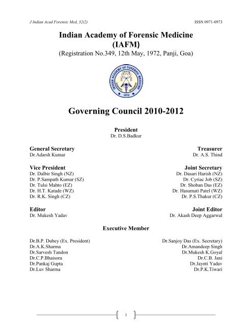
ISSN 0971-0973 J Indian Acad Forensic Med, <strong>32</strong>(2)Journal of Indian Academy of Forensic Medicine(JIAFM)Editor,Dr. Mukesh YadavProfessor & HODForensic Medicine & Toxicology,School of Medical Sciences & ResearchSharda University, Greater Noida, U.P. PIN:201306ResidenceG-216, Parsvanath Edens,Alfa-II, Greater Noida, G.B. Nagar,U.P.-<strong>2010</strong>308Ph. No. 0120-2<strong>32</strong>6060,Mobile No. 09411480753Email: drmukesh65@yahoo.co.inPeer Review GroupJoint EditorDr. Akash Deep AggarwalAssistant Professor,Department of Forensic Medicine,Pt. B.D. Sharma PHGIMS, Rohtak,Haryana-124001ResidenceH.No. 14, Desi Mehmandari,Patiala, PunjabPIN: 147001Mobile No. 9815652621Email:toakashdeep@yahoo.comDr.A.K. SrivstavaProf. & HOD, ForensicMedicine & ToxicologySubharti Medical College,Meerut, U.P.Dr.V.V.PillayProf. & HOD, AnalyticalToxicology,Chief of Poison Control CentreAIMS & R, Cochin-KeralaDr.R.K. GoreaProf. & HOD, ForensicMedicine & ToxicologyGyan Sagar Medical College,Banur, Patiala, PunjabDr.C.B.JaniProf. & HOD, ForensicMedicine & ToxicologyP.S. Medical College,Karamsad, Distt. Anand,Gujarat, PIN: 388<strong>32</strong>5Dr.T.K BoseProf. & HOD, Forensic andState MedicineGovt. Medical CollegeKolkata, West BengalDr.G. PradeepkumarProf. & HOD, ForensicMedicine & ToxicologyKasturba Medical College,Manipal, KarnatkaSharma G.K., (New Delhi)Verma S.K., (New Delhi)Thakur S.D., (Jammu)Kaur Balbir, (Srinagar)Bansal Y., (Chandigarh)Kumar Shantha B., (Tamilnadu)Gupta B.D., (Gujart)Manju Nath K.H, (Karnatka)Das Sanjoy, (U.K.)Bhaisora C.P, (U.K.Advisory BoardMahtoo Tulsi, (Jharkhand)Ravindran K, (Pandicherry)Sabri Imran, (H.P.)Rastogi Prateek (Karnatka)Potwary AJ (Assam)Singh R.K. (Chhatisgarh)Dongre A.P. (Nagpur)Rastogi Pooja (U.P.)Sharma Aditya (H.P.)Khanagwal V. (Haryana)Gupta Pankaj (Punjab)Jani C.B. (Gujrat)Khaja Shaikh (A.P.)Basu R (W.B.)Naik R.S. (Maharastra)Godhikirakar Madhu (Goa)Job Cyriac (Kerala)Vinita K. (U.P.)Yadav B.N. (Nepal)Printed and published by Dr. Mukesh Yadav, Editor, JIAFM and Dr. A. D. Aggarwal, Joint Editor, JIAFM onbehalf of Indian Academy of Forensic Medicine at name of the press [SHIVANI PRINTERS, NOIDA, U.P.]ii
J Indian Acad Forensic Med, <strong>32</strong>(2) ISSN 0971-0973Journal of Indian Academy of Forensic MedicineVolume <strong>32</strong> • Number 2 • April-June <strong>2010</strong>ContentsFrom the Editor‟s Desk 97EditorialUnion Govt., Court, CBI and Members of the MCI/IMA/Medical Fraternity 98on the Dock!Originals and PapersEstimation of Stature by anthropometric mesuarements of Inte-Acromial 101LengthVishal Veer Vasu, Swapnil Sudhir Agarwal, Shashidhar Channamallapa MestriSignificance of sacral index in estimation of sex in sacra of cadavers in 104PunjabAnterpreet Kaur Arora, Pankaj Gupta, Shashi Mahajan, Sonney Singh KapoorAn Epidemiological study of Organophosphorus Poisoning at Manipal Teaching 108Hospital, Pokhara, NepalS. M. Kar, Sidartha Timsinha, Prashant AgrawalStudy of road traffic accidental deaths (RTA) in and around Bastar 110region of ChattisgarhDhaval J Patel, Gopinath AgnihotramEstimation of age in the living municipal employees in the age group of 11<strong>32</strong>5-45 years by physical and radiological examinationA. T. Shendarker , R. Kharat , W.F. Vaz , F.R. Karjodkar , K.R. RedeAge assessment from radiological cranial suture closure in fourth to 120seventh decades: (A Jaipur based study)Rajesh Kumar Verma, Mukesh K. Goyal, Shiv KocharNoble ideas and approaches to learning medical sciences 124Chandra Prakesh, Bhavana Srivastava, Sanjay Gaur, Ajay Kumar SinhaHistopathology examination in medicolegal autopsy: Pros and cons 128Akhilesh Pathak, H.M. MangalPattern of renal pathology in fatal envenomation by Indian Cobra (Naja naja) 1<strong>32</strong>Partha Pratim Mukhopadhyay, Sulekha Ghosh, Tapan Kumar GhoshA study of homicidal deaths by mechanical injuries in Surat, Gujarat 134Pranav Prajapati, M.I. Sheikh, Shyam PatelIncidence of snake bites in Belgaum 139Ashok Kumar Shetty, Prasanna S. JirliDecay in intact DNA recovery in blood samples kept at room temperature 142Imran Sabri, J.A. Usmani, S.A. Hanif, A.U.Khan95
ISSN 0971-0973 J Indian Acad Forensic Med, <strong>32</strong>(2)Pattern of fatal blunt head injury: A two year retrospective/prospective 144medicolegal autopsy studyAmit M. Patil, Walter F. VazProfile of medicolegal cases at Adesh Institute of Medical Sciences & Research, 150Bhatinda, PunjabVishal Garg, S.K. VermaMedical ethics, duties and medical negligence awareness among 153the practitioners in a teaching medical college, hospital-A SurveyShreemanta Kumar DashA comparative study of clinical & autopsy findings (clinical audit) in 100 cases 157died of traumaGeetha O, S. GirishCase ReportsAdult choroid plexus papilloma: Cause of sudden death 160S. Ranjan Bajpai, D.S. Badkur, Reeni Malik, Arneet Arora, Jayanthi YadavSuicide by para-phenylenediamine poisoning 163Sushil KumarAcute Myocardial infarction related to blunt thoracic trauma: 165Review of literature with two case reportsAmit SharmaImportance of scene of crime visit, it may be late but it is never too late 168U.S. Sinha, A.K. Singh, Y.K. PathakFatal multiple painless intussusceptions: A case report 172Nilesh Keshav Tumram, Rajesh Vaijnathrao Bardale, Manish B ShrigiriwarFatal unintentional Carbn Monooxide Poisoning inside a Garage: 174A case reportAvishek Kumar, Ravi RautjiA Rare Case Report: Poland‟s Syndrome with unilateral Amastia in 176alleged victim of RapeKalpsree Bhawmik, Nibedita Shyam, K.C. DasReview ArticlesOperational difficulties in M.B.B.S. 2 nd Professional course due to 179MCI regulations: Is MCI listening?Rajiv Mahjan, Parmod Kumar Goyal, Mukesh YadavThe Medicolegal Autopsy-Its Social and Religious Attitudes 183Putul MohantaCopy Right: No part of this publication may be reprinted or publish without the prior permission of theEditor, JIAFM. Submission of all paper to the journal is understood to imply that it is not being considered forpublication elsewhere. Submission of multi authored papers implies that the consent of each author has beenobtained. In this journal, every effort has been made NOT to publish inaccurate or misleadinginformation. However, the Editor, Joint Editor, Peer Review Group and Advisory Board accept NOliability in consequences of such statements. EDITOR ((JIAFM)Address request for reprint or further information relating to any article may please be made with author and incase of multi authored article, please communicate to Corresponding Author or the First Author96
J Indian Acad Forensic Med, <strong>32</strong>(2) ISSN 0971-0973From Editor‟s DeskJIAFMA Quarterly PublicationVolume <strong>32</strong>, Number 2, April to June, <strong>2010</strong>I feel immense pleasure to present before you the second issue of <strong>2010</strong>. I assure you about thequality of research papers and quality of printing in future issues. Your valuable suggestions arealways encouraging me and I heartily welcome for future suggestions. On behalf of ExecutiveCommittee of IAFM for the years <strong>2010</strong>-2011, I took resolution to further improve the quality andstatus of our Journal. We always learn from mistakes and try to improve upon these. I amthankful to the advertisers who have provided additional financial resources for improving thequality of this issue.Dr. Mukesh YadavEditorSubscription InformationMembers of IAFM will receive the free of cost.Non Members and Institutions (Annual Subscription rates)Personal: In India, Rs. 1000/ (Rest of the world: US$ 200/ or equivalent)Institutions: In India, Rs. 3000/ (Rest of the world: US$ 400/ or equivalent)Subscription orders and payments should be made in favour of “Editor, JIAFM, payable atGreater Noida”We Accept: Bank Cheque / Demand Drafts (Add Rs. 50/- for outstation Cheques)The Scope of the Journal covers all aspects of Forensic Medicine and allied fields, researchand applied.Claims for missing issue:A copy will be sent free to the member / subscriber provided the claim is made within 2 months ofpublication of the issue & self addressed envelop of the size 9” x 12” is sent to the Editor. (Thosewho want the journals to be dispatched by Registered Post must affix Rs. 50/ worth postage stamps).The journal is indexed with IndMed and made available online by Diwan Enterprise (New Delhi) at:1. www.indianjournals.com2. http://www.medind.nic.in3. www.<strong>jiafm</strong>.com4. www.iafm1972.orgEditor97
ISSN 0971-0973 J Indian Acad Forensic Med, <strong>32</strong>(2)EditorialUnion Govt., Court, CBI and Members of the MCI/IMA/Medicalfraternity on the Dock!Current affairs of medical education and health care in India involves important issue of good governanceby the Modern State, discharge of statutory duties by the „public servant‟ and protection of fundamental rights by theCourt, constitutional mandate of division of jurisdiction between Judiciary, Executive and Legislature, three wingsin a democratic setup. Issue of public faith and confidence in a welfare state like India becomes more importantwhen it involves most cherished fundamental right of every citizen i.e. Right to life which includes Right toadequate health care enshrined in the Article 21 of the Indian Constitution.In the cacophony of the IPL saga, a more serious scam has just surfaced, more serious, because, it involveshealth and lives of more than 1.3 billion Indians. In terms of money it does not match up to the IPL rip off, but itseffects, fallout and ramifications are much wider and more somber. The Medical Council of India (MCI) chief andtwo of his cohorts were arrested today by CBI on charges of corruption. This is sure to bring down the prestige ofMCI by quite a few notches, as will it bring shame and self-disgust to every ethical practitioner of <strong>medicine</strong>.Issue of good leadership of IMA:It is an issue of participatory democracy, medical law and ethics vital for maintaining nobility of themedical profession. Issue raises serious challenges before the leadership of India‟s largest body of AllopathicMedical Professionals i.e. Indian Medical Association (IMA). It is also an issue of good leadership among the wholeMedical Fraternity of India.Instead of opposing the move of Union Government to revamp the MCI and defending the actions ofcorrupt and rotten system of MCI prevailing and putting the nobility as stake, leadership of IMA should participatein a constructive way. It is shocking that nowhere IMA office bearers both local and national made any officialstatement against prevailing corrupt practices.If we blame illiterates and rural population for helm of current affairs of the India on the issue ofcorruption, it may sound true but what about the so called intellectual urban population of medical fraternity nottaking lead to improve the situation of their noble profession. It raises serious doubts about the whole educationsystem of India particularly higher education (professional medical education).Reasons for Attraction to Medical Profession:The potential for earnings through the medical profession and the prestige which goes with the professionattracts large number of youngsters towards the profession all over the India. There is a scramble for seats inmedical colleges. Some of the medical colleges had been charging fabulous amounts running into lacs of rupees byway of capitation fee for allotting seats to students. This led to a spurt in the number of medical colleges. Medicalcolleges are seen as money spinning business propositions. This in turn has increased the burden of the MCI. Itsfunctions like granting permission to run medical college, recognition, withdrawal of recognition and the power toregulate the number of seats in medical colleges have put the MCI in a very powerful position.This makes it almost imperative that the MCI should have as its members, persons of integrity and highprofessional standards with values of honesty and probity.Issue of participatory democracy:Unfortunately institutions meant to improve professional standards are passed into hands of unscrupulouspersons day by day. A stage has come when on account of politicking and manipulative tactics of such persons likeKetan Desai in institutions meant to maintain professional standards, no good or eminent person with stature wantsto serve such institutions. This results in institutions being controlled by undeserving persons.Role, Responsibility and powers of MCI:The role of MCI has assumed great importance in course of time. The MCI is charged with theresponsibility of maintaining high levels of medical education and professional standards by the medicalpractitioners. The MCI not only lays down the academic standards for various medical courses including postgraduate and diploma courses but also ensures that proper infrastructure is available in the medical colleges forimparting education and training. The MCI enjoys vast powers for its control over medical colleges; it gets thecolleges inspected through its own teams of inspectors. The inspection reports can lead to refusal of permission tostart medical colleges and withdrawal of recognition to already recognised medical colleges. The MCI regulatesadmissions to medical colleges in as much as if colleges are to increase the number of seats, its approval is required.98
J Indian Acad Forensic Med, <strong>32</strong>(2) ISSN 0971-0973Role of Office bearers of MCI:The Medical Council Act was brought into force with laudable objectives. The MC was envisaged underthe Act as an Apex Body to control and regulate the medical profession. Whether any legislation is able to achieveits objectives depends on persons who occupy important offices under the respective statutes and whoseresponsibility it is to implement the statutes. The role of human beings becomes all important. If human beings whoare to implement the statutes are unfit for the job, they can subvert the spirit behind the statutory provisions.A beneficial statute can be converted into a tool of oppression by incompetent and/or unscrupulous persons.President of the Medical Council plays a pivotal role. It is he who is really responsible for the entire functioning ofthe Council. The role of the President of the Medical Council of India in working out the statute is the main issuebefore us. As per the case of the petitioner in the writ petition, present case is an example of subversion of the IndianMedical Council Act by the present incumbent on the post of President of the Council.Various Issues involved:Failure of the Central Government to constitute the Medical Council of India timely in accordance withsection 3 of the IMA Act of 1956.The eligibility of various office bearers, to seek election as President, Vice-President and Members bothelected and nominated one of the MCI and to hold office as such.Thirdly allegations have been made of misuse of office by office bearers by indulging in corrupt practiceswhich disentitle him to continue to hold office of the MCI.Proper transparent and accountable system of grievance redressal whenever any discrepancy come to thenotice of the anyone interested in the system of quality of medical education.Role of Higher CourtsAlthough the Indian Medical Council (Professional Conduct, Ethics and Etiquettes) Regulations-2002 atpoint 1.7 of Chapter I casts a statutory duty on every registered medical practitioner to expose the unscrupulouspractices indulged by fellow colleague without fear or favour. But there are very few who can dare to follow thisstatutory requirement, reason being not sure of enforcement of these regulations by the regulatory body itself i.e.MCI/ SMC and no hope of reprieve from even higher judiciary at the level of the High Courts or even Apex Courtof the India. Thus, true mandate of establishing the democratic welfare state as envisioned by founder forefathers ofthe Indian Constitution. As it is well said that justice delayed means justice denied. Many cases are pending invarious High Courts in many states and in the Supreme Court for years together. Those who can dare to move thesecourts either oppressed by the authorities by various ways or lose interest after loosing money before unscrupulousadvocate‟s hands in gloves with opposite partiesRole of CBI:This time CBI has put on its website a request to public to let it know any "specific complaint regarding demandof money by MCI officials or giving approval/permission to any such college without having the requiredinfrastructure due to corrupt practices by the Inspection Team or MCI officials".Contents of CBI Public Notice read as “CBI has registered a case related to corruption in Medical Council ofIndia (MCI), which conducts inspections for permission to start new Medical Colleges or to introduce new medicalcourses including Post Graduate Course and also for renewal of permissions, etc. If you have any specific complaintregarding demand of money by MCI officials or giving approval/permission to any such college without having therequired infrastructure due to corrupt practices by the Inspection Team or MCI officials, please contact: Dr. M.M.Oberoi, HoB/AC-III, CBI, New Delhi Tel. No.: 011-24361515 or send email at hobac3del@cbi.gov.in”.How to achieve laudable objectives of statute?Delhi High Court in 2001 observed that:“We are of the considered view that facts of this case call for exercise of power under Article 226 of theConstitution of India to prevent abuse and misuse of statutory office by the present incumbent.We have observed earlier that legislative measures are backed by best of intentions and laudable objectives.It falls on persons who exercise powers under the statutes, as to how they implement the statutoryobjectives.Court concluded in following words that “In other words, ultimately it all depends on persons who arecharged with the duty to act under the respective statutes.”If there are right persons for the jobs, the objectives will be achieved.If there are wrong persons, the statutes will be misused for oppression and corruption.99
ISSN 0971-0973 J Indian Acad Forensic Med, <strong>32</strong>(2)Role of Board of Governors and New MCI Members:It is well said that “A person is known not only by the company he keeps but also by the company heavoids”. This is well suited about the role and conduct of past MCI members well evident from CBI Report, MCIMinutes, court judgments on many previous occasions. Many past members are on pay role of private managementholding double posts in MCI various committees any molding MCI rules and regulations with impunity. RTI use bymany public spirited doctors found that CBI and CVO find many of them indulged in corrupt practices andrecommended various disciplinary actions against them including secretary of MCI and one of the Vice-Chancellorof a Private University and many of them holding posts even after completing 65 years.The Indian Medical Council (Amendment) Ordinance, <strong>2010</strong> (Ordinance 2 of <strong>2010</strong>) was promulgated by thePresident Pratibha Devi Singh Patil on 15 th May <strong>2010</strong>, came into force with immediate effect that empowers theCentral Government to superseded the MCI.Whereas upon the supersession of the MCI and until a new Council is reconstituted within a period of one year, as afirst step, the Central Government exercising its power under Section 3A (4) of the IMC Act, 1956, has now createda six-member “Board of Governors”, to exercise the powers and perform the functions of the MCI under newlyinserted Section 3A (2) of the Ordinance <strong>2010</strong>.Need for restoring the public confidence in the MCI:We all hope and trust that the Board of Governors and present Administrators shall restore the publicconfidence in the MCI and bring the Medical Council back on its feet so that it is able to discharge its statutoryfunctions in accordance with the spirit and object of setting up the MCI.To ensure and restore the credibility of highest medical education regulating body following steps arerecommended:Firstly, Auditing of MCI activities by third parties which can appropriately assess the performance. Secondly,ensuring Transparency in the accreditation and inspection system of educational institutions by publicly displayinginformation of complaints against medical institutions and their effective time bound redressal. Central Governmentshould ensure regular, free and fair elections of key office-bearers especially from the medical teaching category,under the supervision of court‟s observers. The role of the Central and State Governments is immense in ensuringnominated members who are honest, efficient and public-minded especially from medical teaching background bydefining criteria about qualifications and experience through a search committee as in case of appointment of Vice-Chancellors of the University.Central Government intends to separate the two functions of the MCI: medical education and registration ofmedical practitioners and hand them over to new institutions. Medical Education will come under the ambit of theNational Commission for Higher Education and Research (NCHER), which, in turn, will be overseen by theMinistry of Human Resource Development (MHRD), and the registration of medical practitioners under theNational Council for Human Resources in Health (NCHRH), to be overseen by the Ministry of Health andFamily Welfare.The Central Government‟s focus on MCI‟s affairs is, therefore, quite welcome step. But creating new institutions isnot necessarily the best way of correcting the manifold problems of the health sector. The health sector, it is wellknown, requires an overall perspective and accountability that only a single entity devoted to the sector can provide.One should not forget that there was a reason why the MCI was formed as an independent, professional regulator.The health sector requires the creation of in-depth expertise which, if not available, has devastating ramifications.Only health sector experts have the necessary understanding of the professional and technical issues involved inhealth care to design and implement appropriate regulations. This is well recognised and consequently, all over theworld, it is professionally-run medical associations that are in charge of such matters.Also, the NCHER under the MHRD will be working across different sectors; there is generalized apprehension thatIndia‟s bureaucracy will be put in charge of a technical area, so that the cure becomes worse than the originalmalady. The world over, the trend is to provide greater powers to sector experts who have the necessaryunderstanding of their sector and profession.This is the right time before all of us including responsible government authorities, higher judiciary, andleadership of medical fraternity to come forward and play their much desired role in a largest democracy of theglobe in participatory manner to save and serve the humanity.Editor100
J Indian Acad Forensic Med, <strong>32</strong>(2) ISSN 0971-0973Original research paperEstimation of Stature by Anthropometric Measurementsof Inter-Acromial Length*Dr. Vishal Veerbasu Koulapur, MD, **Dr. Swapnil Sudhirkumar Agarwal, MD, DNB, ***Dr. ShashidharChannamallapa Mestri, MDAbstractStature is an important parameter to establish identity of an unknown corpse. It is being used bymedicolegal experts when either complete or parts of human body are recovered. A lot of criteria are available thatcan be used depending upon the part of the body recovered. To add to these criteria, a study was done with theobjective of deriving a regression equation for estimating stature from the inter-acromial length. A cross-sectionalstudy was done during one year period wherein 150 subjects in age group around 23 years were studied. Three setsof regression equations were derived after statistical analysis of the data. The study revealed that there exist apositive and significant correlation between stature and inter-acromial length in both the sexes and that stature canbe estimated with the inter-acromial length when only upper parts of the trunk are available.Key Words: Forensic Anthropology, Identity, Inter-Acromial Length, Stature, Regression EquationIntroduction:Skeletal material present in the mutilated oramputated limbs, trunk etc. has obvious significancein the personal identification in the event of themurders, accidents or natural disasters. Studies onestimation of stature from the skeletal remains orfrom the mutilated limbs, mostly of the long boneshave been reported as indicated by the publishedwork of the Pearson, Trotter and Glesser. The Indianperspective of the problem of stature estimation hasbeen studied by the Athwale et al, Patel et al, Joshi etal, Lal and Lala etc (Jasuja OP and Singh G, 2004).[2]When a complete dead body is found,stature estimation is rather an easy task; but in casesin which only some parts of the body are available,the determination of stature of the individual isdifficult.Corresponding Author:Dr. Vishal Koulapur , *Asst Professor, Deptt. ofForensic Medicine, B. M. Patil Medical College,Sholapur Rd, BIJAPUR- 586103.Ph +91 9916134949E-mail: drvishalk@gmail.com**Associate Professor,Department of Forensic Medicine & Toxicology,Jhalawar Hospital & Medical College,Jhalawar, Rajasthan, India***Professor & HeadDept. of Forensic Medicine & Toxicology,Karpaga Vinayaga Institute of Medical SciencesGST Road, Palayanoor, KanchipuramTamil Nadu, IndiaThus it is necessary to have differentformulae for the determination of stature from thelengths of different body parts in different populationgroups as they vary from population to population(Kroeber AL, 1976). [3]Although a number of studies have beendone on stature estimation by using different bodyparts, very few of them are by using inter-acromiallength. Inter–acromial length is the distance betweentwo bony landmarks, i.e. acromial processes ofscapula on each side. Acromion is the most lateralpoint on the lateral margin of the acromial processwhen the subject stands in normal position with hisarms hanging by the sides (Nath S et. al, 2005). [6]The present study is taken up to fill the above lacuna.In addition, inter – acromial length being a macromeasurement, is easy to measure. In this study, aneffort was done to establish the relationship betweenstatures of different persons of North Karnatakaregion of India and their inter-acromial lengths and todevelop regression equation formulae from these twovariables by simple regression analysis. The formulathus obtained could be used for the determination ofstature of an individual of North Karnataka region ofIndia from his inter-acromial length. Ethical issuesinvolved in the study were minimal and no invasivemethods were used.Material and Methods:A cross-sectional study was done over aperiod of one year from 1 st November 2006 to 31 stOctober 2007. During this period, one hundred andfifty individuals i.e. 75 males and 75 females, bornand brought up in the North Karnataka region of101
ISSN 0971-0973 J Indian Acad Forensic Med, <strong>32</strong>(2)India (Districts included were Belgaum, Dharwad,Bijapur, Gulbarga, Bagalkot, Gadag & Haveri) in theage group 23 years were choosen for the study. Age23 years was chosen for the reason that by this agenearly all secondary centres fuse with the respectiveshafts. Those who were not born and brought up herewere excluded. To minimize error, cases ofdwarfism, gigantism and those having skeletalabnormality of spine and long bones were excludedfrom the study.After taking their written informed consentand recording their full particulars like name, age,sex and place to which they belong, the stature ofeach individual was measured in centimeters with thesubject standing against a vertical backgroundsurface in normal erect position, the shoulders,buttocks and heels lightly touching the background/wall. An anthropometric rod set was used for takingthe above measurements. The measurement from thevertex of head to the ground was taken after bringingdown the adjustable cross-bar to the head and themeasurement was read from the vertical scale. Theinter-acromial length was measured in centimeterswith the person in the same erect position(Momonchand A and Devi TM, 1999). [5]After taking the measurements, statisticalanalysis was done using statistical equations as givenbelow:∑ y = Na + b∑ x∑ xy = a ∑ x + b ∑x 2Where ∑= Sum valuey= Value of statureN= Number of cases studiedx= Value of inter-acromial lengtha= Unit greater than x value by y valueb= Regression coefficientFrom the above equations, regressionformulae, standard errors and co-efficient ofcorrelations were developed to fulfill the aims andobjectives of the study. After statistical analysis ofthe results, three regression equation formulae wereobtained from the relationship between statures andinter-acromial lengths of females, males and males &females combined.Observation and Findings:The maximum, minimum and averagestatures of different sexes along with their maximum,minimum and average inter-acromial lengths werecalculated and tabulated as shown in Table I.The regression formulae, standard errors,standard deviations and coefficient of co-relations ofthe above data were computed using statisticalmethods by presuming X as an independent variableand Y as dependent variable.Results:Then regression formulae, standard errors,coefficient of correlations of the study have beencomputed and presented in Table No. II.Discussion:Population variations in anthropometricdimensions do exist and are attributed to genetic,dietary habits and environmental factors. Thisindicates that specific formulae or regressionequations used in prediction of stature are onlyapplicable to the population from which the datawere collected (Malek AKA et. al, 1990). [4]Various researchers with variable degree ofsuccess have attempted the estimation of stature fromvarious long bones, by using different statisticalmethods such as regression equations andmultiplication factors for a very long time. Thedifficulty in availability of adequate quantities ofbones, in the choice of bones, then cleaning and theneed of trained personnel are encountered whilecorrelating bone dimensions with stature (DikshitPC et. al, 2005). [1] But very little work has beenreported on the use of these statistical methods tocalculate the stature from the inter-acromial length.The standard errors came out to be ± 8 cm(males) and + 5 cm (females) in a study done byMomonchand A and Meera Devi T (Momonchand Aand Devi TM, 1999). These standard errors were + 6(males) and + 4 (females) in our study. There is adifference is of + 2 cm in males and + 1 cm infemales which is better than earlier study.In both the studies, the error is more inmales as compared to females. This can be attributedto the reason that females, being thin and lean; bonyprojections are easily palpable and more accuratelytaken for measurement as compared to muscularmales where it is not so easy to mark out the bonyprojections.Both the studies have been done in India butone can clearly find a notable difference in theregression equations in both the studies. In first studydone by Momonchand A and Meera Devi T(Momonchand A and Devi TM, 1999), [5] theregression equations were y = 1.7x + 84 in femalesand y = 2x + 69 in males, while that in this study, ithas come out to be y = 103.62+1.6x in females and y= 167.50+0.20x. When inter-acromial lengths takenfrom the population study were used to find outstature from regression formulae developed byMomonchand A and Meera Devi T, there was asignificant error in stature calculated. This impliesthat these formulae can be used for estimating thestature in the given population from which they havebeen developed. This also proposes the need to havesimilar studies on different populations so that similarequations can be evolved for different populationgroups and the differences can be studied further.102
J Indian Acad Forensic Med, <strong>32</strong>(2) ISSN 0971-0973Conclusion:There exists a significant correlation ofheight with the inter-acromial length of an individualin both the sexes and that stature can be estimatedwith the inter-acromial length when only upper partof the trunk is available. The correlation matrix of thepresent study shows that the use of a singleregression equation to predict stature from themeasurements of inter-acromial length does not makea great difference from individual measurementequations based on sex. In the present study, theregression equation developed for stature estimationfrom the inter-acromial length by regression equationformulae y = 167.50+0.20x in males and y =103.62+1.6x in females can be useful in estimatingstature of the population of North Karnataka regionand whether these equations are useful with otherpopulation needs to be researched.Similar studies need to be done on deadbodies in mortuary, where it is easier to mark bonyprojections accurately as they can be felt moreprominently, even by rough manipulation withoutpain factor, which is a hindrance in living subjects.Also no ethical issues are involved here. May be inthese studies the standard of error can be minimizedand these regression equations can be made moreuseful.More studies need to be conducted toestimate the stature from inter-acromial length amongTable No. Iother racial groups and of different geographicalareas, as it can be extremely useful to estimate thestature when mutilated upper part of trunks areavailable.AcknowledgementAcknowledgments are due to Mr. M. D.Mallapur [M.Sc.], Lecturer in Statistics, Departmentof Community Medicine, KLE University‟sJawaharlal Nehru Medical College, Belgaum for hisvaluable help in the statistical analysis of this studyand all the subjects, who have whole heartedlyparticipated in the study.References:1. Dikshit P.C., Aggarwal A., Mukta R. (2005) Estimationof stature from hand length. Journal of Indian Academyof Forensic Medicine. 27 (4), 219-221.2. Jasuja O.P., Singh G. (2004) Estimation of stature fromhand and phalange length. Journal of Indian Academyof Forensic Medicine. 26 (3), 100-106.3. Kroeber A.L. (1976) Kroeber Anthropology, 3 rd ed.New Delhi, Oxford and IBH Publishing Company, p.56.4. Malek A.K.A., Ahmed A.M., Sharkawi A.A.S., HamidA.M.A. (1990) Prediction of stature from handmeasurements. Forensic Science International. 46, 181-187.5. Momonchand A., Devi T.M. (1999) Determination ofStature from the inter-acromial length. Journal ofForensic Medicine and Toxicology. 16 (1), 72-73.6. Nath S., Sehgal V.N., Bhasin M.K. (2005) Forensicanthropology - Science and Medicine, 1 st ed. NewDelhi, Kamal-Raj Enterprises, p. 175.Characters Males FemalesMaximum stature 184.2 167.9Minimum stature 160.2 140.4Average stature 170.3 157.3Maximum inter-acromial length 40.2 33Minimum inter-acromial length 30.2 25.6Average inter-acromial length 34.2 29.9The above table shows sex wise distribution of maximum, minimum and average statures and inter-acromiallengths in cmsTable No. IICharacters Regression formulae Standard error Coefficient of correlationMales & Females together y =103.82+2.1x ± 5 cm 0.7Males only y =167.50+0.20x ± 6 cm 0.5Females only y =103.62+1.6x ± 4 cm 0.6The above table shows regression formulae, standard errors, and coefficient correlations; where „y‟ is statureand „x‟ is inter-acromial length in cms103
ISSN 0971-0973 J Indian Acad Forensic Med, <strong>32</strong>(2)Original research paperSignificance of Sacral Index in Estimation of Sex in Sacra ofCadavers in Punjab*Dr. Anterpreet Kaur Arora, **Dr. Pankaj Gupta, ***Dr Shashi Mahajan, ****Dr Sonney Singh KapoorAbstractIn the identification of sex in human skeletal remains, Sacrum is an important bone for identification of sexin human skeletal system. Since it is a component of axial skeleton and because of its contribution to the pelvicgirdle and in turn to the functional differences in the region between the sexes, it has an applied importance indetermining sex with the help of measurements carried upon it. Over the years different authors had carried varioustypes of measurements on human sacra of different races and regions. A study for sexing of sacra was carried on 40sacra (20 male & 20 female sacra) in Punjab. The method used was sacral index. The measuring instrument usedwas sliding vernier calliper. All the sacra taken were normal. The sacral index of sacra its mean and standarddeviations were calculated. Then calculated range (mean + 3S.D.) and demarking points (DP) of both the parametersand the percentage of bones in which sex could be identified by them was also calculated. The results werecompared with the available literature. It was found that D.P of sacral index was very reliable in sexing of sacra.Keywords: Sacra, Sacral Index, Demarking Points, AnthropometryIntroduction:The best indicators of sex in the skeleton areto be found in the pelvis. This is because one of themajor biological differences between men andwomen, that of having babies, largely determines theshape of that part of the body. This can be seen thatfrom the sacral index, and sex can usually bedetermined even if part of the pelvis is destroyed. Theability to determine sex from unknown skeletalremains is vital, and methods to do this on the variousbones of the human skeleton have been researchedextensively.Many researchers have emphasized the needfor population specific data for methods which arebased on measurements, as there are vast differencesin body size in various populations.Corresponding Author:*Dr. Anterpreet Kaur Arora244, Medical Enclave, Opposite Kahlon, Diagnostics,Circular Road, Amritsar.Ph No: 0183-2570301; 09814975545,Email:doctor_neeru_preet@yahoo.com**Associate Professor,Department of Forensic Medicine***Assistant professor, Department of Physiology****Senior Resident, Department of PaediatricsSri Guru Ram Das Institute of Medical Sciences andResearch, Vallah, Sri AmritsarThe well known method for determinationof male type sacrum or female type sacrum hasalways ideally been the Sacral Index method.The formula for Sacral Index is; SacralIndex = Width of Sacrum X 100 / Height of Sacrum.Materials and Methods:The materials for the present study consistedof 40 human adult sacra obtained from Dept. OfAnatomy, SGRDIMSAR, Amritsar. The use ofsliding vernier calliper was incorporated to determinethe sacral index of sacrum. Therefore sacral indexwas measured by taking the breadth and length ofindividual sacrum with the help of vernier calliper.The stem of calliper was applied to middle ofpromontory and middle of anteroinferior border offifth sacral vertebra for the length (height)photograph- 2 and measurement of maximum breadthwas taken across the greatest expanse of lateralmasses of the bone as shown in Photograph1 (a) (b).Photograph-1(a, b) to measure the width ofsacrum with Sliding Vernier Caliper(a)104
J Indian Acad Forensic Med, <strong>32</strong>(2) ISSN 0971-0973(b)Anterior straight breadth of sacrum:measured with sliding calliper as the distancebetween the anterior projection of left and rightanterior surfaces. Instrument used is sliding calliper.Photograph No. 2(To measure the height of sacrum)The maximum height or length wasmeasured by applying the sliding calliper to themiddle of promontory and middle of antero-inferiorborder of fifth sacral vertebra as shown inPhotograph-2. Instrument used is sliding calliper.Sacral Index:Thus, sacral index was calculated as Width(Maximum Breadth) X 100 / Maximum HMean and standard deviations were calculated for theranges of each parameter of both the sexes. Usingthese values „calculated range‟ was arrived at by theformula „meant± 3SD‟. For a given male, calculatedrange of „a to b‟ and female calculated „x to y‟,values of „a‟ (minimum in male range) and ‟y‟(maximum in female range) were chosen as„demarking points‟ (DP) [3], [4], [5] for females andmales respectively. The range, mean, calculatedrange (mean + 3S.D.) and demarking points (DP) ofvarious parameters and the percentage of bones inwhich sex could be identified by them, are given intable-2Results:The mean value of the width was higher infemale sacra than that in the male sacra and the meanof maximum height was higher in male sacra thanthat in the females. The mean value of sacral indexwas higher in females.The differences in the mean values of malesand females were analyzed statistically and theirsignificance is shown in Table-1.Table-1 shows that the width of sacrum andsacral index are important parameters as far as thesex determination of sacrum is concerned because45% of male bones and 40% of female bones couldbe identified by using the D.P. for the sacral index.The observations showed that in case of thesacral index method; the range for males was 58.9-128.38 and in case of females it was 90.94- 159.76;mean for males was 93.685 and for females it was125.35, as shown in Table-1. Thus by usingmean±3S.D. the demarking point for males was128.38. The presentstudy had found 9 readings of male falling within thedemarking point and 8 readings of female fallingwithin the demarking point. Therefore, the percentagebeyond demarking point for males was 45% and forfemales was 40% with an accuracy of 99.75% asshown in tableThe„t‟ value for sacral index was 3.68 and„p‟ value was < 0.0001 and it was considered highlysignificant.Discussion:It has widely been recognized that skeletalcharacteristics vary among populations [6-8], and dueto this regional variability that each populationshould have specific standards to optimize theaccuracy of identification. Several studies using avariety of measurements and characteristics of thepelvic bones have therefore been conducted from allover the world, with varying degrees of accuracy.The sacral index is lower in males than infemales, but in poorly preserved series they arevirtually useless, since these parts of the pelvis aremost susceptible to post-mortem erosion. In the past,many workers have evolved various metricalparameters and indices for sexing. [9] MacLaughlinand Bruce in 1986 attempted to improve methods ofskeletal identification through development of newmethods of determining sex or fine-tuning of existingmethods on various parts of the skeleton so that it canbe admissible in court. [10][11]The reports concerning results andaccuracies of studies in the metric characteristics ofmale and female sacra also seem to be quite different,105
ISSN 0971-0973 J Indian Acad Forensic Med, <strong>32</strong>(2)with some authors finding it usable and others less.[12] [13] [14] [15]Singh and Singh [16] have shown thatdemarking point should be calculated separately fordifferent regions of population because the mean of aparameter differs in values in different regions. [12]Other researchers also had also emphasisedthat to be certain in identification, calculated rangehas to be considered, which is worked out by addingand subtracting 3 X standard deviations (SD) to andfrom the mean of any parameter. Jit and Singh [4]have called the limiting point of such calculatedrange as demarking points, which identify sex with100% accuracy [18] from any given region.[5] Thedemarking points of various parameters, if crossed byany sacrum will identify the sex with certainty, whichis of paramount importance in medicolegal cases.However, it is not necessary for any bone to cross theD.Ps of all the parameters before sex could beidentified. Any single D.P. for any of the parameters,if crossed would detect the sex with 100% accuracy)[3]Flander [13] had showed the univariate andmultivariate methods for sexing the sacrum. She hadused numerous new osteometric dimensions (around15 dimensions), the method she had followed wasrather complex. Flander's study was useful becauseshe had developed a technique to assess sex and racesimultaneously by using sacra from American Blacksand Whites (50 each sex-race).Two discriminantfunctions were developed by her. The first oneassumed that race was known. The accuracy ofdetermination based on a total of six measurementsranged from an average of 84% for Whites to 91%for Blacks. The most discriminating variables werethe anteroposterior dimension of the S1 body andtransverse breadth of the S1 body for both races inknown races. In the second function, she hadassumed race to be unknown. Classification accuracyranged from 54% to 78%. Weisbach in his essay“REPORT ON THE BONES OF THE HUMANSKELETON”, reported [18] measurements of sixteenpelvis. The sacral index in his specimens, on hismode of measurement, is as follows :-Czechs 1026,Italians 1009, Ruthenians 1008, Magyars 99l, Gipsies973, South Slays 965, Germans 9.5, Poles 949,Roumanians 94.5, Slowa.ks 90. [19]Stradalova, [14] had also shown a complexmethod for sexing of sacra using 15 dimensions andher sample females were from Charles University,Prague. The accuracy ranged from 86.5% to 88.5%,depending on the number of measurements taken.The mean sacral index of the male sacra ofthe present series 93.68 falls under dolichohiericgroup (narrow sacrum). Similar observations werereported by Jana et al. [20] in their study of sacra ofWest Bengal (mean sacral index of male being 95.7)and Singh et al. [21] in Jammu region. However,Davivongs [22] and the Raju et al. [5] reported thatthe male sacra of their study fall under subplatyhiericgroup. The mean sacral index of thefemale bone of the present series (125.35) falls underplathyhieric group, which is similar to theobservations of Raju et al. [5] and Davivongs [22].Martin [23] reported that in the European sacrumboth male and female means fall into the plathyhiericgroup, being 112.4 in the male and 114.8 in thefemale. Any way an attempt to use the sacral indexfor ethnic discrimination is very doubtful(Davivongs, [22]However, its importance in sexdetermination cannot be denied since the differencesbetween the males and females are highly significant,statistically.Kimura [15] had presented a base-wingindex and his samples included Japanese sacra (52males and 51 females) from the Yokohama cityMedical school, American Whites (50 males and 50females), and American Blacks (49 males and 48females) from the Terry collection.Measurements and the index obtained fromthese collections included the transverse width of thesacral base (i.e. the transverse width of superiorsurface of first sacral vertebra), and transverse widthof the wing (lateral margin of the base to the mostlateral border of the wing i.e. ala of sacrum) and theindex was calculated as width of the wing X 100 /width of base i.e. Kimura's index = Width of wing X100 / Width of base.Mishra et al [12], showed in their study thatwhile using sacral index method, 39.2% of male sacrawere identified and 80.1% of female sacra wereidentified by demarking point. The percentage ofbone identified by demarking point by using sacralindex according to Mishra et al was shown asgraphical representation and also percentage of boneidentified by demarking point by using sacral indexand according to the present study was alsorepresented in the form of graphs.Similarly thePresent study showed that according to sacral indexmethod; 45% of male sacra were identified(demarking point) and 40% of female sacra(demarking point) were identified. Thus 9 readingsout of 20 males sacra confirmed male type and 8reading out of 20 female sacra confirmed female typeby using Sacral index method. In this study also thefemales are classified with more accuracy than males.Conclusion:The present study therefore revealed that forsexing of sacrum, the readings obtained by sacralindex method were relevant and more significant.106
J Indian Acad Forensic Med, <strong>32</strong>(2) ISSN 0971-0973Histograms:References:1. M. Steyn and M.Y. İşcan. Metric sex determination fromthe pelvis in modern Greeks. Forensic Science International.2008; 179 (1): 86.e1-86.e62. Richard A. Borst. Whose Skeleton is in Your Closet?Modified by Sims from "Bring Your Skeleton to Life".TheScience Teacher. 1986; pg.42-46.3. Singh, S. and Raju, P.B. Identification of sex from the hipbone-demarking points Journal of Anatomical Society ofIndia 1977; 26: 111- 117.4. Singh J, Jit I. Sexing of adult clavicles. Ind.J.Med. Res.1966; 54: 551-571.http://www.indmedica.com/journals.php?journalid=8&issueid=54&articleid=670&action=article5. Raju P B, Singh S. Padamnabhan R. Sex determinationand sacrum. Journal of Anatomical Society of India. 1981;30: 13-15.6. MacLaughlin S M, Bruce M F. Sex determination from thepelvis in a Dutch skeletal series. J. Anat. 1985; 140: 5<strong>32</strong>.7. DiBennardo R, Taylor J V. Multiple discriminant functionanalysis of sex and race in the postcranial skeleton. Am. J.Phys. Anthropol. 1983; 61: 305-314.8. King C A, Loth S R, İşcan M Y. Metric and comparativeanalysis of sexual dimorphism in the Thai femur. J. ForensicSci. 1998; 43: 954-958.9. Derry De. On sexual and racial characters of human ilium.J.Anat. 1923; 58: 71-83.10. Rogers T L. Determining the sex of human remains throughcranial morphology. J. Forensic Sci. 2005; 50 (3): 493-500.11. Williams B A, Rogers T L. Evaluating the accuracy andprecision of cranial morphological traits for sexdetermination. J. Forensic Sci. 2006; 51 (4): 729-735.12. Mishra S R, Singh P J, Agrawal A K, Gupta R N.Identification of sex of sacrum of Agra region, J. Anat. Soc.Ind. 2003; 52 (2): 7-12.13. Flander L B. Univariate and multivariate methods for sexingthe sacrum. Am. J. Phys. Anthrop. 1978; 49: 103-110.14. Stradalova V. Sex differences and sex determination fromthe sacrum. Anthropologie. 1975; 13: 237-244.15. Kimura K. A base-wing index for sexing the sacrum, JAnthrop Soc. Nippon, 90 suppl 1982; 153-162.16. Singh S P and Singh S. Identification of sex from thehumerus. Indian Journal of Medical Research. 1972; 60:1061-1066.17. Rao C R. In advanced statistical method in biometricresearch. Johnwiley & Sons, Inc. London, 1962; pp. 291-296.18. http://www.bookrags.com/essay-2006/6/20/144525/764.Accessed on 2/2/10.19. http://19thcenturyscience.org/HMSC/HMSC-Reports/Zool-47/PDFpages/0047.pdf. Accessed on 2/2/10.20. Jana T K., Koley T K., Saha S B., Basu D., Basu S K.Variation and sexing of adult human sacrum. Journal ofAnatomical Society of India (Proceeding of the AnatomicalSociety of India). 1988; 37: pp II-III.21. Singh H, Singh J, Bargotra R.N. (1988). Sacral index asobserved anthrpometrically in the region of Jammu. Journalof Anatomical Society of India (Proceeding of AnatomicalSociety of India). 1998; 37: p.1.22. Davivongs V. The pelvic girdle of the Australian aborigines-Sex difference and sex determination. American Journal ofPhysical Anthropology. 1963; 21: 443-455.23. Martin Cited by Comas. J. Manual of PhysicalAnthropology. 2nd Edn. Charles. C. Thomes Springfield,Illnois U.S.A. (1960). 1928; 415Sr.No.Parameter1 Length ofSacrum(mm)23Width ofSacrum(mm)SacralindexSexTable IParameters of Sacrum for Sexing N = 40 (20+20)MeanCalculated rangeS.D. 't' value 'p' valueMean ± 3 S.D.Male 109.74 11.66 6.235 With 110.24 60Female 91.22 6.34838 degreesof freedom72.2- 110.24
ISSN 0971-0973 J Indian Acad Forensic Med, <strong>32</strong>(2)Original research paperAn Epidemiological study of Organophosphorus Poisoning atManipal Teaching Hospital, Pokhara, Nepal*Dr. S. M. Kar, MD, **Dr. Sidartha Timsinha, MBBS, ***Dr. Prashant Agrawal, Ph.D.AbstractOrganophosphorus compounds are chemical compounds containing carbon-phosphorus bonds (apart fromphosphate and phosphite esters), primarily used in pest control and are often persistent organic pollutants. Acutepoisoning by organophosphorus (OP) compounds is a major global clinical problem, with thousands of deathsoccurring every year in Nepal. Most of these pesticide poisoning and subsequent deaths occur due to deliberate selfingestion of the poison.Sixty five patients with severe organophosphorus poisoning were admitted to the emergency ward ofManipal Teaching Hospital, Pokhara, Nepal from January 2008 to December 2008. History of ingestion, clinicalsigns & symptoms and survival time in case of death was also recorded to diagnose the OP poisoning.The mean age of patients was about 27 years. Most of the admitted cases were of suicidal in nature andwomen are the main victim.Suicidal deaths due to ingestion of organophosphorus compound are very common in Nepal especially inwomen. The reason may be the increasing stress in the family and economic constraints. Further study should beneeded by government and NGO to evaluate it.Key Words: Organophosphorus, Poisoning, Nepal, Carbon-Phosphorus, SuicideIntroduction:Acute poisoning by organophosphorus (OP)compounds is a major global clinical problem, withthousands of deaths occurring every year. Most ofthese pesticide poisoning and subsequent deathsoccur in developing countries following a deliberateself ingestion of the poison. Metacid (Methylparathion) and Nuvan (Dichlorovos) are commonlyingested OP pesticides; Dimethoate, Profenofos, andChlorpyrifos are other less frequently ingestedcompounds in Nepal.Thirty one percent of all suicidal deaths inthe country in 1999- 2000 were due to poisoning. [1]Hospital- based studies from five majorhospitals across the country in 1999-2000 showed OPcompounds were the most common form ofpoisoning comprising 52% of total cases. [2]Various isolated hospital-based studies alsoclearly demonstrate that OP compounds occupy thegreatest burden of poisoning related morbidity andmortality in Nepal. [2]Corresponding Author:*Dr. S. M. KarProfessor & HOD, Forensic Medicine,Manipal College of Medical Sciences,Pokhara, NepalEmail: drsuvarna.mkar@yahoo.co.in**MD 3 rd year resident***LecturerMaterials and Methods:In this study 65 cases of organic phosphorus(OP) intoxication in the Western region of Nepalwere investigated. Patients with OP intoxicationadmitted to the Emergency department of ManipalTeaching Hospital, Pokhara, Nepal during January2008 to December 2008 were evaluated. Thisprospective cohort study included 21 male (M) and44 female (F) consecutive patients.History of ingestion, availability of bottlesand typical clinical symptoms and signs help todiagnose the OP poisoning. Manyorganophosphorous agents have a characteristicpetroleum or garlic – like odour, which may behelpful in establishing the diagnosis. The age, sex,cause of ingestion, compound involved, time elapsedbetween ingestion and admission to the hospital,duration of hospital stay, need for assistedventilation, cardiac manifestations at the time ofpresentation, and during the in-hospital stay wererecorded.Results and Discussion:Table No. 1Distribution of Sex amongst victims of OPpoisoningSex Number %Male 21 <strong>32</strong>.31Female 44 67.29Total 65 100108
J Indian Acad Forensic Med, <strong>32</strong>(2) ISSN 0971-0973Table No. 1 describes that out of total 65studied cases females were dominant with 67.2%.The number of male victims was 21 (<strong>32</strong>.31%).Female predominance was also observed in Nepal byprevious workers. [3-8] In Nepal females are mainworking group both indoor and outdoor field, henceprone for stress.Table No. 2Distribution of Age amongst victims of OPpoisoningAge Number %0-15 3 4.6216-30 43 66.1531-45 14 21.5446-60 5 7.69Total 65 100Table No. 2 depicts that OP poisoning wasvery common in age group of 16-30 years (66.13%)followed by 31-45 years (21.54%). The reason maybe that this is the main working age group and havethe whole responsibility of their family and alsoexposed to OP compounds while working incultivation.Table No. 3Seasonal variation in incidences of OP poisoningSeason Number %January-April 11 16.92May-August 37 56.92September-December 17 26.16Total 65 100Table No. 3 describes the seasonal variationin the incidences of OP poisoning. Results indicatethat the incidences are very large in the month duringMay to August (56.92%). The reason may be that thisis the main harvesting season in Nepal and the majorinsecticide used in field is OP compounds. Thereforedue to the easy availability the material increases theincidence. No previous study has recorded till date onthe seasonal incidences of insecticide poisoning inNepalTable No. 4Types of different OP compounds consumedOP compounds Number %Metacid (Methyl42 64.62Parathion)Baygon Spray 11 16.92Malathion 3 4.62Dichlorovos 6 9.23Unknown 3 4.61Total 65 100As per the availability and use of differentOP compounds in Nepal incidences of poisoning bythose insectides seen commonly. Methyl Parathion(patent name been Metacid) shows highestconsumption (64.62%) for suicide.Manner ofdeathTable No. 5Manner of poisoningNumberMale % Female %Accidental 1 4.76 - -Suicidal 20 95.24 44 100Homicidal Nil - Nil -Total 21 100 44 100Table No. 5 clearly indicated that thevictims of OP poisoning used it for suicidal purpose(n=64). No homicidal case was recorded. Only onemale child (6 years) was treated with a history ofaccidental poisoningConclusion:As Nepal is an agricultural country. Nowdays they have introduced OP pesticides duringcultivation and there is no restriction on its use inNepal. Therefore the incidences of poisoning due tothese compounds suddenly increase during harvestingseason. Females are the main working mass in thesociety and face the stress more from daily activitiesin comparison to male. Therefore these pesticides arebecoming main suicidal agents.Further detailed study is needed in different zones ofNepal to evaluate and control the incidence ofpoisoning cases by OP compounds.References:1. His Majesty‟s Government of Nepal. Central Bureau ofStatistics, National Planning Commission. Statistical YearBook of Nepal. 2001.2. Gupta SK, Joshi MP. Pesticide Poisoning Cases AttendingFive Major Hospitals of Nepal. J Nep Med Assoc 2002; 41:447-56.3. Paudyal BP. Organophosphorus Poisoning. J Nepal MedAssoc 2008;47(172):251-84. Bhattarai MD., Singh DL., Chalise BS., Koirala P. A casereport and overview of organophosphate (OP) poisoning.Kathmandu University Medical Journal (2006), Vol. 4, No. 1,Issue 13, 100-1045. Karki P., Ansari JA., Bhandary S., Koirala S. Cardiac andelectrocardiographical manifestations of acuteorganophosphate poisoning. Singapore Med J 2004 Vol 45(8): 3856. Joshi S., Biswas B., Malla G. Management ofOrganophosphorus Poisoning. Update in Anaesthesia7. Karki P., George HS., Bhandari S., Shukla A., Koirala S.A clinico-epidemiological study of organophosphoruspoisoning at a rural-based teaching hospital in eastern Nepal.Tropical doctor. 2001, vol. 31, no1, pp. <strong>32</strong>-34.8. Bhattarai N, Rauniyar A., Chaudhary D., Jaiswal S.,Banthia P., Rana BB. Patterns of organophosphorouspoisoning attending a teaching hospital. J Nepal Med Assoc.2006 Apr-Jun; 45 (162):228-<strong>32</strong>.109
ISSN 0971-0973 J Indian Acad Forensic Med, <strong>32</strong>(2)Original research paperStudy Of Road Traffic Accidental Deaths (RTA) in and AroundBastar Region of Chhattisgarh*Dr.Dhaval J. Patel, **Gopinath AgnihotramAbstractTo analyze the magnitude of head injury in fatal RTA cases, present study was conducted in the departmentof Forensic Medicine Toxicology, Govt. Medical College, Jagdalpur (Chhattisgarh) in 2009 calendar year. Thepresent study was undertaken on 105 victims of RTA who died due to head injuries, which autopsied at GMC,Jagdalpur (C.G). Most of the accidents occurred in the afternoon hours (12: 01 - 18:00). There was a clear maledominance (88.57%). The most affected age was middle age (21-40 yrs) & most commonly affected age group is21-30 yrs. Vehicular occupants were commonly affected (63.80%) & amongst them two wheeler occupants mostcommonly involved. Fissure fracture of the skull was commonest (45.71%) & parietal region of head was mostlyinvolved region of the head (27.61%). Among the intracranial hemorrhages, subdural hemorrhage (SDH) wascommonest (31.42%). In relation duration of survival time 59.04% of victims died within 24 hrs of fatal accident.Key Words: Road Traffic Accident (RTA), Head injury, HemorrhageIntroduction:Accident constitutes a complex phenomenonof multiple causations. The causative factorsclassified into human & environmental (1). During1990 RTA was 9 th leading cause of death in the world.RTA may become 2 nd commonest cause around 2020(2). The total annual deaths due to road trafficaccidents has crossed 1.18 lakh, according to latestreport of National Crime Records Bureau, NCRB (3).Total no. of deaths were only 83,430 in 2003, itcrossed 1.18 lakh in 2008, an increase of nearly 40%.RTA had the maximum (37.10%) share ofunnatural causes of accidental deaths in country. In2009 rate of road deaths is 13 persons per hour.According to NCRB maximum cases reportedbetween 15:00 – 18: 00 hrs in Asian countries 60-80% road traffic accidents occurs in urban & semiurbanregions. In India rate of RTA is 7.5 accidentsper 1000 vehicles. [5]In developing countries like India, all theabove mentioned figures are due to lack of goodquality roads, increase number of vehicles, carelessdriving by young people, unsafe traffic environmentand lack of knowledge regarding traffic rules. So thepresent study is smallest attempt to understand themagnitude of problem.Corresponding Author:*Assistant Professor, Department of ForensicMedicine, Govt. Medical College, Jagdalpur(Bastar), Chhattisgarh -494 001M – 09617660480 / 09617660563E-mail: agn_bio@ yahoo.com**Demonstrator, Department of BiochemistryMaterials and Methods:The present study was conducted indepartment of Forensic Medicine & Toxicology,Govt. Medical College, Jagdalpur (C.G). It includesthe retrospective study & analysis of 105 cases inwhich cause of death was head injury due to RTA,over a period of one year from 01/01/2009 to31/12/2009. The epidemiological data such as age,sex, time of accident, survival time, type of victim,type of vehicular occupants & all pathologicalfeatures as type & site of skull fracture, type ofintracranial hemorrhage were noted at the actualautopsy examination with related history as well.Observations:Total 105 cases studied during one yearperiod. Male comprised (88.57%) and female(11.42%) makes M: F ratio almost 8:1. Age groupsdivided in to 10 years period ranging from 0 -70years. The youngest victim was 10 years old boy andoldest was 69 years male. Highest victims of RTAfound in 21 – 30 years group (<strong>32</strong>.38%) and least inboth extreme age group, 0 – 10 and 61 – 70, onevictim each. [Table No.1]Highest no. of RTA cases occurred during12:01 -18:00 hrs, (43.80%) and least no. of casesduring 00:01- 06:00 hrs (10.47%). [Table No.2]In present study pedestrians (36.19%) andvehicular occupants (63.80%) involved and make aratio of 1:1.75. [Table No. 3]Two wheeler occupants are mostly(65.67%), involved in RTA death, which is very highin comparison to deaths by LMV (14.92%) and HMV(19.40%). [Table No.4]The study shows fissure fracture wascommonest (45.71%), where as comminuted,110
J Indian Acad Forensic Med, <strong>32</strong>(2) ISSN 0971-0973depressed and multiple type (>1) were seen in19.04%, 14.28% and 20.95% respectively. [TableNo.5]Most commonly affected site is parietalregion (27.61%), which is followed by multiple site>1 involvement (23.80%). The least affected site isoccipital region (7.61%). [Table No.6]Ours study shows SDH was commonest typeof intra cranial hemorrhage (31.42&), which isfollowed by multiple type (23.80%). The leastobserved hemorrhage was intraventricular (3.80%).[Table No.7]Most of the RTA victims died within 24hrs(59.04%) and only 7.61% can survive for more than aweek. [Table No.8]Discussion:Head injury is still the major cause of deathin RTA cases. The reason behind this may beurbanization, more industrial growth in smaller townsas well as population growth & increasing number oftwo wheelers. These factors causes tremendous overcrowding of vehicles on roads which eventually leadsto more accidents.In present study, male dominance with M: Fratio 8:1, because males are more mobile due togoing to work, studies etc. and so more prone toaccident. [6, 7, 12] In present study, third and fourthdecades were commonly affected. This correspondswith other studies. [7, 9, 11] This age group mainlyconsists of working people and students, who usuallytraveled by own vehicles or other publictransportation. This leads to involvement of this agegroup more commonly in RTA.Vehicular occupants were more involvedthan pedestrians. This corresponds with other studies.[10, 12] It is due to careless driving mainly byyounger age group. Some of the studies also showedthe more involvement of pedestrians than vehicularoccupants. [8, 9, 10]In our study most of the accidents were inafternoon hours (12:01-18:00hrs) followed bymorning hours (06:00-12:00hrs). It may be due toheavy traffic during peak hours.Most common type of intra cranial hemorrhage wassubdural hemorrhage (31.42%). This correspondswith other study. [6] This is followed by multipletype of hemorrhage (>1 type), where as in otherstudies second most common type of hemorrhagewas subarachnoid hemorrhage. [10]Extra dural, intra cerebral and intraventricular hemorrhages found in very less number ofcases, which corresponds with other studies. [9, 10]In present study, 62% of RTA victims diedon the spot or within 24 hours of accident. Thiscorresponds with other studies. [13, 14]Conclusion:From present study males of middle agegroup (21- 40yrs) are involved in accidents duringafternoon hours (12:01-18:00). More accidents in twowheeler occupants than other vehicles and subduralhemorrhage (SDH) were commonest. The almostsame type of conclusion observed in so many otherstudies due to using the same parameters for thestudy. It‟s necessary to do much more studies onRTAs and strict implementation of the alreadyexisting rules.References:1. Norman LG. Road Traffic Accidents Epidemiology,Control & Prevention. World Health Org. Geneva,1962; 7-18.2. WHO, The world Health Report 1995, Report of theDirector General WHO.3. NCRB, Annual Report on Road Accidents.4. WHO (2002), Injuries in South East Asia Region,Priorities for Policy & Action. SEA/ Injuries.5. The Government of India, Ministry of Home Affairs,National Crime Record Bureau, Accidental Deaths &Suicides in India 1999.6. Menon A et al: Pattern of Fatal Head Injuries due toVehicular Accidents in Manipal, JIAFM. 2005; 27(1),19-22.7. Pathak et al : Profile of Road Traffic Accidents &Head Injury in Jaipur (Rajasthan), JIAFM. 2007. 30(1),6-9.8. Patel NS. Traffic fatalities in Lusaka, Zambia. Med.Sci. Law 1979; 19(1): 61-65.9. Chandra J et al: Pattern of Cranio-cerebral injuries inFatal Vehicular Accidents in Delhi 1966-76. Med. Sci.Law 1979; 19(3): 186-194.10. Akang et al: Pattern of Fatal Head Injuries in Ibadan-A10 year Review. Med. Sci Law 2002; 42(2): 160-166.11. Gautam Biswas et al. Pattern of Road TrafficAccidents in North-East Delhi.JFMT 2003; 20(1): 27-<strong>32</strong>.12. Nilambar Jha et al. Epidemiological study of RoadTraffic Accident cases: Study from South India. IndianJournal of Community Medicine 2004; 29(1):20-24.13. Singh H. Pattern & Distribution of Injuries in FatalRoad Traffic Accidents in Rohtak. JIAFM. 2004; 26(1):20-23.14. Singh YN et al. An Epidemiological Study of RoadTraffic Accident victims in Medico legal Autopsies.JIAFM. 2005; 27(3): 166 – 169.Table No. 1Age and Sex distributionAge (years)No. of casesMale Female Total0 – 10 01 00 0111 – 20 16 02 1821 -30 30 04 3431 – 40 28 02 3041 – 50 13 03 1651 – 60 04 01 0561 – 70 01 00 01Total 93 12 105111
ISSN 0971-0973 J Indian Acad Forensic Med, <strong>32</strong>(2)Table No. 2Time of AccidentInterval(hrs) No. of cases (%)00:01 – 06:00 11 (10.47%)06:01 – 12:00 <strong>32</strong> (30.47%)12:01 – 18:00 46 (43.80%)18:01 – 24:00 16 (15.23%)Total 105 (100%)Table No. 3Distribution according to Type of VictimType of victim No.of cases (%)Pedestrians 38 (36.19%)Vehicular occupants 67 (63.80%)(Two Wheeler, LMV,HMV)Total 105 (100%)Table No. 4Distribution according to Type of VehicularOccupantsType of vehicle No. of cases (%)Two wheeler 44 (65.67%)Light motor vehicles (LMV) 10 (14.92%)Heavy motor vehicles 13 (19.40%)(HMV)Total 67 (100%)Table No. 5Distribution according to Type of Skull FractureType of skull fracture No. of cases (%)Fissured 48 (45.71%)Comminuted 20 (19.04%)Depressed 15 (14.28%)Multiple type (>1) 22 (20.95%)Total 105(100%)Table No. 6Site of Skull FractureSite No. of cases (%)Frontal 20 (19.04%)Temporal 23 (21.90%)Parietal 29 (27.61%)Occipital 08 (07.61%)Multiple(>1 site) 25 (23.80%)Total 105(100%)Table No.7Distribution according to Type of IntracranialhemorrhageType of hemorrhage No. of cases (%)Extradural (EDH) 21 (20.00%)Subdural (SDH) 33 (31.42%)Subarachnoid (SAH) 16 (15.23%)Intracerebral 06 (05.71%)Intraventricular 04 (03.80%)Multiple(>1) 25 (23.80%)Total 105(100%)Table No.8Duration of Survival timeDuration No. of cases (%)0 – 24hrs 62 (59.04%)24 -48hrs 13 (12.38%)2 – 7days 22 (20.95%)> 7days 08 (07.61%)Total 105 (100%)112
J Indian Acad Forensic Med, <strong>32</strong>(2) ISSN 0971-0973Original research paperEstimation of Age in the LivingMunicipal Employees in the Age Group of 25-45 Years by Physicaland Radiological Examination*Shendarkar A.T., **Kharat R, ***Vaz WF, ****Karjodkar F.R., *****Rede K.R.AbstractEmployees from government and local self-government bodies come to Departments of Forensic Medicinefor age estimation for the purpose of permanent employment. According to the rules and regulations of theMunicipal Corporation of Greater Mumbai (MCGM) and court orders, they are confirmed as permanent employeesafter a specified duration of temporary service. The age criterion is very important as regards their dates ofretirement and service benefits. But because of low socioeconomic status & illiteracy, most of these employees haveno documentary evidence of their birth dates. Their age, therefore, remains a mystery to be solved.The human body develops very fast in the first 20 years of life, but growth slows thereafter. There isprecious little information on the estimation of age in the later years, resulting in the lack of reliable methods for thispurpose.A sincere attempt to arrive at a fairly conclusive range of age with respect to changes in physical features,especially graying of hair, wrinkling of skin and radiological evaluations of fusion of components of the sternumand changes in the teeth and mandible with respect to mandibular canal seen in an oral orthopantomogram wascontemplated.Key Words: Orthopantomogram, Sternebral Fusion, Xiphisternum, Manubrio-Sternal Fusion, Regression FormulaIntroduction:The current study was conducted in theDepartment of Forensic Medicine and Toxicology ofa municipal medical institution in Mumbai, on 64male municipal employees. Of these, <strong>32</strong> weretemporary hands, randomly selected as the studygroup from a among 140 sent by the Section Officer,Solid Waste Management, G North, MCGM for thepurpose of estimation of their ages prior to conferringpermanent status upon them.Corresponding Author:*Lecturer, Deptt. of Forensic Medicine &Toxicology, NDMVPS‟s Medical College, Nashik,Email: ajay_fmt@yahoo.co.in**Lecturer, Deptt. of Forensic Medicine &Toxicology, Bharati Vidyapith, Pune***Professor & Head, Deptt .of Forensic Medicine,Seth GS Medical College & KEM Hospital, Mumbai****Professor & Head, Deptt. of Oral Radiology,Nair Dental College & Hospital, Mumbai*****Statistician & Lecturer, Deptt. of Preventive &Social Medicine, TN Medical College & BYL NairCharitable Hospital, MumbaiAnother <strong>32</strong>, whose ages were known fromlegitimate birth certificates, were chosen as controls.The project was undertaken with the objectives ofsimultaneously fulfilling the requirement by theMCGM of certification of age of its temporaryemployees and the development of a simple, yetreasonably accurate, and reliable method forestimation of age in the living between the ages of 25and 45 years.All the subjects were physically examined,with special reference to graying of scalp, body andpubic hair and wrinkling of skin of the forehead,temporal region and below the eyes (all of whichwere assessed subjectively). They were thensubjected to routine radiological examination forfusion of the sternal components as seen in a rightlateral view of the chest, and an oralorthopantomogram (OPG) to view parameters thatwere anticipated to be useful in the estimation of age.Fusion of the components of the sternum could beeasily determined by naked-eye scrutiny of theroentgenograms of the chest, while the OPGs wereseparately assessed by three independent observers.Materials and Methods:1. Study Group:Thirty two temporary municipal employeessent by the Section Officer, Solid Waste113
ISSN 0971-0973 J Indian Acad Forensic Med, <strong>32</strong>(2)Management, for examination andcertification of age, divided into four agegroups, viz, 25 + to 30 years (Sa), 30 + to 35years (Sb), 35 + to 40 years (Sc) and 40 + to45 years (Sd), on the basis of their ownstatements.2. Control Group:Thirty two permanent municipal employeeswhose age is known from documentaryproof, belonging to the four age groups asdescribed above (denoted as Ca, Cb, Cc andCd).Express written informed consent, dulywitnessed by a disinterested third party, wasobtained from all subjects (both sample andcontrol groups).3. Equipment / Instruments:a. Orthopantomogram (OPG) machinePLANMECA Proline CC PM 2002 CCequipped with standard positioning device),using variable kVp 60-80 at 4-12 mA andconstant exposure time of 18 seconds,Kodak Ektavision (USA) 6” x 12” cassette,Rare Earth regular extra-oral imagingscreen, O-matt 6” x 12” OPG filmb. X-Ray machine : Hiliophos D,500 mA, kVp30-120 Siemens, India, Film Indu,Hindustan Photoprints, 12”x15”, Kiranregular screen, O Matt 12”x15”.c. X-Ray view boxd. Tracing papere. Faber-Castell glass scale graduated in mm &cmf. Faber-Castell acrylic protractor graduated indegreesg. Magnifying lens1. Physical Examination:a. Visual examination of colour of scalp, bodyand pubic hair.b. Visual assessment of wrinkling of skin overthe forehead, temporal regions and belowthe eyes.2. Radiological Examination:a. Measurement by scale and protractor of thefollowing parameters in tracings oforthopantomographic plates of both jaws(Figure 1):I. Perpendicular distance (D1) of themental foramina from tangentsdrawn along the lower borders ofthe body of the mandible.II. Least perpendicular distance (D2)of the mandibular canals fromtangents drawn along the lowerborders of the body of themandible.III. Angle between tangents drawnalong the posterior borders of therami of the mandible and the lowerborders of the body of the mandible(A).b. Visual assessment of the fusion (or not) ofthe xiphoid process and manubrium sterniwith the body of the sternum on right lateralradiographic views of the chest (Figure 2).FIGURE 1OPG PLATE OF BOTH JAWS, AND TRACINGOF MANDIBLE FOR MEASUREMENT OF D1,D2 & A)FIGURE 2LATERAL VIEW OF CHEST FOR FUSION OFMANUBRIUM & XIPHOID PROCESS WITHTHE BODY OF THE STERNUMAnalysis of Results:a. Method of selection of subjects (both studyand control groups): Simple randomsamplingb. For graying of hair, wrinkling of skin andfusion of xiphoid process and manubriumwith the body of the sternum and formeasurements of D1, D2 and A the AM(The American Institute for Research andJohn Cohen) Statistical Software Beta114
J Indian Acad Forensic Med, <strong>32</strong>(2) ISSN 0971-0973Version – 0.06.02 was used for theexecution of the regression command andfor preparation of a prediction equation forthe age estimation.Observations and Results:The observations made in the <strong>32</strong> subjects ineach group were recorded in a master chart. Groupwiseobservations of each parameter are displayed inthe Tables.It was seen (Table No.1) that the scalp hairof 2 subjects in the control group of age group 25-30years (Ca) had grayed, while this was seen in only 1subject from this study group (Sa). None of thesubjects from either control or study groups hadgraying of hair over the body or pubic area at thisage.In the age group of 31-35 years, graying ofscalp hair was noticed in 2 subjects each in thecontrol group (Cb) and study group (Sb). Neithergroup had any subject with gray body or pubic hair.7 subjects in the control group in the age range of 36-40 years (Cc) had gray scalp hair and 2 showedgraying of body hair, but their pubic hair was stillblack in colour. In the study group (Sc), there were 7subjects with gray scalp hair, 3 with gray body hairand none with gray pubic hair.In the last age group (41-45 years) 8 subjectsin the control group (Cd) unmistakably had grayscalp hair and 6 of them had gray body hair.However, none of them had graying of pubic hair. Inthe study group (Sd), 8 subjects had evident grayingof scalp hair, 8 had gray body hair and only one ofthem showed graying of pubic hair.The first composite variable, i.e.,“Combined Hair Score” (CHS) was derived fromTable No. 2 by allocating a linear addition of 1 pointeach for the presence (0 for absence) of graying ofhair at the three different sites, viz., scalp, body andpubis. Thus the total score ranged from a minimum of0 to a maximum of 3.Table No. 2 shows the observations ofwrinkling of the skin. None of the subjects in thecontrol group (Ca) in the age group of 25-30 yearshad wrinkles on either the forehead or over thetemporal region, but only one had wrinkles below hiseyes. In the study group (Sa) none of the subjects hadwrinkles in any of these locations.In the second age group (31-35) years, noneof the subjects in the control group (Cb) had wrinklesover the forehead, 2 of them had overt wrinkles overthe temporal region and none of them had wrinklingof skin below the eyes. In the study group (Sb), 1subject was spotted with wrinkles over the forehead,two with wrinkles over the temporal region and onewith wrinkles below the eyes.In the age group of 36-40 years, fivesubjects in the control group (Cc), had wrinkles overthe forehead, seven wrinkles over the temporal regionand two wrinkles below the eyes. In the study group(Sc), wrinkles over forehead were observed in sixsubjects, wrinkles over the temporal region in seven,and wrinkles below the eyes in five.Eight subjects from the control group (Cd)in the age range of 41-45 years undeniably hadwrinkling of skin over the forehead, eight over thetemporal region and six below the eyes. In the studygroup (Sd), wrinkling of skin was noted in eightsubjects each at the three sites.A “Combined Skin Score” (CSS) wascalculated from this data, also by linear addition ofone point each for presence (0 for the absence) ofwrinkled skin at three different sites, viz., forehead,temporal region and below the eyes, with a total scorerange from 0 to 3.The combined skin score was found to be2.562, with a standard error of 0.613 and astatistically significant p > I t I value of 0.000. It was,therefore, used for the equation for estimation of agein a prediction model.It is evident from Table No. 3 that neitherthe xiphoid process nor the manubrium had fusedwith the body of sternum in either control group (Ca)or study group (Sa) in the age group of 25-30 years.Similarly, in the age group of 31-35 years, the studyof skiagrams did not display such fusion in any of thesubjects in the control group (Cb). In the study group(Sb), however, the xiphoid process of one subject hadfused with the body of the sternum. Fusion of themanubrium with the body of the sternum was notseen in any of them.Fusion of neither the xiphoid process normanubrium with the body of the sternum hadoccurred in any subject in the control group in the agegroup of 36-40 years (Cc). On the other hand, in thestudy group (Sc), the skiagrams of five subjectsmanifested fusion of xiphoid process with the body ofthe sternum, but manubrio-sternal fusion had nottaken place in even one of them.The xiphoid process had fused with the bodyof the sternum in eight subjects from the controlgroup (Cd) in the age group of 41-45 years; in noneof them had the manubrium fused with the body ofthe sternum. In the study group (Sd), eight subjectshad radiological evidence of xiphoid-sternal fusion,but in none of them had the manubrium fused withthe body of sternum.From this data, a Combined Bone FusionScore (CBFS) was calculated by linear addition of 1point each for the presence (0 point for the absence)of the fusion to the body of the sternum of its twoother components, viz., the manubrium and thexiphisternum. Thus the total score of the CBFS couldrange from a minimum of 0 to a maximum of 2.115
ISSN 0971-0973 J Indian Acad Forensic Med, <strong>32</strong>(2)The combined bone fusion score was foundto be 3.514 with a standard error of 1.284, and a p > It I value of 0.01. It was found to be statisticallysignificant, and was used for the equation forestimation of age in the prediction model.The oral orthopantomograms (OPGs) wereexamined for changes in the mandible, namely theperpendicular distance of the mental foramen fromlower border of body of the mandible (D1), the leastperpendicular distance of the mandibular canals fromthe tangents drawn along the lower borders of thebody of the mandible (D2), and the angle between thetangents drawn along the posterior border of the ramiof the mandible and the lower borders of the body ofthe mandible.The mean values of each measurement oneach side of each group (control and study) were thencalculated, and are shown in Table 5, above.Linear Regression:Using the relevant data of cases, viz., thenine independent variables of Combined Hair Score,Combined Skin Score, Combined Bone Fusion Score,Mandible Right D1, Mandible Right D2, MandibleRight Angle, Mandible Left D1, Mandible Left D2,and Mandible Left Angle were linearly regressedagainst the known dependent variable „Age‟ todevelop a prediction Model for Age estimation.AM (The American Institute for Research and JohnCohen) Statistical Software Beta Version – 0.06.02was used for the execution of the regressioncommand.The <strong>32</strong> cases of the control group, whoseages were known, were used for the Regression(Prediction Model).Adjusted Wald TestF(9,23)=46.2231P(F> f) = 1.2913e-012Extremely SignificantDependent Variable: AgeR=0.936R-Square = 0.877Ratio of the mean square, i.e., the F Statistic,was very high, and was extremely significant, as canbe seen from the p value (< 10 -11 ) in the Wald test.There was very high correlation between thepredicted values and the observed values of thedependent variable (Age) given by the regressionmodel (Correlation Coefficient = 0.936)Around 88% of the variation in the dependentvariable (Age) can be explained by the independentvariables.This showed very high fitness of this predictionmodel (88%).The following variables proved to be significantlyassociated with, and more important in the estimationof age:1. Combined Skin Score2. Combined Bone Fusion Score3. Mandible Right D24. Mandible Right Angle5. Mandible Left AngleThe standardized estimates clearly demonstrated thatthe most important of these were the Mandible RightAngle and the Combined Skin Score.The model thus yielded the following regressionequation:Age=2.562 (combined skin score)+3.514 (combined bone fusion score)+8.687 (mandible right D2)+0.436 (mandible right angle)-0.263 (mandible left angle)-+8.442 (constant)As the model used the data from subjects between25 to 45 years of age, this equation can be used forthis age group, and holds true with 88% accuracy.Using this regression equation, the age of the <strong>32</strong>subjects in the study group was estimated as shown inTable 6.Discussion:Hair on the head tends to become grayusually after 40 years of age and silvery white inadvanced old age. [1, 2] Graying of the hair wasfound to be of no statistical significance for theestimation of age. This was due to the fact that thepatterns of graying of hair are very unreliable. Manyfactors have been held responsible for this, amongthem nutrition, shock, [3] cosmetics and black or redcoloring for concealment of age, and artificialcoloring according to the dictates of current fashion(either darkened by using henna, phenylenedioamine,metallic salts of lead, bismuth or silver, or bleachingwith chlorine, hydrogen peroxide, dilute nitric acid ornitrohydrochloric acid). Colour changes due tooccupation have also been observed, e.g., a greenishhue in ebony turners and copper smelters, and abluish tinge in indigo workers. [1] A plethora ofhereditary factors (autosomal dominant conditionssuch as progeria, Werner‟s syndrome, myotonicdystrophy, Book‟s syndrome, Fisch‟s syndrome,Rothmund-Thomson syndrome, Waardenburgsyndrome, ataxia telangiectasia, Seckel‟s syndrome,cri-du-chat syndrome, multiple lentigines syndrome),autoimmune disorders like pernicious anaemia,hypothyroidism, hyperthyroidism, sympatheticconditions (facial hemiatrophy), infections (e.g.,HIV), certain metabolic defects like oculocutaneousalbinism or albinoidism, Hemansky-Pudlacksyndrome, Cross-McCusick-Breen syndrome, Tietz116
J Indian Acad Forensic Med, <strong>32</strong>(2) ISSN 0971-0973syndrome (all of them are generally due to defectivemelanin production) and last, but not the least, drugssuch as chloroquine, triparanol, flurobutyrophenone,mephenesin and dixyrazine, all play a greater orlesser role in the early or delayed graying of hair. [4]The statistically significant variables, asalready mentioned in the results, are skin changes,fusion of the components of sternum, position of themandibular canal and the angle of the mandible onthe right side, and the left angle of the mandible. Ofthese, the skin changes are of least reliable, becauseof the variety of factors affecting it, viz. heredity(aging genes, cellular senescence, telomereshortening, longevity genes, and nonenzymaticglycosylation), nutrition, oxidative stress, amino acidracemisation, exposure to sunlight, (Photoageing --one of the most crucial factors in the Indiansubcontinent), smoking, treatments likephotoprotection, trans-retinoic acid, alpha-hydroxyacid, antioxidants or Hormone therapy, caloricrestriction, application of cosmetics and, morerecently, injection of <strong>medicine</strong>s into the skin andmuscles (Botox / Dysport / Myobloc -- the Botulinumtoxin A and Botulinum toxin B preparations) [5] as aremedy for of wrinkles of the skin.The manubrio-sternal joint, which liesbetween the manubrium and sterna body is usually asymphysis, the bony surfaces are covered by hyalinecartilage and are connected by a fibrocartilage whichmay ossify in the aged. [6, 7, 8] The fusion of thecomponents of the sternum, [6, 9] as visualized inright lateral views of X-rays of the chest, were foundto be useful enough for the estimation of age in thisparticular age group.The first practical method for estimation ofage at death of individuals from their teeth waspresented by Gustafson. [10] The mandibular canaland mental foramen are standard anatomicallandmarks. [7] Changes in the mandible, [2, 6, 8, 11]as seen in oral orthopantomogram, particularly on theright side, were determined to be of greater value forage estimation than those on the left side. The reasonsfor such a difference are thought to be a missing tooth/ missing teeth on either side, artificial dentures oneither side, chewing habits, malocclusion of a tooth /teeth, non-eruption of a tooth / teeth, and thehandedness of the person. (The handedness of theperson is related to the dominance of the cerebralhemisphere. Therefore, in a right-handed person, as isthe case with majority of the population, [12, 13] useof the dominant hand, e.g., the right hand forbrushing the teeth will exert more pressure on theright side of the mandible while brushing the teeththan a left-handed person, who will induce more agerelatedchanges on the left side than the right.Conclusion:The observations and results of the studyconfirmed the presumption made before it wascommenced that subjective assessment of graying ofhair was of little or no help in the estimation of agebecause of the host of factors that were involved inwhen it commenced or why it never appeared at all.This was emphasised by statistical analysis of aCombined Hair Score (CHS) that had beendeveloped.Although a roughly equivalent number offactors played a role in changes in the skin, wrinklingat the three locations mentioned above proved moresignificant in that regard when converted into aCombined Skin Score (CSS).Of all the parameters studied, a review ofliterature suggested that the least criterion affected byexternal and internal factors was fusion of themanubrium and xiphoid process with the body of thesternum, however uncertain the ages at which thesephenomena occur might be. The degree of theirreliability in the estimation of age was borne out bystatistical analysis of the data, which yielded a highsignificance and highest coefficient of a CombinedBone Fusion Score (CBFS) in the equation for theassessment of age in the later years.Among the age changes in the mandible, theright and left angles (MRA & MLA) and the leastperpendicular distance between the mandibular canalon the right side and a tangent drawn along the lowerborder of the body of the mandible (MRD2) turnedout to be the most reliable parameters in theassessment of age in the later years, and weretherefore incorporated into the equation developedfor that purpose.Using statistical analysis of the data, linearregressions graphs were prepared on a speciallydeveloped software program by plotting fivesignificant variables (CSS, CBFS, MRD2, MRA andMLA) against the dependent variable of age Thefollowing equation for estimation of age between theages of 25 and 45 years was derived from thesegraphs:Age = 2.562(CSS) + 3.514(CBFS)+ 8.687(MRD2) + 0.436(MRA)- 0.263(MLA) + 8.442From a statistical standpoint, the fitness ofthe formula (i.e., its accuracy) was very high (88%).Using this formula, the ages of the <strong>32</strong> subjects in thestudy group were predicted, and were found to tallyalmost exactly with the ages as claimed by 22 (69%)of them.In conclusion, it may be said that thisparticular study has succeeded in most instances inpredicting the ages of the study group, and in arrivingat a formula for age estimation between the ages of25 and 45 years without using any invasive, costly,117
ISSN 0971-0973 J Indian Acad Forensic Med, <strong>32</strong>(2)time-consuming, or troublesome method, therebyfilling the lacuna in previous studies of a definitivemethod of estimation. However, as the sample sizewas small, more detailed research using a largersample, a variety of populations, and adequateconsideration of the numerous factors affecting theparameters employed is called for. This pioneeringeffort was a plunge into the sea of knowledge in thesincere hope of bringing up a pearl. We anticipatethat the ripples created by it will widen with time, sothat more studies will be conducted in this neglectedfield.References:1. Subrahmanyam B.V. Modi‟s “Medical Jurisprudence andToxicology”, 22 nd Ed., Butterworth (2001) p 59, 70.2. Reddy K.S.N. “The Essentials Of Forensic Medicine AndToxicology,” 20 Ed. Suguna, Hyderabad (2001), p 65-67.3. Medicolegal Journal (MJ), Vol.49, No.2, 19<strong>32</strong>, 504. Walia R.G. IADVL Textbook and Atlas of Dermatology, 2 ndEd., pp 751-752.5. Freedberg IM et al. Fitzpatrick‟s Dermatology in GeneralMedicine, 6th Ed. McGraw Hill Publications (2003),pp.1697-1706.6. Gabriel, Williams P, et al. Grey‟s Anatomy, 38th Ed..Churchill Livingstone (2000), pp 537 – 539, 576-578.7. McMinn. Last‟s Anatomy -- Regional and Applied, 9th Ed.,Churchill Livingstone (2001), pp.49-50, 289-2918. Chourasia B.D. “Human Anatomy”, CBS Publishers andDistributors, New Delhi & Bangalore, 4 th Ed. (2004), Vol. I:194-198, III,Vol. IIII, pp31-34.9. Simpson et al. Taylor‟s Principles and Practice of MedicalJurisprudence, 12 Ed Churchill Livingstone (1985), pp 142-143.10. Gustafson G. Age Determination of Teeth. J AmericanDental Association, 41, July 1950, at pp 45-54.11. Polson C.J. The Essentials of Forensic Medicine, 4 th Ed.Churchill Livingstone (1985), p 1<strong>32</strong>.12. Guyton and Hall. “Textbook of Medical Physiology,” 10thEd., Harcourt Asia Pvt. Ltd. (2001), pp 667-668.13. Snell R. Clinical Neuroanatomy. Lippincott Williams andWilkins (2001), pp.292-293.Table No. 1Greying of HairFEATURE CONTROL GROUP STUDY GROUPCa Cb Cc Cd Sa Sb Sc SdScalp 2 2 7 8 1 2 7 8Body 0 0 2 6 0 0 3 8Pubic 0 0 0 0 0 0 0 1Table No. 2Wrinkling of SkinFEATURE CONTROL GROUP STUDY GROUPCa Cb Cc Cd Sa Sb Sc SdForehead 0 0 5 8 0 1 6 8Temporal Region 0 2 7 8 0 2 7 8Below eyes 1 0 2 6 0 1 5 8Table No. 3Fusion in the SternumFEATURE CONTROL GROUP STUDY GROUPCa Cb Cc Cd Sa Sb Sc SdXiphoid Process 0 0 0 8 0 1 5 8Manubrium 0 0 0 0 0 0 0 0118
J Indian Acad Forensic Med, <strong>32</strong>(2) ISSN 0971-0973Table 4Mean Values of Changes in the Mandible as Observed in an OrthopantomogramCONTROL GROUPSTUDY GROUPCa Cb Cc Cd Sa Sb Sc SdRIGHTLEFTD1 1.612 1.450 1.612 1.512 1.487 1.475 1.562 1.487D2 1.087 1.012 1.125 0.950 0.925 1.000 0.975 0.875A 116.25 121.87 124.62 120.12 118.25 119.37 116.87 125.12D1 1.637 1.337 1.550 1.512 1.562 1.437 1.500 1.487D2 1.112 0.975 1.025 0.962 0.987 1.137 0.975 0.875A 118.12 120.37 123.12 118.87 120.50 120.62 116.87 125.12Parameter nameEstimateStandardErrorTable 5CoefficientsStandardizedestimatet-StatisticP > ItI8.442 9.764 ---- 0.865 0.394Combined hair score 1.397 0.731 0.187 1.912 0.065Combined skin score 2.562 0.613 0.527 4.181 0.000Combined bone fusion 3.514 1.284 0.257 2.738 0.01scoreMandible Right D1 -0.827 2.626 -0.03 -0.315 0.755Mandible Right D2 8.687 1.721 0.352 5.049 0.000Mandible Right angle 0.436 0.122 0.61 3.562 0.001Mandible Left D1 -1.976 2.805 -0.072 -0.704 0.486Mandible Left D2 -5.128 2.567 -0.178 -1.998 0.055Mandible Left angle -0.263 0.095 -0.369 -2.783 0.009Mean Square Error 4.305 -- -- -- --Table 6Serial Numbers of Subjects Predicted AgeSerial Numbers of Subjects Predicted Agein the Study Groupin the Study GroupSa1 35.4497 Sc1 44.6703Sa2 37.0137 Sc2 42.3354Sa3 37.8923 Sc3 41.972Sa4 37.875 Sc4 47.2286Sa5 39.5504 Sc5 44.1653Sa6 37.7127 Sc6 44.4044Sa7 41.7598 Sc7 47.5464Sa8 35.3663 Sc8 44.9016Sb1 37.1033 Sd1 48.3966Sb2 38.1376 Sd2 45.2616Sb3 38.685 Sd3 47.2686Sb4 40.0517 Sd4 48.7904Sb5 39.107 Sd5 48.783Sb6 46.11<strong>32</strong> Sd6 49.352Sb7 37.5856 Sd7 46.03Sb8 38.0723 Sd8 51.193119
ISSN 0971-0973 J Indian Acad Forensic Med, <strong>32</strong>(2)Original research paperAge Assessment from Radiological Cranial Suture closure in Fourthto Seventh decades (A Jaipur Based Study)*Dr. Rajesh Kumar Verma, **Dr Mukesh K Goyal, ***Dr Shiv. KocharAbstractReasonably a correct estimation of age in important in Legal, Medical, Social and Administrative mattersi.e. regularization of temporary employee, retirement from service or superannuation, re-employment in services,settlement of pension cases, distribution of old age pension, potency certification, for relaxation in imprisonment onthe grounds of old age and good behavior of a prisoner, to provide senior citizen benefits etc.In the dead person the age estimation at the time of autopsy is done to help the Investigating Officer. InIndia and other countries the task of scientific confirmation of disputed age issue of civil and criminal nature is thedomain of Forensic Expert. This study is done to determine the age of individual in the fourth decade to seventhdecades by correlating radiological finding of various views of skull along with the other physical findings.Maximum Number of cases was in the age group of 51-55 yrs (17%). Female to male ratio was 1:2.2, 66% caseswere urban and 34% were rural, 72% Hindu and 73% were from Middle class.Key Words: Cranial Coronal and Squamosal Suture, Age Estimation, Fourth to Seventh decadesIntroduction:The assessment of age is done byanthropologist, archeologist, anatomist and personengaged in medico-legal or <strong>forensic</strong> case works.Among all these, the work of <strong>forensic</strong> expertsrequires special attention because his findings aredirectly related to the administration of law, and hisconclusions are debated in court of law. The needs ofage determination vary from intra-uterine life to oldage for different purpose. Sometimes even when theage of person is known by horoscope, hospitalrecords and birth certificate, but still its scientificconfirmation is required by court of law and certainadministrative department. In India and many othercountries the task of scientific conversion of disputedage issue of civil and criminal nature is the domain ofForensic expert.The scientific estimation of age is not aneasy task especially in adult age group.Corresponding Author:Dr Rajesh Kumar Verma,*Assistant Professor,Department of Forensic Medicine,SMS Medical College Jaipur (Rajasthan)Mobile No +91 9413622969**Associate Professor, Department of ForensicMedicine, People‟s College of Medical Sciences,Bhanpur, Bhopal.(M.P.)***Professor, Department of Forensic Medicine,SMS Medical College, Jaipur, RajasthanUsually the age estimation up to 25yrs isdone by physical examination, appearance ofsecondary sexual characters, data from dentaleruption, and maturity of bones, appearance andfusion of various ossification centers. However thesedata to some extent are influenced by heredity,climate, race, diet, hormone level, disease process,etc.After 25 yrs of age, other scientific methodslike tooth microscopy, Gustafson‟s methods(applicable to dead persons only), study of pubicsymphysis, study of union of parts of sternum,lipping of joints and closure of cranial suture areconsidered for age estimation of the individual.Suture are analogus to the epiphysio-diaphysis planein which both are loci of age on radiological changesin cranial suture may be done scientifically in thepost adult age group and elderly persons.Aims and Objectives:The Present study was undertaken with aview to draw a define criteria of estimation of age onthe basis of radiological closure of various suture ofskull of living persons in the known age group of 40-70 yrs. This study was done with following aims andobjectives:1. To study the patterns of closure of various sutureof skull.2. To find out the average age of fusion of varioussutures of skull.3. To find out the earliest age of fusion ofindividual sutures of the skull.4. Comparative study of patterns of closure of skullsuture region wise with available data of120
J Indian Acad Forensic Med, <strong>32</strong>(2) ISSN 0971-0973previous work of different workers in India andabroad.Methodology:This prospective study was conducted indepartment of Forensic Medicine and Toxicology ofS.M.S. Medical College & Associate Hospital,Jaipur, i.e. Feb-2000 to March-2002. The personselected for the study were in the age group 30-35yrs, 36-40yrs, 41-45yrs, 46-50yrs, 51-55yrs, 56-60yrs,61-65yrs, 66-70yrs, 71-75yrs, 76-0nwards.Aftertaking proper history in relation to determination ofage ,availability of certificate indicating date of birth ,horoscope or any such document, physicalexamination, skiagraphy were carried out afterobtaining their informed consent and the cases wereclassified into two groups.Group I: Subject with confirmed age as per birthcertificate/ horoscope/service records.Group II: Subject without any age proof, whoseage was labellled from their statement withcorrectness within six month to two years.Cases that had any major endocrinological ormetabolic diseases were excluded from thestudy.Observations:All findings which were recorded inspecially designed Performa which were than reducedto tables and subjected to computer aided statisticalanalysis.Discussion and Conclusion:In this study we have used radiologicalobliteration of cranial suture for age estimation alongwith other accessory criteria viz. greying of hair,appearance of lines and furrow on face (Facialaging), arcus senilis and appearance of menopause infemales. These criterion were not considered byYadav S.S. and Puri P.R. (1971) [1] Patil T.L. (1981),[2] Bhagwat S.S.(1983) [3] and Chandrashekharan P.(1985). [4] The fusion process of cranial suture atautopsy has been modified as 0, 1 and 2 by MoondraA.K.(2000), [5] in contrast to Fredric rating scale 0,1, 2, 3, 4. Since our study is radiological study inliving subject. Four Cranial Sutures-Saggital,Coronal, Lambdoid and Squamosal have been studiedand their progression towards fusion has been dividedin three categories: 1- Not commenced, 2- In processof fusion and 3-Fused.The development and consolidation of thebones of the skeleton, which ossify in cartilagesoccurs , as a rules, about two yrs earlier in femalesthan in males ,but obliteration of sutures of vault ofskull sets in a little later and proceeds slowly infemales than in males i.e. the obliteration of vaultsuture occur earlier in males than in females Rentouland Smith, H.(1963), [6] similar observation werealso made by Yadav S.S., Puri R.R. (1971), Vyas,P.c.(1996), [7] Moondra A.K.(2000). [5] We too arein agreement with their observation. Nandy A.(1986), [8] has also found that ossification activityoccur earlier in western population, than Indianpopulation, which is attributed to racial difference,food habits and nutritional status, in the present studyFindings are also in accordance. The workerwho has studied the autopsy specimen for study ofprocess of ossification and suture fusionKrogman(1978). [9] Rentoul & Smith (1973) [6] hasconcluded that the study of ectocranial fusion is lesssignificant than endocranial fusion viz. Dwight(1890), [10] Patil (1981) [2]. Robert Shapiro (1960) ,[11] because suture along the outer table are more orless serrated while at inner table they are combativelystraight, whereas the process is speedy and moreuniform and complete in the endocranial surface. Thephenomenon of lapsed union is more common in theectocranial surface, Todd & Lyon (1924). [12] Ourstudy is based on the radiological facts of the fusionprocess where the difference between ecto andendocranium is not possible on x-ray of skull.Conclusion:In this study we have Important Inferenceregarding Male subjects (67%) are that CoronalSuture the fusion occurred in the lower part of this ofsuture at the age of 42-44 yrs and then proceed toupper 1/2 part at the age of 52yrs. Squamosal Suture -This suture is visualized in lateral view of skull x-rayonly, it get fused at the age of 80 yrs. ImportantInference regarding Female subjects (33%) are thatCoronal Suture - the lower ½ part of this suture getfused at the age of 42-44 yrs and is followed by upper½ part, which is found fused at the age of 56 yrs.Squamasol Suture-appreciable fusion occur at the ageof 81 yrs.References:1. Yadav, S.S. and Puri, R.R. Jr. Indian Academy ForensicScience January. 11: (1971).2. Patil,T.L.,Bhargava and Qureshi, A.A. The study ofcranial suture closure of the vault. J.anat.Soc.of Ind.30-38,(1981).3. Bhagwat S. S., Pal G.P., Outal R.V.R. Patel V. A study ofsutural bones in Gujrati Crania. J. Anat. Soc. India 33 (1): 33,(1984).4. Chandrashekhran, P. Skull suture – Forensic Science as iswhat is: The Forensic Science Society of India, Madras: 166,(1985).5. Moondra A.K. Age assessment from vault suture closure inelderly person(An Autopsy study in Haroti Region):Dissertation for M.D. : University of Rajasthan (2000).6. Rentoul, E. and Smith H. Glaister‟s Medical Jurisprudenceand Toxicology: 13 th ed. Churchill Livingstone, Edinburghand London, 80, (1973).7. Vyas P.C. and Sischer, H. Age estimation by closure ofsuture of skull in individuals of 25-45 yrs of age of Jaipurarea. Dissertation for M.D. (Forensic Medicine) University ofRajasthan (1996).8. Nandy, A. Principles of Forensic Medicine. Ist ed., Newcentral book Agency (P) ltd., Calcutta 77, 78, 83 (1995).121
ISSN 0971-0973 J Indian Acad Forensic Med, <strong>32</strong>(2)9. Krogman, W.M. The human skeleton in Forensic Medicine.3 rd ed Charles C. Thomas Publisher U.S.A., 76-91, (1978).10. Dwight T. The closure of the cranial suture as a sign of age:boston.Med.Surg.J. 122:389-392, (1890).11. Shapiro,R. and Janzen,H.A. The normal skull-A roentgenstudy, Paul B hober Inc., Medical division of harper andbrothers. 161, (1960).12. Todds,T.W. and Lyon, Jr.D.W. Endocranial suture closure,its process and age relationship Part I, Adult males of whitestock. A.J.P.A. 7(3): <strong>32</strong>5-384, (1924).Table No. 1Age and Gender wise Distribution of CasesAge in years Male Female TotalNumber (%) Number (%) Number (%)30 – 35 9 (9) 2 (2) 11 (11)36 – 40 3 (3) 4 (4) 7 (7)41 – 45 11 (11) 5 (5) 16 (16)46 – 50 7 (7) 2 (2) 9 (9)51 -55 11 (11) 6 (6) 17 (17)56 – 60 6 (6) 5 (5) 11 (11)61 – 65 9 (9) 6 (6) 15 (15)66 – 70 2 (2) - (-) 2 (2)71- 75 3 (3) 2 (2) 5 (5)76 Onwards 3 (3) 2 (2) 5 (5)Total 67 (-) 33 (-) 100 (100)Table No. 2Fusion process of Coronal Suture in MaleAge No ofUpper ½Lower ½casesNotIn FusedNot In ProcessFusedCommenced ProcessCommencedNo % No % No % No % No % No %30-35 9 9 100 9 10036-40 3 3 100 3 10041-45 11 10 90.1 1 9.9 1 9.9 10 90.146-50 7 7 107 100051-55 11 1 9.9 10 90.1 11 10056-60 6 6 100 6 10061-65 9 9 100 9 10066-70 2 2 100 2 10071-75 6 6 100 6 10076- on 3 3 100 2 100Table No. 3Fusion process of Coronal Suture in FemalesAge No of cases Upper ½ Lower ½Not In Process Fused Not In ProcessFusedCommencedCommencedNo % No % No % No % No % No %30-35 2 2 100 2 10036-40 4 4 100 4 10041-45 5 5 100 5 10046-50 2 2 100 2 10051-55 6 1 16.6 5 83.3 6 10056-60 5 2 40 3 60 5 10061-65 6 - 6 100 6 10066-70 - - - - - -71-75 1 - 1 100 1 10076- on 2 - 3 100 3 100122
J Indian Acad Forensic Med, <strong>32</strong>(2) ISSN 0971-0973AgeNo ofcasesTable No. 4Fusion process of Squamosal SutureMaleFemaleNot Commenced In Process Fused Not In ProcessFusedCommencedNo % No % No % No % No % No %30-35 11 9 100 2 10036-40 7 3 100 4 10041-45 16 11 100 5 10046-50 9 7 100 2 10051-55 17 11 100 6 10056-60 11 6 100 5 10061-65 15 9 100 6 10066-70 2 2 100 - -71-75 7 4 66.6 2 33.3 1 100 1 10076- on 5 3 1 33.3 2 66.6 0 0 1 50 1 50Table No.5Closure of the sutures reported by various authorsS. No. Workers/ Authors Coronal Squamosal1. Todd &Lyon (1924) 26-50--23-252. Reddy (1990) 40 --3. Parikh (1990) 40-60 --4. Apurba Nandy(1995) 25-45 --5. Robert Shapiro & Janzen (1960) 24-38 --6. Werner & Fisher, Text book 25 --7. Vyas P.C MF45-60 --50-60 --8. Moondra A.K M46-50 --EndocraniumF56-60 --MEctocranium51-55 --F56-60 --F 50-60 Above 75123
ISSN 0971-0973 J Indian Acad Forensic Med, <strong>32</strong>(2)Original research paperNovel Ideas and Approaches to Learning Medical Sciences*Dr. Chandra Prakash, **Dr Bhavana Srivastava, ***Dr Sanjay Gaur, ****Dr Ajay Kumar SinhaAbstractMedicine is a progressive discipline for medical students who are going to be future doctors. Therefore todetermine the opinion of students regarding the 2 nd Professional M.B.B.S. subject, teaching methodology, reforms tobe introduced, computer and Internet use and its application in teaching learning process this study was done.475 students were given a questionnaire which consisted of 2-5 options. Questionnaire consisted of 2 parts.First part was about demographic characteristic, second part was about the subject 465 students in the age group 19-24 yr with male female ration 1:1, mostly from northern India were included in the study. Forensic Medicineemerged as the favorite subject followed by Pharmacology, Pathology and Microbiology. 40.86% (190) wantedmore of clinically oriented classes, problem based learning and use of audiovisual aids. 72.04% (335) wantedintegrated teaching curriculum. So in our opinion we can adopt a curriculum which is clinically oriented, integrated,revised pattern with computer application and internet use. This will make the teaching learning process in themedical sciences more effective and relevant to the health of the society.Key Words: Problem Based Learning, Integrated Curriculum, Teaching Methodology, Computer, InternetIntroduction:Medicine is a progressive discipline formedical students who are going to he future doctors.It is important that medical students have properorientation towards signs and symptoms, cases offoul play, various forms of suspicious or unnaturaldeath, pharmacological principles, and toxicologicalknowledge and apply them in the practice of<strong>medicine</strong>. [2]In many medical colleges the teaching isvery teacher and student centered, with emphasis onacquiring detail knowledge about the signs andsymptoms and that does not train the students onclinical aspects, therapeutics and hidden evidences. Inthe past decade or so a number of educationprogrammes have been developed in differentinstitution.Problem Based Learning (PBL) has beenwidely used and some medical colleges havecompletely switched over to PBL while others usePBL in conjunction with Lecture Base Learning(LBL). [3]Corresponding Author:**Associate Professor, Department of PharmacologyUFHT Medical College, Haldwani (U.K.)Email: bhavanaufht@ yahoo.co.in* Associate Professor, Forensic Medicine***Assistant Professor, Pharmacology****Assistant Professor, AnaesthesiologyAt the newly established U.F.H.T. MedicalCollege, Haldwani, India, we have 475 students whohave gone through 2 nd professional. Teaching takesplaces through didactic lectures, PBL, spotting, groupdiscussion, seminars, post mortem examination,histo-pathological examination, and demonstration ofprocedures, poison detection and its management.Each subject has 2-3 lectures and 1-2practical sessions of two hours each every week.Students are divided into small batches of 20 andthen further divided into groups of five. [4]Each group is given a specific case and thendiscussion on important aspect is done. The studentsolve problem using the concept taught in lecture andwith the help of books from the library. A projectwork is allotted to each student and they are expectedto submit it within two months based on case reports,postmortem examination histo-pathologicalexamination, poisoning pharmacovigilance,toxicology, adverse drug reactions etc.The conventional undergraduate practicalare deficient and suggestions regarding clinicallyuseful procedures and research orientation have beenemphasized by the clinician experts and medicalcouncil of India.To obtain opinion and suggestion of the students onthe teaching and learning, to make appropriatechanges in the methodology and introduce reformsthis study was done.124
J Indian Acad Forensic Med, <strong>32</strong>(2) ISSN 0971-0973Material and Method:Study area:The study was carried out in the UttarakhandForest Hospital Trust, Medical College, Haldwani,Nainital.Subject:This included 475 students who have done IIprofessional MBBS course. There were 237 girls and238 boys.Method of Data Collection:Using the retrospective method, 475questionnaires were distributed to students of which10 were not properly answered and so rejected. Thequestionnaire included questions about demographiccharacteristic, 2 nd Professional subjects, teachingmethodology and suggested reforms to be introduced.Each question had 2-5 options and students wereasked to select the option which they felt was thebest. The questionnaires were collected immediatelyafter completion to minimize interpersonalcommunication amongst the subjects and to preventthe influence of friends on individual responses.The informed consent of the student was alsoobtained.Statistically Analysis:The result were analysed and percentagecalculated and tabulated.Result:Demographic Characteristic:Out of the 465 students who answered thequestions there were 237girls and 238 boys in the agegroup of 19-24 years 80.64% (375) were fromEnglish medium schools and rest 19.35% (90) fromnon English schools. 55.55% (50/90) from non-English schools did not find any difficulty inunderstanding the subject while 5.55% (5/90) founddifficulty in writing and 16.66% (15/90) founddifficulty in understanding. No were ready to takeany classes in English either writing or speaking.Most come from educated middle classbackground 80% (372) of the students were fromUttarakhand state and rest 20% (93) from otherstates. 64.51% (300) of the students joined themedical college out of their interest and the rest tookadmission because of the parents or social pressure.Opinion regarding 2 nd professionalsubjects50.53% (235) considered the subjects useful,22.58% (105) considered it interesting and 22.58%(105) found it difficult whereas 5.31% (20) found ituseless.Opinion regarding teaching methodologyand reforms: (Table 1)36.98% (172) found practical to be mostinteresting 18.27% (85) found lecture to beinteresting 29.98% (158) found group discussion asinteresting. Surprisingly it was found that only 5.31%(20) students considered the students seminars to beof use.59.13% (275) found the duration of thecourse sufficient while 40.87% (190) found it notsufficient and wanted few more months. 82.79%(385) students wanted more emphasis on objectivestructured practical examination and problem basedlearning than didactic lecture.Opinion regarding learning strategies:(Table 2)60.21% (280) of the students studied the subject fromtext book and lectures. <strong>32</strong>.25% (150) studied thesubject regularly because it was interesting, <strong>32</strong>.25%(150) studied during exam while 26.88% (125)studied only occasionally. 55.91% (260) of thestudents found their grasping power fair, 27.95%(130) considered it good while 16.12 % (75) found itpoor.Opinion regarding examination:81.72% (380) students were satisfied withthe marks allotted to the subject and the restunsatisfied. 55.91% (260) considered the assessmentsystem fair and <strong>32</strong>.25% (150) considered itsatisfactory whereas 11.84% (55) considered itunfair. 33.33% (155) of the students felt very anxiousduring examination while 29.03% (135) feet itmoderately and another 29.03% (135) felt littleanxious, nearly all felt anxious.Regarding cheating 52.68% (245) said thereis no cheating in the examination; while 23.65%(110) said it occurs sometimes in practical 15.05%(70) said it occurs sometimes in theory.Opinion regarding teachers:89.24% (415) of the students affirmed thatthe teachers inculcate in them a process of selflearning through problem based learning, groupdiscussion tutorial, seminars, post mortemexamination and histo-pathological examination.They were of the opinion that the goodteacher should be friendly who can motivate andguide the students, remove fear from their minds,knowledgeable that could make the subject easy,interesting and comprehensive.Discussion:In India students enter medical college aftertwelve years of schooling. The 2 nd ProfessionalM.B.B.S. students at UFHT medical College,Haldwani (UK) are in the age group of 19-24 years.80.64% (375) of the students from English mediumschools had fair grasping power of the subject.Students from non English medium school had someproblem in writing and understanding the subjectbecause medical education is in English.Hopefully by final professional this problemwill may be overcome. 64.51% (300) of the students125
ISSN 0971-0973 J Indian Acad Forensic Med, <strong>32</strong>(2)joined the course because of their interest in medicalsciences and the rest took admission because ofparents or social pressure.. Forensic Medicineemerged as the favourite subject followed byPharmacology, Pathology and Microbiology (Fig.1).The postmortem examination, case report studies,photographs, poison identification, fire armsidentifications , Problem Based Learning where thefavourite with the students. [5], [6]72.04% (335) of the students wanted somesort of integration of the subject with the finalprofessional course (Fig.2.). In the study done byWoodman OL et al, they proposed features of anintegrated curriculum that facilitate the learning of ina situation where problem based learning andintegration set the curriculum framework. [7]40.86% (190) opined for clinically orientedlectures with PBL and the use of audiovisual aids.While the use of blackboard as teaching medium isalso liked by substantial number of students (Fig.3).Studies have shown that problem based curriculumencourages a shift to deeper approaches and reduceslearning pathologies. [8]Authors opined that the students not onlyfound the PBL interesting but they activelyparticipated by way of discussion and casepresentation. Unnecessary burden of memorizingdetail can be avoided. Clinically oriented study isthus fast catching up and students are thus interestedin clinical and applied aspect of subjects. A reportfrom the University of Arkansas, College ofMedicine (USA) outlines changes in studentattendance from poor to high following changes inthe teaching style . [9]These changes if included will, encourageindependent learning, reduced lecture time andincreased problem solving exercises. 72.04% (335)wanted total integrated methodology with clinicalsubject taught in 3 rd professional. So a change to thispattern will be welcome by the student Surprisinglyonly 5.31 % (20) students found seminars to be ofany use so it was felt by the faculty to make it moreclinically oriented and interesting . [9] It is interestingto note that most of the students 50.53 % (235)considered the subject useful and important 41%(190)60.21% (280) of students studied fromtextbooks and lectures regularly for gainingknowledge and had good grasping power. Thusregularity and dedication does bear fruits and thesefacts cannot be ignored. Students were satisfied withthe marks allotted to the subject and considered theassessment system fair 55.91% (260) (Fig. 4). [10]Students felt some sort of anxiety duringexamination, so making a friendly interventionbetween students and teachers were recommended.In a study regarding student‟s exam showed that90.9% of the students had anxiety. To relieve anxietyconsultation intervention by specialist, relaxation,counseling and using comic and jokes in exams willbe useful . [11]A considerable amount of students 38.70%(180) said there were some sorts of cheating inexamination. In our opinion there is no such thing butpaying attention to this aspect is very essential.Finding the cause, eliminating it and positiveguidance will help in removing this problem. Thiswas also stressed by few studies. [12, 13]Maximum number of the students 89.24%(450) said that the teacher inculcated in them aprocess of self-learning by case report study,postmortem examination, problem based learning,group discussion and seminars. They considered theirteachers at par or above other subject teachers andwanted them to be friendly, imbibing confidence,knowledge, impartial, motivating and understanding.Conclusion:Students wanted more of clinically orientedclasses, postmortem examination, case report andPBL. They advocated the use of audiovisual aids,integrated methodology and more frequent use ofcomputer and internet. So in our opinion we canadopt a curriculum which is clinically oriented,integrated, revised pattern with refresher courses incomputer application and internet use. [1] This willmake the teaching learning process in the medicalsciences more effective and relevant to the health ofthe society.References:1. Khoo, H.E. Implementation of problem-based learningin Asian medical schools and students' perception oftheir experience. Medical Education. 2003; 37(6): 401-409.2. Rangachari, P.K. Basic sciences in an integratedmedical curriculum: the case of pharmacology.Advances in health Science Education. 1997; 2:163-171.3. A Chandly MP. Forensic Medicine in Tunisea Journalof Clinical Forensic Medicine, June,1998; 5 (2): 69-71.4. Steinert, Y. Student perceptions of effective smallgroup teaching. Medical Education. 2004; 38: 286-293.5. Philip S.L. Beh. “undergraduate education PBL and theteaching of Forensic Medicine at the university of HongKong” Journal of Clinical Forensic MedicineSept.1999; 6 (3): 207.6. Shankar PR, Dubey AK, Binu VS, Subish R,Deshpande VY. Learning styles of preclinicalstudents in a medical college in Western Nepal.Kathmandu University medical Journal 2006; 4(15):390-395.7. Woodman OL, Dodds AE, Frauman AG, MosepeleM. Teaching pharmacology to Medical students in anintegrated problem based learning curriculum: anAustralia perspective. Acta pharmacol Sin. 2004 Sep;25(9): 1195-203.8. De Vries, T.P.G.M. Presenting clinical pharmacology:a problem based approach for choosing and prescribingdrugs. British Journal of Clinical Pharmacology. 1993;35: 581-586.126
% ageJ Indian Acad Forensic Med, <strong>32</strong>(2) ISSN 0971-09739. MC Millan DE, Wengen GR. Effects of curriculumand format changes in a medical pharmacology course.Acad Medicine 1987; 62: 836-41.10. Kaufman, D.M., Mann, K.V. Comparing students'attitude in problem-based and conventional curricula.Academic Medicine. 1996; 71: 1096-1099.11. Zohra Moa L deli MS. A Survey on the student‟sexam anxiety in the Fatemeh (P.B. V.H.) college onNursing and Midwifery. Journal of Medical EducationDevelopment Centre 2005 1 (2): 65-72.12. Nozar nakhaee MD. Investigation of medical studentsopinions on teaching and its frequency. Studies inDevelopment of Medical education. Journal of MedicalEducation Development Centre 2005; 1(2) 57-64.13. Duffield, K.E., Spencer, J.A. A survey of medicalstudents' views about the purposes and fairness ofassessment. Medical Education. 2002; 36: 879-886.37.63%21.5%11.76%Fig.1. 2 nd Professional Course21.5%30.11%PathologyFig. 2. Integration in clinical subjectPharmacologyMicrobiologyForensic Medicine8.46% Total70.04%PartialNot at allTable1.Opinion regarding teaching Methodology andreformsParameter % ofstudents(n)Most interesting1. Lecture2. Practical3. Seminars4. Tutorial5. Group discussionDuration of the course Sufficient1 Yes2 NoObjective structured practicalexamination1 Yes2 No18.27 % (85)36.98 % (172)5.31 %(20)8.46 % (30)29.98 % (158)59.13 % (275)40.87 % (190)82.79 % (385)17.21 % (80)Table 2Opinion regarding learning StrategiesParameter% of students (n)Study subjects from1. Lecture only6.53 % (35)2. Textbook only 21.50 % (100)3. Textbook + Lecture 60.21 % (280)4. Own notes11.76 % (50)Pattern of study1. Regularly2. Occasionally3. Only during exams4. Do not studyGrasping power of thesubject1. Good2. Fair3. Poor<strong>32</strong>.25 % (150)26.88 % (125)<strong>32</strong>.25 % (150)8.62 % (40 )27.95 % (130)55.91% (260)16.12 % (75)Fig.4. Method of Assesment35Fig. 3. Lectures to be done302525.80%40.86%LCD projectorsBlack board20151052.16%Over headprojectorNo medium tobe used0MultiplechoiceShortanswerLonganswerQuestionwithreasoningskillClinicalcase study31.18%127
ISSN 0971-0973 J Indian Acad Forensic Med, <strong>32</strong>(2)Original research paperHisto-Pathology Examination in Medico-legal AutopsyPros & Cons*Dr. Akhilesh Pathak, ** Dr. H.M. MangalAbstractMedico legal autopsy examination is performed by the <strong>forensic</strong> expert with a view to searching primarilyfor the cause of death. It is performed on the instructions of legal authority in circumstances relating to suspicious,sudden, obscure, unnatural, litigious or criminal deaths and the information so derived, to be applied for legalpurpose to assist the course of justice. Medico legal autopsies differ in the purpose and procedure from pathologicalautopsies but sometimes they overlap with each other at various levels. Histopathology examination is commonlyasked by autopsy surgeon to establish the cause of death when he recognizes any morbid anatomical changes intissues and suspect that it may be the reason for cessation of vital functions of deceased. During the period of threeyears from 2005-2007, a retrospective study was conducted at PDU Medical College, Rajkot to know weatherroutine histo-pathological examination is essential in medico legal autopsies or it is creating only an extra burdenover the pathologists, autopsy surgeon and law enforcing agencies in any way.Key Words: - Autopsy, Histopathology, Cause of death, MedicolegalIntroduction:An Autopsy, literally meaning self study ofa dead body, is carried out for clinical as well asmedico-legal purposes. Clinical autopsy, looselytermed as pathological autopsy, is carried out todiagnose the disease which has caused the mortalitywhen ante-mortem efforts have failed. Many a timesclinical autopsy is done despite the cause of deathhaving been established ante mortem, to study thedisease process in situ, thus enriching medicalknowledge. Medico-legal autopsy is performed withthe aim of providing answers to questions about theidentity, cause of death, time of death, circumstancesof death, etc. thus helping the law enforcing agenciesto solve the crime.Although the procedure of both theautopsies is same, they differ from each other inmany aspects. Usually the clinical autopsy isperformed by the pathologist and Medico-legalautopsy by a Forensic expert._______________________________________Corresponding Author:Dr. Akhilesh Pathak*Associate Professor,Forensic Medicine Department, Govt. MedicalCollege,Vadodara. (Gujarat)E-mail: dr.akhilesh_pathak@yahoo.co.in** Professor, Forensic Medicine Department,PDU Medical College, RajkotHowever on occasions, especially in ArmedForces Medical Services (AFMS) setup and inMalaysia, the pathologist may have to perform aMedico-legal autopsy. On the other hand an autopsystarted as Medico-legal autopsy by a <strong>forensic</strong> expertmay turn out to be a purely clinical autopsy, e.g. incases of sudden death [1].Forensic histopathology is a very importantbranch of Forensic Medicine. It deals with themicroscopic analysis of various changes atcellular/tissue level throwing light on cause of death,solving a crime mystery. It is microscopic study oftissues of the deceased. To be complete histopathologist,one has to be competent in handlingmicroscopes and histo-techniques [2].The utility of histopathological findings indeath due to electrocution and poisoning has beenreported by various authors[3,4,5] while the otherauthors[6,7] believe that histopathologicalexamination is not of much useful modality in casesof medicolegal postmortem examination. Gupta et al[6] and Jani et al [7] have reported in their studiesthat histopathology examination in postmortemexamination should be used in a manner which ismore rational and not defensive. Mollina et al [8]have reviewed brain, heart, and liver, kidney, andlung sections on 189 routine <strong>forensic</strong> cases andcompared the results to the gross anatomic findings.They have also reported almost same conclusion inher study in which microscopic examination affectedthe cause of death in only 1 case out of 189 studiedcases, while in no case the manner of death wasaffected by it.128
J Indian Acad Forensic Med, <strong>32</strong>(2) ISSN 0971-0973Material and Method:Rajkot is a district in Gujarat withpopulation of 10 lakes as per 1991 census in an areaof 13,582 Sq. Km. We have conducted aretrospective study in the Department of ForensicMedicine, P.D.U. Medical College Rajkot, during theperiod of 3 years from 1 st January 2005 to 31 stDecember 2007. All cases of autopsy, in which histopathologyexamination was requested, were selectedfor the present study and routine viscera werepreserved for histo-pathology examination in 10%formalin solution, which were sent to pathologydepartment of same institute for further proceedings.We reviewed the histo-pathological findingsof five major organs e.g. Brain, Heart, Lungs, Liver,Kidneys and compared the results with grossanatomical findings observed during post-mortemexamination. We tried to find out whether histopathologyexamination is affecting the cause of deathand legal status of the case in any way or it can beavoided in routine autopsy examination. Thecomparison was carried out with other such similartypes of studies (Indian and foreign authors) andfruitful conclusions were hence drawn.Results:Age and sex wise distribution of casesshows that the incidences were higher in 3 rd and 4 thdecades of life. Males were more prone to death bydiseases (63.33%) as compared to females (36.67%).Our study shows that in maximum cases thepathology was detected either in Cardio-VascularSystem (40% cases) or in Respiratory System (30%cases). The other systems were less commonlyinvolved as shown in Table-2. Table-3 shows thecomparative study of the pathological findingsobserved during autopsy examination and histopathologicalexamination.On comparison of these findings, congestionwas observed in maximum number of cases duringautopsy which was also noticed duringhistopathology examination in different ratio asshown in table three. Atherosclerosis with narrowingof coronary arteries was observed during autopsy in23.33% cases which was also confirmed byhistopathology in almost same number of cases(25.55%). Lungs edema was noticed during autopsyin 37.78% cases and confirmed by histopathology in33% cases.Tubercular changes in lungs were noticed in8.89% cases and confirmed by histopathology in13.33% cases. Pneumonic changes were observed inlungs during autopsy in 31.11% cases whileconfirmed by histopathology in only 24.44% cases.After conducting autopsy we could make out thecause of death in 82.22% cases by combining effectsof history, inquest papers and postmortemexamination, while in 17.78% cases the cause ofdeath could not be revealed by all efforts. In all thesecases the sample of routine viscera were sent for thehisto-pathology examination.After receiving the histopathologyexamination report no discrepancy was foundregarding the cause of death in 94.44% while in only5.56% cases it was found. Table-5 shows the effect ofhisto-pathology examination on legal status of thecase. It shows that even after receiving the histopathologyexamination report, the legal status of allautopsy cases weather it was Natural or UN-Natural,remained same as it was during the autopsyexamination.Discussion:Medicolegal autopsies are commonlyconducted in cases of sudden and unexpected deathsprimarily to establish the cause of death in caseswhere such deaths have occurred in apparentlyhealthy individuals under suspicious circumstances.The outcome may quite often reveal some naturaldisease, the presence of which may trigger issues likeassociation of the disease with trauma, work, crimeetc. and its relative contribution towards death [9].About 1800-1900 postmortems per year aregoing on in Mortuary of P. D. U. Medical College,Rajkot. During the span of 3 years (2005-2007)histopathology examination was requested byautopsy surgeon in total 90 cases and all these caseswere selected for the present study. Age and sex wisedistribution of cases show that the incidences werehigher in Males as compared to female during 3 rd and4 th decades of life, similar to others [6,7]. Inmaximum cases major pathology was detected eitherin Cardio-Vascular System (40% cases) or inRespiratory System (30% cases) as compared to othersystems, which was also mentioned by other authors[9, 10].We reviewed the histopathological findingsof five major viscera (Brain, Heart, Lungs, Liver andKidneys) and compared the results with grossanatomical findings of autopsy examination, whichshows that the morbid anatomical features observedin viscera during autopsy examination were alsonoticed in majority of the cases during histopathologyexamination. Table-4 shows that during autopsy wecould reveal the cause of death in 82.22% cases bycombining effects of history, inquest papers andpostmortem examination of the deceased while in17.78% cases the cause of death could not berevealed by all efforts during autopsy.Aftercomparison of histopathology report with grossfindings of autopsy no discrepancy was foundregarding the apparent cause of death in 94.44%while in only 5.56% cases some discrepancy wasfound.129
ISSN 0971-0973 J Indian Acad Forensic Med, <strong>32</strong>(2)It shows that in very few cases (5.56%)histo-pathology examination was helpful to affect theapparent cause of death in autopsy. Mollina et al [8]has also reported almost same conclusion in her studydone on 189 routine Forensic cases. Table-5 showsthat histopathology examination did not affect thelegal status of all studied cases by any means weatherit was a Natural or UN-Natural death. It shows thatthe legal status of the cause of death in any of thecase is not changing even after the histo-pathologyexamination, which is well supported by otherauthor‟s also. [6, 7, 8]In few of the cases studied, we have noticedthat morbid anatomical findings observed duringautopsy examination were not supported by histopathologyexamination further, which creates anunwanted contradiction in the opinions of two expertsand it also provides a valid ground to get some legalbenefit by the accuse party.Because of such bitter experiences in courtand advanced knowledge in postmortem to diagnosethe pathology may help the <strong>forensic</strong> experts to avoidthe histo-pathology examination in routine autopsycases. In most of the pathological deaths legalauthorities prefer to conduct the medicolegal autopsyof the deceased to rule out the causes of death otherthan pathology and once it is established duringautopsy that it is a case of pathological death (Naturaldeath) then they are also not interested to know theexact pathology and its significance as it will notaffect the legal status of case in future.There may be number of reasons of thiscontradiction between autopsy and histopathologyexaminations, few of them are discussed here:‣ Improper sampling/ preservation of tissuesduring autopsy.‣ Autolysis of the tissues is quite common.‣ Sections for histo-pathological examination maybe taken from the site where lesion is notpresent.‣ Necrosed tissues which are visible during grossexamination in autopsy may slough out duringpreservation or during processing of tissues formicroscopic examination.‣ Most of the tissues received for histopathologicalexamination show non specificfindings e.g. congestion, cloudy swelling,inflammatory cells etc., which may not behelpful to establish the cause of death.‣ Insufficient priority given to histo-pathologicalexamination of autopsy specimens by technicalstaff and pathologist already burdened withincreasing workload of surgical resections,biopsies and cytology.‣ Some pathologists do not want to indulgethemselves in medicolegal complications and toavoid legal queries further they may not writeany specific opinion about the pathology found.Conclusion:An autopsy can be helpful to the family andrelatives to understand about the cause of death oftheir loved one and it might also benefit futuregenerations of the family, if deceased person diedbecause of an inherited disorder. On the other hand itfacilitates doctors and law enforcing agencies to replyon various facts of the death especially when it is acase of sudden death, where the deceased is usually ahealthy person and died because of some unknownreason. Our study shows that the autopsy should becarried out to a much higher standard and it should bedirected to answering the specific questions asked bythe law enforcing agencies in relation to the legalmatters not about the pathology.Therefore, now the trends are changingregarding the utility of histopathology examinationwhere the autopsy surgeons are inclined to addressthe specific questions asked by legal authorities inregard to cause of death not to know the exactpathology and its future use. Autolysis of tissues,improper sampling and non-specific or incompleteinformation in histopathology report etc. may be thereasons to support them further. Thus, we feel thatroutine microscopic examination in <strong>forensic</strong> autopsyis avoidable and should rather be used rationally asand when the circumstances are indicating its worthand demand.References:1. Kotabagi RB, Charati SC and Jayachandar MD. Clinicalautopsy Vs Medicolegal autopsy. MJAFI. 2005; 61: 258-63.2. Rao NG. Practical Forensic Medicine. 3 rd Edition, 2007.149.3. Aggrawal A. Histopathological changes in electrocution.Anil Aggrawal's Internet J Forensic Med and Toxicol [serialonline], 2002; 3(2):11.4. Job C, Revi NG and Chandran MR. Regional study ofparquet poisoning during 1997. Indian Acad Forensic Med.2000; 23(1): 63-68.5. Sutay SS and Tripude BH. Pattern of histopathologicalchanges of liver in poisoning. J Indian Acad Forensic Med.2008; 30(2): 63-68.6. Gupta BD & Jani CB. Status of histopathologicalexamination in medicolegal postmortem examination: Indianscenario. J Forensic Med Toxicol. 2003; 20(2): 15-18.7. Jani CB, Gupta S, Gupta M, Patel K and Shah M.Forensic Histopathology: Bane or a Boon. J Indian AcadForensic Med. 2009; 31(3): 222-29.8. Molina DK, Wood LE and Frost RF. Is routinehistopathological examination beneficial in all medicolegalautopsies? Am J Forensic Med Pathol. 2007; 28: 1-3.9. Vij K. Textbook of Forensic Medicine and Toxicology. 3 rdEdition; 2005: 149.10. Reddy KSN. The Essentials of Forensic Medicine andToxicology, 27 th Edition. K.Saguna Devi, Hyderabad, 2008,133.130
J Indian Acad Forensic Med, <strong>32</strong>(2) ISSN 0971-0973Table-1Age and sex wise distribution of casesAge Group Male Female Total0-10 04 01 0511-20 04 04 0821-30 11 12 2331-40 17 07 2441-50 08 03 1151-60 07 01 0861&above 06 05 11Total 57 33 90Table-2Distribution of cases according to major systeminvolvedSystem InvolvedNumber of CasesNervous System 04 (4.44%)Cardio-Vascular System 36 (40.0%)Respiratory System 27 (30.0%)Gastro-Intestinal System 06 (6.67%)Renal System 01 (1.11%)Genito-Urinary System 09 (10.0%)Not Known (Negative Autopsy) 07 (7.78%)Total 90Table-4Effect of histo-pathology examination on cause ofdeathCause of Death No. of CasesAfter Autopsy Apparent 74 (82.22%)Not Apparent 16 (17.78%)After HistoPathology Discrepancy Found 05 (5.56%)ExaminationDiscrepancy Not 85 (94.44%)FoundTotal 90 (100%)Table-3Distribution of cases according to pathologyobservedOrgansPathologyObservedDuring Histo-PathologyExaminationNo. ofCases (%)No. ofCases (%)Brain Congestion 70 (77.7%) 38 (42.2%)Oedema 09 (10.0%) 03 (03.3%)Lungs Congestion 72 (80.0%) 52 (57.7%)Oedema 34 (37.7%) 30 (33.3%)Pneumonia 28 (31.1%) 22 (24.4%)Tuberculosis 08 (8.89%) 12 (13.3%)Heart CoronaryAtherosclerosis21 (23.3%) 23 (25.5%)Valvular Heart 04 (4.44%) 04 (04.4%)DiseaseMyocardial 00 (00%) 08 (08.8%)InfractionLiver Congestion 63 (70.0%) 44 (48.8%)Fatty Changes 05 (5.55%) 08 (08.8%)Necrosis & 02 (2.22%) 06 (06.6%)InflammationCirrhotic 01 (1.11%) 02 (02.2%)ChangesKidneys Congestion 63 (70.0%) 42 (46.6%)Coagulative 04 (4.44%) 38 (42.2%)NecrosisChanges of 03 (3.33%) 03 (03.3%)Renal FailureSample AUTOLYSED 11 (12.2%) 11 (12.2%)Table-5Effect of histo-pathology examination on legalstatus of cause of deathLegal Status of No. of CasesCaseAfter Autopsy Natural 66 (73.33%)After Histo PathologyExaminationUN-Natural 24 (26.67%)Natural 66 (73.33%)UN-Natural 24 (26.67%)Total 90 (100%)131
ISSN 0971-0973 J Indian Acad Forensic Med, <strong>32</strong>(2)Original research paperPattern of Renal Pathology in Fatal Envenomation by Indian Cobra(Naja naja)*Dr. Partha Pratim Mukhopadhyay M.D., Ph.D., **Dr.Sulekha Ghosh D.C.P, M.D., ***Dr.T. K.Ghosh, D.C.P.AbstractDeaths due to poisonous snakebite are a significant health related problem especially the rural heartland ofin tropical countries. Renal involvement in snakebite is well documented especially so in bites by the Viperidaegroup. The Elapidae family consisting of cobra and kraits among other varieties are mainly considered neurotoxic.The venom of neurotoxic variety predominantly has direct depressing action on the respiratory center andneuromuscular junction. We investigated the renal changes at autopsy and histology of fatal cobra bites.This series included autopsy examination of 14 cases of fatal cobra bite in our hospital-based study.Dissected kidneys were sectioned, stained with hematoxylin & eosin stain and histological examination was doneunder light microscope. Five cases from head injury subject were used as control. The study reveals renalinvolvement in 64.28 %of fatal bites by Indian cobra (Naja naja) primarily considered neurotoxic. The major renalchanges were tubular necrosis 1(7.14%), cortical necrosis 3 (21.42%) and interstitial nephritis 3(21.42%). This factis worth giving due consideration during management and monitoring of cases of envenomation by cobra.Key Words: Snake Bite, Renal Pathology, Indian Cobra, Cortical Necrosis, Glomerulopathy, AutopsyIntroduction:Death due to poisonous snakebite is asignificant health related problem especially in therural heartland of South Asian countries. [1, 2] Theincidence of renal failure in poisonous snakebitevaries from 13% to 22%. [3, 4] Renal involvement insnakebite is well documented. [5, 6, 7] Bites by theViperidae group are the main cause of renal failure.[8, 9] The Elapidae family consisting of cobra andkraits among other varieties are mainly consideredneurotoxic.For clinical convenience poisonous snakesare broadly grouped into haemotoxic (Vipers) andneurotoxic (Cobra and Kraits). [10] The venom ofneurotoxic variety predominantly has directdepressing action on the respiratory center andneuromuscular junction. Snake venom contains theenzymes and non –enzyme proteins that mediate thetissue reactions. The treatment however involvescommonly polyvalent anti-serum. The present workwas designed to examine the incidence, pattern andspectrum of renal lesion in snakebite by Indian cobra(Naja naja) from the autopsy sample.Corresponding Author:Dr. Partha Pratim Mukhopadhyay*Associate ProfessorDepartment of Forensic and State MedicineBurdwan Medical College, Burdwan, West Bengal,India, PIN -713104Email: drpartha99md@gmail.com**Associate Professor, Department Of Pathology***Demonstrator, Department of PathologyMaterials and Methods:In India snakebites are unnatural deaths thatneed to be examined at autopsy (medico-legal)according to statutory rules. The series includedautopsy examination of 14 cases of fatal cobra bite inour hospital-based study. All the cases were from thesurrounding area under the jurisdiction of the localpolice administration. The sample consisted of allfatal cobra bite cases examined at complete <strong>forensic</strong>autopsy by the first author in 2009 during his routinemedico legal postmortem work The cases included allknown deaths due to cobra bite. The subjects werefrom those admitted to the hospital and receivedtreatment at Emergency ward.The snake type was retrieved from records, policeinquest, history or the offending snake captured/killed and examined. Unknown bites were excluded.Brought dead subjects with history of snakebite werealso excluded from the study. Autopsy wasconcluded within six hours of death. Any case withovert local necrotic changes and evidence ofcouagulopathy were summarily excluded.At complete autopsy all findings wererecorded in a standard protocol.Only themorphological changes in the kidneys wereconsidered for the present study. Dimensions andweight of the kidneys were recorded. Macroscopicand microscopic studies were done.Dissected kidneyswere sectioned, stained with hematoxylin & eosinstain and histological examination was done underlight microscope. Five cases from head injury subjectwere used as control.1<strong>32</strong>
J Indian Acad Forensic Med, <strong>32</strong>(2) ISSN 0971-0973The pathologists noted histopathologicalfindings. The slides were coded and blinding wasdone to keep the type of snake unknown to theobservers (other authors). The results were pooledand analyzed.Observation / Results:In the present series of 14 cases of fatal bitesby Naja naja the mean and standard deviation of ageat death was 26.5 and 8.92 respectively. Themacroscopic examination of the kidneys showedbilateral congestion in all the 14 cases. Bilateral subcapsular pinpoint hemorrhages were noted in onefemale subject. On section (longitudinal) all thekidneys showed congestion of cortex and medulla.Intense medullary congestion was seen in five cases.Cortical hemorrhage (pin point) was found in onecase (7.14%).The pattern of renal changes(histopathological findings) is seen from Table 1.64.28% of cobra bites had some renal lesion apartfrom congestion. The major renal changes athistology found in the neurotoxic group were tubularnecrosis 1(7.14%), cortical necrosis 3 (21.42%) andinterstitial nephritis 3(21.42%). Focal corticalnecrosis was seen in 2(14.28%) cases. Glomerularchanges (glomerular collapse and fibrin deposition)were seen in 2(14.28%) cases of fatal cobra bite.Discussion:In the present study 9 (64.28%) of the cobrabites showed distinct renal lesions at autopsy(confirmed and classified by histopathology). Itclearly indicates acute renal failure as the leadingmechanism of death in snakebite. Our findings arenot in consonance with earlier documented studies.[3, 4, 5] The spectrums of renal lesions were also notconsistent with previous published works. [6, 7, 8]This finding of the present work is distinct from earlyworks [8, 9] where haemotoxic bites were onlyreported to cause nephropathy.The study highlights the renal involvementin cobra bite cases contrary to the known fact thatthose are mainly neurotoxic. This observation can beexplained by two possible mechanisms. Firstly allsnake venoms are mixed toxins that have intrinsicactivity on the kidneys. Secondly there is a highpossibility of immunologically mediated nephropathyin snakebite. The interstitial nephritis (21.42%)observed in this series supports our contention (Fig1). Some earlier workers [7] however have refutedthis. Another mechanism of renal involvement incommon neurutoxic bites is ischaemia inducednephropathy especially cortical and tubular lesions.This work is our preliminary observation based onour experience of the prominent renal morphologicalchange at autopsy in fatal cases of cobra bite.This fact is worth giving due considerationduring management and monitoring of cases ofenvenomation by cobra. Also there is ample scope ofspecial investigation on the biochemical andimmunological components of the venom of this.This prompts further large-scale investigations withmore detailed pathology of cobra bite withultrstructural and molecular studies. The predominantmechanisms mediating the changes in kidney may bedifferent but the organ damage found at autopsy issignificant for planning management. Polyvalentantivenom is still the mainstay of therapy in mostcountries. The major organ failure needs closemonitoring and support. Renal involvement that hasbeen less frequently reported in known neurotoxicbites is indeed a problem to reckon with inemergency care. This is particularly important inrural areas of South Asian countries whereprevalence of snakebite is alarmingly high.Table 1:Major histo-pathological changes found in thefatal cases of snakebite (Naja naja naja) (n=14)Sr. No. Lesions No. of cases (%)1. No Renal change 5(35.7%)2. Glomerular change 2(14.28)3. Tubular necrosis 1(7.14)4. Cortical necrosis 3(21.42)5. Interstitial nephritis 3(21.42)References:1. Warrel DA. Snake bite. The Lancet <strong>2010</strong>; 375(9708): 77-88.2. Hati A K, Mondal M, De M K, Mukherjee M, Hati R N.Epidemiology of snakebite in the district of Burdwan, WestBengal. Journal of Indian Medical Association.1992; 90:145-47.3. Chugh K S, Pal Y, Chakravarty R N, Dutta B N, MehtaR, Sabhiya R, Mondal A K, Sommers S C et al. Acuterenal failure following poisonous snake bite. Am J KidneyDisease. 1984; 4: 30-38.4. Mittal B V. Acute renal failure following poisonoussnakebite. Journal of Postgraduate Medicine.1994; 40(3):123-26.5. Ali G, Kak M, Kumar M, Bali S K, Tak S I, Hassan G,Wadhwa M B. Acute renal failure following Echis carinata(saw scaled viper) envenomation. Indian Journal ofNephrology. 2004; 14: 177-181.6. Sakhuja V, Kohli H S. Snake bite and acute renal failure.Saudi Journal of Kidney Diseases and Transplantation. 2003;14(2): 165-176.7. Sitprija V. Snake bite nephropathy. Nephrology. 2006;11(5): 442-448.8. Dutta T K, V Mukta. Snake Bite. Journal of Indian MedicalAssociation.2006; 104(5): 250-52.9. Bandyapadhyay S K, Ghosh S, Bandyapadhyay R, DuttaA. Prognostic factors in haemotoxic viper bite; analysis ofdata from a referral hospital. J Indian Med Assoc. 2009;107(10): 12-13.10. Sant S M, Purandarg N M. Autopsy study of cases ofsnakebite with special reference to renal lesions. J PostgradMed. 1992; 18: 181-183.133
ISSN 0971-0973 J Indian Acad Forensic Med, <strong>32</strong>(2)Original research paperA study of homicidal deaths by mechanical injuries in Surat,Gujarat*Dr. Pranav Prajapati, MD, **Dr. M. I. Sheikh, MD, DNB, ***Dr. Shyam Patel, MBBSAbstract:Violence has been always an integral part of the human civilization since its inception. Human beings havebeen progressively become expert in producing various type of weapons both for offensive and defensive purposes.This study was aimed to evaluate the mechanical injuries in homicidal cases of different motives and also to find outthe most vulnerable portion of the body so as to minimize the loss of human life and common weapon used in suchcases.The present study was carried out on 166 cases of homicidal deaths due to mechanical injuries during theyear 2004 -05. Incidence of homicidal death due to mechanical injuries was averagely 83 cases per year. Mechanicalinjuries were quite common in homicidal deaths involving 130 males and 36 female victims. Majority of victimswere in the age group of 21-40 years include 104 cases (62.65%). Stab injuries were commonly seen on theabdomen and chest due to sharp cutting weapons. Blunt force was the most commonly employed method in 80 cases(48.19%). Incidence of firearm was quite rare. Lungs were commonly involved internal organ. Male group wasdominant over female group in defensive mechanism.Key Words: Violence, Defense Wound, Homicidal Injury, Hard, Blunt WeaponIntroduction:Homicide is the death of one human beingas a result of the conduct of another. Homicide is anexpression of aggression in its most extreme form.Amongst the strongest factors that predict aggressionand violence in a poor family atmosphere as reflectedin is rejection, punitiveness, hostility, permissivenessand aggression. Factors weakly related to aggressionare a male gender, neuropsychological deficits,having a muscular physique, high plasma testosteronelevels and being raised in an urban environment.In addition to factors that trigger or provokeaggression. There are other elements of a potentiallyaggressive situation that facilitate the expression ofthe behavior. These facilitators include the presenceof weapons and seeing other people acting violently;which may either simply increase arousal or they maysuggest to the observer that violence is an acceptableoption. [1, 2]Corresponding author:Dr. Pranav Prajapati*Assistant Professor,Forensic Medicine &Toxicology,Surat Municipal Institute of Medical Education &Research,Surat – 395010 IndiaMobile: +91- 9426362474Email: pprajapati79@gmail.com**Professor & Head***TutorInjuries are a leading cause of death in allage groups and both sexes (sex). Violence andinjuries account for 9% of the global mortality and12% of all disability adjusted life years (DALYs). [3]It is estimated that 5.8 million people worldwide dieeach year as a result of some form of injury. [4]Globally, around 520,000 people die each year as aresult of interpersonal violence, which equates to1400 deaths every single day. [5]The crime is now days propagatedthrough the most advanced audio-visual apparatusthrough television transmission, modern motives andcrime friction stories without any inhibition andcontrol over quality and variety of material in them.Killing of human being is very common; a deathbeing flashed in the columns of the popularnewspapers every day. It is still a challengeable taskfor investigating agency to reveal the mystery and forjudiciary to award a deterrent sentence to the guilty.The circumstances become more complex when suchexercise also involves socio-economic factors inaddition to medico legal ingredients of a case.Homicide investigation can never becomplete without a detailed postmortem examination.The detailed analysis and scientific interpretation ofautopsy finding is imperative to reconstruct thecrime. The medical criterion of homicide isconclusive demonstration that death was violent, i.e.caused by injury. Demonstration that the lethalviolence arouse from an unlawful act, agency,134
J Indian Acad Forensic Med, <strong>32</strong>(2) ISSN 0971-0973procurement or culpable omission of another person,satisfies the legal requirement. [5]Material and Methods:The present study was carried out onhomicide victims, consisting of 193 cases brought formedico legal postmortem examination to ForensicMedicine & Toxicology Department of GovernmentMedical College and New Civil Hospital, Surat fromJanuary 2004 to December 2005 and Surat MunicipalInstitute of Medical Education and Research(SMIMER) from June 2005 to December 2005(fivepolice station area were being authorized toSMIMER for doing postmortem examination from 1 stJune 2005).The study was conducted for two year periodduring which 4680 medico legal postmortem wereconducted. Out of them 193 cases were of homicide.166 cases showed homicidal mechanical injuries. Allcases of homicidal victims either confirmed byinvestigating police officer or suspected or found tobe homicide at postmortem.Every case of this study was examined todetermine age, sex, type of mechanical injury, type ofweapons to be used, involvement of organs and bloodvessels, and type of defense wound.Observations:Scientifically significant data of medicolegal nature are highlighted in table mentioningbelow. The figure in bold letters in tables highlightsthe higher incidence or important parameters.In this study, 78.31% of total cases were of male andonly 21.69% were of female. The percentage ofhomicide by mechanical injuries among males was3.61times more than females. (Table 1)In homicidal deaths, male group receivedmore mechanical injuries than the female group. 18male victims showed more than 20 injuries. (Table3)Sharp cutting injuries (Chop and incisedwounds) as well as blunt injuries were commonlyseen over head. Head (22.65%) was the target regionfor mechanical injuries followed by chest (21.48%)and abdomen (16.15%). Most of the stab woundswere due to single edged sharp cutting weapon andtarget region for stab injuries were abdomen andchest as evident from (Table 4).Majority of the victims were of age group21-30 years (37.95%) followed by age group 31-40years age group (24.70%). The least incidences wereseen in above 61 years of age groups. (Table 2)Amongst 166 cases of homicide due tomechanical injuries, 74 victims showed stab injuries.55 cases (74.<strong>32</strong>%) were of male while only 19victims (25.68%) were female. Three stab woundswere found in 25 cases (33.76%). (Table 5)In 80 cases (48.19%), hard & bluntweapons/objects were likely to be used formechanical injuries in this study which was followedby sharp cutting weapons (39.16%). Incidence ofusing firearms (4.22%) was quite rare. (Table 6)On examination of mechanical injuries overbody, 93 cases (56.02%) showed both superficial anddeep injuries followed by deep injuries (24.10%)alone. (Table 7)Internal injuries affecting the viscera andblood vessels were produced by sharp cuttingweapons (66.00%). 147 cases showed involvement ofinternal organs and blood vessels. Lungs were mostfrequently involved internal organ seen in 34 cases(23.13%) followed by major blood vessels, heart andsmall intestines. (Table 8)Only 54 cases (<strong>32</strong>.53%) showed defenseinjuries over right and left arm, forearm and hand.Males (88.89%) were eight times more defensivethan female (11.11%). Most defensive group is 20-29years (40.74%) followed by 30-39 years (27.78%).(Table 9)The most common type of injury in defensewounds was incised wound (48.30%) followed bycontusion (18.52%) and lacerated wound (16.67%).(Table 10)Discussion:Day by day, incidences of homicides areincreasing in Surat city due to industrialization,urbanization and migration of people. As it is crimeagainst human by human, various epidemiological,social and geographical factors of human life affectthe pattern. Only in two year, 193 cases (4.12%) ofhomicide which were lower than observed by Dikshitet. al [6]– 28%, Khanagwal & Paliwal [7]– 10%,Tosayonand[8] – 7.7% but higher than O.Ghambhir[9] -2.89%.Male victims were commonly involved inhomicides due to mechanical injuries. In this regardthe present study is in the same line with most of theauthors. The male predominance may be explainedby the fact that males by nature indulge in moreviolent activities as compared to females. In society,revenge is also usually aimed at males, women andchildren being generally spared. Higher incidence ofhomicide was seen in the age group of 21–40 years,104 cases, (62.65%). Persons in this age group aremore active, violent, and more vulnerable to the fastchanging social trends and culture, and usually theyget married by this age. Most importantly they are themain earning member of the family. However,Kominato et al [10] reported 46–55 years to be themost commonly involved age group.Male victims also showed presence of moremultiple injuries as compared to the female victims.As the majority of the external injuries were due toblunt force, they appear mostly either superficial orsuperficial and deep. There are many reasons formultiplicity of injury. One of them is firm135
ISSN 0971-0973 J Indian Acad Forensic Med, <strong>32</strong>(2)determination on the part of the perpetrator to makedoubly sure that the victim is dead or will not recoverlater on. Another reason may be the theory of anextreme hatred and frenzy resulting into over killing.It may also be simply due to involvement of multipleassailants or just because the victim goes on fightingfor a longer duration.This study reflects that the commonestweapon of choice for mechanical injuries is hard andblunt weapon like stone, wooden stick, iron pipe, axe,spade in the majority of cases because when anyperson comes in heat of passion at any place, he findall these types of hard and blunt objects from concernfield work which are easily available without anypreparation. This finding is consistence with findingof Kominato et.al.[10] Dikshit et.al. [6] but contrastto the findings of Moh. Zahir basir et al [2], SheikhM [11], Upadhyay et al, [12] in which, sharp cuttingweapon occupied the top most position. Murphy [13]Sinha et.al [14] and Fimate et.al [15] observed firearms as the most commonly used weapons inhomicide with a similar explanation of easyavailability of license and non license fire arms intheir respective study areas.In this study maximum numbers of externalinjuries were seen over the head (174 externalinjuries, 22.65%). It was followed by the chestbearing as many as 165 external injuries (21.48%).Most of the external injuries over head werecontusion, lacerated wounds by hard and blunt forcefollowed by chop and incised wounds by sharpcutting weapons. Modi [16] also quoted a similarobservation that in India most of the scalp injuries aregenerally produced by hard and blunt weapon/objects. Adelson [17] gives some sound reasons forthis dominance of head injuries.1. The head is the target of choice in the greatmajority of assaults involving blunt trauma.2. When the victim is pushed or knocked to theground, he often strikes his head.3. The brain and its coverings are vulnerabledegrees of blunt trauma that would rarely belethal if applied to other areas.Stab injuries were the most commonwounds. Out of the 219 stab injuries, 204 injurieswere due to single edged sharp cutting weapons.Among them, 95 stab injuries were over abdomenfollowed by 88 stab injuries over chest. The reasonfor choosing abdomen and chest to be the electivesites for stabbing may be explained by the generalcommon believe perpetuated amongst people thatchest and abdomen contain vital organs of the bodyand so chances of death of the person is almost sure.At the same time it is easier for a sharp weapon topenetrate the chest or abdomen with a fatal outcome.The third common site for the stabbing in the presentstudy was the neck region (18 cases). It was usuallyassociated with stab injuries at other sites on thebody. External jugular vein, carotid artery or airpassage was commonly involved with death on thespot. Though the site is narrow and short, it isprotected by the chin.Involvement of viscera and/or major bloodvessels was seen in 147 cases (88.55%). Sharp andpointed weapons were the first to affects the internalvital organs in 97 cases (66%) while only in 11 cases(7.48%) where both sharp cutting and hard & bluntweapons were used. Different type of injuries foundover body suggesting that possibility of more thanone accused can not be ruled out and hence suchcases need real expertise to quantify and correlateaccused and their respective culpability at the time oftrial. Lungs were involved in as many as 34 cases(23.13%) followed by injury to the major bloodvessels (20.41%), heart (12.93%) and small intestine(10.88%). Aim of the accused to kill the person atany cost, make him to injure the person over mostvital organs of the body. Vital organs like heart,lungs, liver are the target of sharp cutting weapons inthis study to end the life of victims.Defense wounds are the results of theimmediate and instinctive reaction of the victim tosave himself. Defense wounds in homicidal deathsresulting from raising the arm to ward off the attackfrom the assailant or by grasping the weapon. Out of166 cases, only 54 cases (<strong>32</strong>.53%) showed defensewounds. Male victims were more defensive than thefemale victims. Both in male and female, 20-39 yearsgroups were more defensive because of younger ageand more willpower. In the present study, incisedwounds were most commonly found over upper andlower extremities as defense wound which arepossible only by sharp cutting weapon. From this itappeared that when assailants attack with sharpweapon, victim known the risk of life and try to seizethe weapon.Conclusions:1. Total 193 cases of homicidal deaths in this study,166 (86.01%) cases belong to deaths due tomechanical injuries.2. Younger age group (3 rd and 4 th decade) showedmore involvement in violent activity and moredefensive.3. Involvement of male group was 3.6 times higherthan female group in homicidal mechanicalinjuries.4. Male group was eight times more defensive thanfemale group.5. Hard and blunt objects were the commonestweapon to be used for mechanical injuries.6. Vital organs like lungs, heart and major bloodvessels were the most affected in this study bysharp cutting weapons.136
J Indian Acad Forensic Med, <strong>32</strong>(2) ISSN 0971-09737. Abdomen and chest were the target regions forstabbing by the accused using sharp cuttingweapons.8. For homicidal purpose, Head is the target region.9. Most of the stab injuries were possible by usingsingle edged sharp cutting weapons.In court of law, the <strong>forensic</strong> expert will haveto answer certain questions concerning mechanicalinjuries found over the body of victim as well asaccused. Most of the questions are about the type ofweapon used by assailant, position of assailant andvictim, number of assailants involved, nature ofinjuries, time of injury etc. In the present study wehave tried to explain various relevant and scientificissues concern with homicide deaths, but still we feelthat continuous research in this field is need of hoursto constitute strategies to foil unlawful humankillings.References:1. Mason J.K. The Pathology of Trauma. 3 rd edition London:Arnold; 2000: 462-42. Muhammad ZD, Ahmed Saeed, D.Khan. A study ofpattern of homicidal death in Faisalabad. 2001-20023. Krug E. Message of Support: Summary Annual Report 2003.South Africa: Medical Research Council.4. Holder Y, Peden M, Krug E et al (Eds.). InjurySurveillance Guidelines. Geneva: World HealthOrganization; 20015. Preventing Violence. A guide to implementing therecommendations of the world report on violence and health.Geneva: World Health Organization, 2004.6. Dikshit P.C., Dogra T.D., Chandra J.. Comprehensivestudy of homicides in south Delhi, 1969-79, J Med Sci Law1986: 26:3 pp. 230-234.7. Khanagwal V.P., Paliwal P.K.. Evaluation of homicidalvictims in Haryana, Paper presented at XIII AnnualConference of IAFM, 1991.8. Tosayaround S..Homicide, A study at Siriraj Hospital,Bangkok, Med. Sci. Law, 1984: 24:3, p.2229. O. Gambhir, B.D.Gupta. Evaluation of mechanical injury inhomicidal deaths; JIAFM; Vol: 23, No.3; 2007, p.18-22.10. Kominato Y., Shimada I., Hata N., Takizawa H.HomicidePatterns in the Toyama Prefecture, Japan. Med. Sci. Law,Vol. 37 No. 4, Oct. 1997, 316–<strong>32</strong>0.11. Moh.iliyas Sheikh. Study of homicide in Surat with specialreference to chanching trends. A dissertation Submiited toThe South Gujarat University for PG. 199412. Upadyay P, Tripathi CB. Homicidal deaths in Varanasiregion, JFMT 2 (2); 2004.13. Murphy G.K. “Beaten To Death-An Autopsy series ofhomicidal blunt force injuries”. Am. J. Forensic Med. Path.Jan. 12(2), 1991, 98–101.14. Sinha U.S., Kapoor A.K., Pandey S.K. Pattern ofhomicidal deaths in SRN Hospitals Mortuary at Allahabad,Journal of Forensic Medicine and Toxicology; Vol. 20, No.2, Jul.-Dec. 2003, 33–3615. Memchaubi Ph., omonchand A., Fimate L. Homicides inand around Imphal; JIAFM, Vol. 25, No. 1, Jan.-Mar. 2000,13–15.16. Mathiharan, K. and Patnaik A. Modi‟s MedicalJurisprudence and Toxicology, 23 rd Ed. New Delhi, LexisNexis Butterworths, India, 200117. Adelson. L. Homicide by blunt violence; The Pathology ofhomicide; spring field: Charles C Thomas, 1974.Table: 1: Sex wise distribution of homicidal casesYear Male Female Total (%)2004 68 20 88 (53.01)2005 62 16 78 (46.99)Total 130 (78.31%)36(21.69%)166 (100)Table: 2: Age and sex wise distributionAge(years)Male Female Total (%)0-10 1 6 7 (4.21)11-20 19 4 23 (13.86)21-30 59 4 63 (37.95)31-40 31 10 41 (24.70)41050 11 6 17 (10.24)51-60 4 2 6 (3.61)61-70 0 4 4 (2.40)>70 5 0 5 (3.01)Total 130 36 166 (100)Table: 3: Distribution of mechanical injuriesaccording to numberNo. ofmechanicalinjuries overbodyNo. of cases Total (%)Male Female1 8 2 10 (6.02)2 18 4 22 (13.25)3 23 3 26 (15.66)4 12 7 19 (11.45)5 15 5 20 (12.05)6-10 33 9 42 (25.30)11-15 2 Nil 2 (1.20)16-20 1 2 3 (1.82)>20 18 4 22 (13.25)Total 130 36 166 (100)Table: 5: Sex wise distribution of stab injuriesStabinjuryMale Female Total (%)1 10 6 16 (21.63)2 12 7 19 (25.68)3 21 4 25 (33.76)>5 12 2 14 (18.93)Total55(74.<strong>32</strong>)19 (25.68) 74 (100)Table: 6: Year wise distribution of weapons to beusedType ofweapon2004 2005 Total (%)Sharp 33 <strong>32</strong> 65 (39.16)Blunt 43 37 80 (48.19)Sharp and Blunt 9 5 14 (8.43)Firearm 3 4 7 (4.22)Total 88 78 166 (100)Table: 7: Gross appearances of woundsGross appearance No. of cases PercentageSuperficial 33 19.88Deep 40 24.10Superficial anddeep 93 56.02Total 166 100137
ISSN 0971-0973 J Indian Acad Forensic Med, <strong>32</strong>(2)Table: 9: Age and sex wise distribution of caseswith defense woundsAge groupCases withDefense wounds Total (%)Male Female0-9 1 0 1 (1.85)10-19 5 0 5 (9.25)20-29 20 2 22 (40.74)30-39 12 3 15 (27.78)40-49 8 0 8 (14.81)50-59 2 1 2 (3.70)60-69 0 0 070-79 1 0 1 (1.85)Total48(88.89)6 (11.11) 54 (100)Table: 10: Analysis of type of defense woundwoundsType of defensive injury No. of cases %Contusion 10 18.52Incised wound 25 46.30Lacerated wound 9 16.67Stab wound 2 3.70Contusion+ Incised Wound 2 3.70Contusion +Lacerated Wound 2 3.70Stab W + Incised Wound 3 5.56Stab W +Lacerated W +Contusion1 1.85Total 54 100Table: 4: Distribution of injuries according to the region of bodyA- Abrasion, Co- Contusion, LW- Lacerated wound, M- Multiple.(For accuracy of calculation, abrasions were excluded which were multiple in number)RegionHeadFaceNeckChop/incisedSharp injurySingle edgeStabDouble edgeTotal(%)Blunt injuryA Co LW CrushTotal(%)GrandTotal (%)76 - - 76 (16.93) - 56 36 6 98 (30.72) 174 (22.65)<strong>32</strong> 4 - 36 (8.02) M 16 47 - 63 (19.75) 99 (12.89)27 16 2 45 (10.02) M 29 2 - 31 (9.72) 76 (9.90)ChestViscera & Bld.vessel23 81 7 111 (24.72) M 42 12 - 54 (16.93) 165 (21.48)Abdomen 7 90 5 102 (22.72) M 22 - - 22 (6.90) 124 (16.15)Upperlimbs60 3 - 63 (14.03) M 8 10 - 18 (5.64) 81 (10.55)Lowerlimbs 4 10 1 15 (3.34) M 30 3 - 33 (10.34) 48 (6.25)Genital1 0 0 1 (0.22) - - - - 0 1 (0.13)Total 230 204 15 449 (100) M 203 110 6 319 (100) 768 (100)Sharp cuttingweaponTable: 8Involvement of viscera and blood vesselsHard & Bluntweapon/objectSharp &Blunt weaponsFirearm Total (%)Brain 1 7 1 0 9 (6.12)Lungs 22 6 2 4 34 (23.13)Heart 15 0 1 3 19 (12.93)Liver 7 3 1 2 13 (8.84)Spleen 4 3 1 0 8 (5.44)Kidney 5 1 1 0 7 (4.76)Stomach 0 0 0 0 0S. Intestines 10 3 2 1 16 (10.88)L. Intestines 2 0 0 0 2 (1.36)M. Blood vessels 26 1 2 1 30 (20.41)O. Generation 0 2 0 1 3 (2.05)Vertebral Column 5 0 0 1 6 (4.08)Total 97(66.00) 26 (17.68) 11(7.48) 13 (8.84) 147(100)138
J Indian Acad Forensic Med, <strong>32</strong>(2) ISSN 0971-0973Original research paperIncidence of Snake Bites in Belgaum*Dr. Ashok Kumar Shetty, MBBS, MD, **Dr. Prasanna S Jirli, MBBS,MDAbstractBelgaum is surrounded by rivers and ever-green dense forests and is home for many wildcreatures. People here usually come in contact with these wild creatures, specially snakes. Hence aprospective study was undertaken to find out incidence of snake bites which were admitted to DistrictHospital, Belgaum over a period of one year from September 2000 to August 2001. District Hospital,Belgaum is a referral hospital which caters to the needs of public within a radius of 100 km.Out of 290 poisoning cases, 40 were snake bites. Among them 25 cases were poisonous snakebites. Themaximum number of patients were in the age group of 21 to 40 years and least were above 40 years.Males predominated, females in the ratio 3: 2. The bites were most commonly found on the lower limbs. Maximumsnake bite cases were observed in monsoon. Maximum victims were agricultural workers who work in out-fields.The maximum victims belonged to rural areas.Key Words: Snake bite, Agricultural Workers, Rural Areas, MansoonIntroduction:Snakes are long, thin reptiles which donot have legs and slither along the ground. Thereare about 2400 species of snakes in the world. Theylive almost everywhere, in deserts, forests,oceans, streams and lakes. They cannot survivein places where the ground stays frozen all theyear round like the polar regions or at highmountain elevations. Snakes serve an importantrole as a predator in the ecosystem and snakesexisting is a sign of healthy ecosystem. Theyhelp in maintaining population of rodents whichare a curse for a farmer‟s yield.Indian sub-continent boasts of housingapproximately 10 percent of the total snakespecies found in the world. One can find snakesin almost all the habitats in India. In India, thereare 216 species of snakes, of which only fourare venomous snakes namely, the cobra, krait,Russell‟s viper and saw-scaled viper. Snake-bitesare the common cause of morbidity and mortalityin tropical countries.It is a serious problem in many parts ofthe world, especially in South Asian countries.Snake-bites are the common cause of morbidityand mortality in tropical countries.Corresponding Author:Dr. Ashok kumar Shetty*Associate Professor, Dept. of Forensic Medicine,J N Medical College, Belgaum – 590010.Mobile No. 9448266972E-mail: drakshetty@rediffmail.com**Associate ProfessorIt is a serious problem in many parts ofthe world, especially in South Asian countries. InIndia only, almost 2,00,000 persons fall prey tosnake bites per year and 35,000 to 50,000 ofthem die. West Bengal, Maharashtra, UttarPradesh, Tamil Nadu and Kerala record thelargest number of snake bites in India.Belgaum is situated in the foothills ofthe Western Ghats and is surrounded by rivers,hills and dense evergreen forests. People aremostly into agricultural occupation and coming incontact with snakes is a natural scenario. Therehas been an incredible increase in snake bitepoisoning.Material and Methods:All snake bite cases admitted at DistrictHospital, Belgaum from September 2000 toAugust 2001 form the material for the study.Out of 290 poisoning cases, 40 were snake bites.The data was obtained from hospital case recordsand also by direct interrogation from relatives,friends and others accompanying the deceased. Aproforma was evolved to get uniform informationfrom all the above mentioned sources.The presence of two definite puncturewounds with progressive swelling, tenderness withpersistent bleeding was taken as a poisonous bite.Inverted „U‟ shaped or multiple teeth marks,non-progressive swelling with minimal bleedingwas taken as a non-poisonous snake bite.Haematoxic and neurotoxic signs and symptomswere also considered for differentiating poisonousand non-poisonous snake bites.139
ISSN 0971-0973 J Indian Acad Forensic Med, <strong>32</strong>(2)Observation and Results:Out of 290 poisoning cases, 40 were snakebites which constitutes 13.79 %Non-poisonous snake bites constitute 37.5 %and poisonous snake bites 62.5 %Maximum number of patients were in theage group of 21 to 40 yearsMales predominated females in the ratio 3: 2The bites were most commonly found on thelower limbsMaximum snake bite cases were observed inmonsoon 62.5 %Maximum victims were agricultural workerswho work in out-fieldsThe maximum victims belonged to ruralareasTable No. IIncidence of Snake BitesSnake Bite No. Of cases PercentageNon-poisonous 15 37.5Poisonous 25 62.5Table No. IIIncidence of Age & SexAge (years) Male Female Total (%)0 – 20 6 4 10 (25)21 – 40 17 11 28(70)41 – 60 0 1 1(2.5)> 60 1 0 1(2.5)Table No. IIISite of BiteSite No. Of cases PercentageUpper limbs 7 17.5Lower limbs 30 75Others 3 7.5Table No. IVSeason-Wise IncidenceSeasonNo. of PercentagecasesSummer (Feb. to May) 5 12.5Monsoon (June to 25 62.5Sept.)Winter (Oct. to Jan.) 10 25Table No. VOccupational IncidenceOccupation No. of cases PercentageAgricultural workers 31 77.5Others 9 22.5Table No. VILocality-Wise IncidenceLocality No. of cases PercentageRural 29 72.5Urban 11 27.5Discussion:Snake bites can be deadly if not treatedquickly. It is considered as a serious healthhazard in many countries around the world,specially tropical and sub tropical countries wherethere is limited access to health care andantivenoms.According to Ganneru Brunda andR.B.Shashidhar [1], majority of snake bite victimswere in the age group of 21 to 50 years. Malespredominated, females in the ratio 3: 1. The study isconsistent with the present study.As per study by D.P.Punde [2], majoritypeople affected by snake bites were farmers andmaximum were bitten by poisonous snakes duringmonsoons which is consistent with the presentstudy.Hansdak SG, Lallar KS, Pokharel P,Shyangwa P, Karki P, Koirala S [3] in theirstudy showed that snake bites were 2.5 % morein males than in females and the body areamaximum exposed for snake bites was the lowerlimbs and maximum incidence occurred duringmonsoons which is consistent with the presentstudy, but the age group affected maximum bysnake bites was 11-20 years which is contrast tothe present study where maximum affected werein the age of 21-40 years.The present study shows that maximumcases of snake bites were from rural areas andmales predominated females which is consistentwith the study of Hayat AS, Khan AH, Shaikh TZ,Ghouri RA, Shaikh N [4].According to the study done by Sanjib K.Sharma, Francois Chappuis, Nilhambar Jha, PatrickA. Bovier, Louis Loutan and Shekhar Koirala [5],majority of the victims were males, maximuminto agricultural occupation residing in rural areasand these incidences occurred most during themonsoons which is consistent with the presentstudy.As per the study by Albuquerque H.N.,Fernandes A., Albuquerque I. C. S. [6], males wereaffected in majority, maximum victims were ofage between 30 -39 years, lower limbs was themaximum affected area, victims belongedmaximum to rural areas and snake bite caseswere seen mainly during the rains which isconsistent with the present study.According to Pandey Deb Prasad [7],males were the maximum victims, bite markswere seen mainly on the lower extremeties andthe victims were mostly into agriculturaloccupation which is consistent with the presentstudy. But the age of victims maximum affectedwas 10-20, maximum were bitten by non-140
J Indian Acad Forensic Med, <strong>32</strong>(2) ISSN 0971-0973poisonous snakes and during summer which iscontrast to the current study.As per the study done by Eric K. I.Omogbai + , Zuleikha A. M. Nworgu, Michael A.Imhafidon, Anwakang A. Ikpeme, David O. Ojo andCharles N. Nwako [8], males were the maximumvictims and the body area commonly bitten wasthe lower limbs which is consistent with thepresent study but the peak age range of victimswho got affected were mostly the youth whowere in their early twenties which is a contraststudy.Panna Lal, Srihari Dutta, S. B. Rotti, M.Danabalan, Akshay Kumar [9], in their studypointed that the maximum affected victims weremale, majority were agricultural workers and theseason was monsoon which is consistent to thepresent study.According to Nuchhi Udaykumar C,ShahRajan K, Reddy K. S. Narayan [10], maleswere affected the most, common age group was20-29 years, the body site most bitten was thelower limbs, maximum number of bites werecaused by poisonous snakes, maximum victimswere agricultural labourers and maximumbelonged to rural areas. This study is consistentwith the present study.Conclusion:The above study clearly showed thatsnake bites were more common in rural areasand among people who were engaged inagricultural works. Most common occurrences wereduring the monsoons. The lower limbs wereaffected the most and majority victims weremales mostly bitten by poisonous snakes.Avoiding snake bites specially during thepeak season when agricultural works are in itsfull swing is a very tedious job. But some carecan be taken like keeping the surrounding of thehouse premises clean, avoiding children play inbetween woods or leafy areas, avoid walking inareas of tall grass, do not disturb snakesintentionally, etc.References:1. Ganneru Brunda, R. B. Sashidhar. Epidemiologicalprofile of snake-bite cases from Andhra Pradeshusing immunoanalytical approach. Indian J Med Res125, May 2007, pp 661-668.2. D.P. Punde. Management of snake-bite in ruralMaharashtra : A 10 yr. Experience. The NationalMedical Journal of India Vol. 18, No. 2, 2005.3. Hansdak SG., Lallar KS., Pokharel P., Shyangwa P.,Karki P., et al. (1998). A clinic-epidemiological studyof snake bite in Nepal. Trop Doct 28: 223–226.4. Hayat AS., Khan AH., Shaikh TZ., Ghouri RA.,Shaikh N. Study of snake bite cases at LiaquatUniversity Hospital Hyderabad/Jamshoro. J AyubMed Coll Abbottabad. 2008 Jul-Sep;20(3):125-7. PMID: 19610536 [PubMed - indexed forMEDLINE].5. Sanjib K. Sharma, Francois Chappuis, NilhambarJha, Patrick A.Bovier, Louis Loutan and ShekharKoirala. Impact of snake bites and determinants offatal outcomes in Southeastern Nepal. The Americanjournal of tropical hygiene and <strong>medicine</strong>.www.ajtmh.org/cgi/content/full/71/2/2346. Albuquerque H.N., Fernandes A., Albuquerque I.C. S. Snakebites in Paraíba, Brazil. J. Venom. Anim.Toxins incl. Trop. Dis. V.11, n.3, p.242-251, 2005.7. Pandey Deb Prasad. Epidemiology of snake bitesbased on field survey in Chitwan and Nawalparasidistricts, Nepal. Journal of Medical Toxicology.Springer New York. Vol. 3, No. 4 / December 2007:164-168.8. Eric K. I. Omogbai ., Zuleikha A. M. Nworgu,Michael A. Imhafidon, Anwakang A. Ikpeme, DavidO. Ojo and Charles N. Nwako. Snake bites inNigeria: A study of the prevalence and treatment inBenin City. Tropical Journal of PharmaceuticalResearch, June 2002; 1 (1): 39-44.9. Panna Lal, Srihari Dutta, S. B. Rotti, M. Danabalan,Akshay Kumar. Epidemiological Profile of Snakebitecases admitted in JIPMER Hospital. Indian Journal ofCommunity Medicine. Vol. 26, No. 1(2001-01-2001-03).10. Nuchhi Udaykumar C., ShahRajan K., Reddy K. S.Narayan. A study of snake bite poisoning in Gulbargaregion (a five year study). Indian Journal of ForensicMedicine & Toxicology Year: 2009, Volume: 3, Issue:2.141
ISSN 0971-0973 J Indian Acad Forensic Med, <strong>32</strong>(2)Original Research PaperDecay in Intact DNA Recovery in Blood Samples kept at RoomTemperature*Sabri Imran, **Usmani J.A., ***Hanif S.A.,****Khan A.U.AbstractBlood Samples were kept at room temperature for a period of 3-6 months at room temperature to know theamount of quantitative DeoxyriboNucleic Acid [DNA] recovery from these samples. We are able to recover goodamount of DNA for about first 3-6 weeks after which the DNA is decreased drastically and after two months therehardly any chance of intact DNA recovery from these samples. It has been concluded that blood samples recoveredfrom scene of crime after about 1-2 months is a waste. The samples must be recovered as early as possible torecover intact DNA from them. The samples must be collected within 1-2 months from scene of crime until andunless the climate is cold enough to increases decay time. This study is very useful for the investigating authoritieswhich can make errors while collecting blood samples for DNA analysis.Key Words-Blood Sample, DNA, Scene of CrimeIntroduction:Blood is the most important and mostcommon Forensic Sample recovered from the sceneof crime however the time of recovery may differfrom fresh blood to many months. Most of the timeblood was sent to the Forensic Laboratory for DNAisolation and the investigation agencies thought that itis a minor work to do, which is not so. Our study isbased on the fact that the DNA yield decreases withtime [1] but what is that time period after which weare unable to isolate the intact DNA from thesesamples.Although there is no hard and fast rule thatthis is the time after which we are unable to recoverintact DNA from the samples but our study just showchances of recovery. In the first phase of our studywe took only one type of sample [liquid blood] so asto avoid any confusion. In normal conditions blood isfound in various states like dried blood, clottedblood, liquid blood etc.Corresponding Author:Dr.Imran Sabri*Assistant ProfessorDepartment of Forensic Medicine,Dr Rajendra Prasad Govt. Medical College, TandaKangra, H.P.E-mail imransabri@gmail.comCell +919805788755, 987830036** Professor & ***Associate Professor,Department of Forensic Medicine,J.N. Medical College, AMU Aligarh*** Interdisciplinary Biotechnology Unit, A.M.U.AligarhBut only liquid blood is taken so as to guidethe investigating agencies about the chances ofrecovery of intact DNA from blood samples. A totalof sixteen samples were taken to study.Material and Methods:The Blood samples were obtained fromblood bank of J.N. Medical College Hospital wherethey were stored at 4 Centigrade Positive [3]. ZeroTime was the time when the first recovery was donewhich was same or different from time of Samplecollection from the subject or scene of crime. Beforethis time the time was be calculated as negative valuei.e.: minus one hour or minus one day etc. 2.5 ml ofblood was taken to isolate the DNA. After storage, itwas processed for DNA analysis using phenolextraction method [2]. The optical density of isolatedDNA was done by spectrophotometer.Volume of Blood Taken: 2.5 mlConc. of DNA (µg/ml) = OD260 X 50µg/ml XDilution Factor X Total Vol. in mlFormula: OD260 x conversion factor = µg/ml ofnucleic acidOD260 Unit = 50µg/ml for dsDNAConc. = Concentration OD = Optical DensityNm = Nano-Meter 50 = constantDilution Factor (Note: This is a ratio hence do nothave unit) = Amount of water or buffer used fordilution / Amount of Sample (Isolated DNAdissolved in TE Buffer ).Observations and Discussion:The yield of DNA decreases with time aslysis of DNA occurs in the samples as a result ofenzymatic action so the amount of recovery isexpected to be decreased with time. The question142
Amount of Recovery in µg/mlJ Indian Acad Forensic Med, <strong>32</strong>(2) ISSN 0971-0973arises is that what is the time [in Indian environment]after which we are unable to recover DNA from thesesamples. After a period of 12 weeks there is fewerchances to recover DNA from blood samples kept atroom temperature. The curve becomes parallelshowing some amount of recovery even after 12weeksSummery and conclusion:The recovery of DNA in blood kept at roomtemperature decreases with time. After one monththere is less chance of recovery of intact DNA fromthese samples. The study shows marked decrease inRecovery of intact DeoxyriboNucleic Acid [DNA]after a period of 1-2 months so we can conclude hereis that there are hardly any chances of intact DNArecovery after a period of 2 months in samples foundat room temperature by phenol-chloroform method[2] However there are protocols available to recoverDNA from blood even after 3-4 months which are notincluded in our study. In the light of above study ithas been concluded that blood samples recoveredfrom scene of crime after about 1-2 months is awaste. The samples must be recovered as early aspossible to recover intact DNA from them. Thesamples must be collected within 1-2 months fromscene of crime until and unless the climate is coldenough to increases decay time.References:1. Bomjen G, Raina A, Sulaiman IM, Hasnain SE, DograTD, Effect of storage of blood samples on DNA yield,quality and fingerprinting: a <strong>forensic</strong> approach, Indian J ExpBiol. 1996 Apr;34(4):384-6.2. Sambrook, J., et al. Molecular Cloning, 2nd ed.: ALaboratory Manual (Cold Spring Harbor Laboratory Press,Plainview, NY, 1989).3. Ra ina A, Do g ra TD, Methods of Collection,Preservation and Forwarding of Biological material for DNAfingerprinting, Indian Internet J For Med & Tox. Vol.1. No.2.2003.4. Bruce A. Roe, Judy S. Crabtree, Akbar S. Khan,Protocols For Recombinant DNA Isolation, Cloning andSequensing Published by John Wiley & Sons(1996), Isbn 0-471-97<strong>32</strong>4-0.5. Sabri I., Usmani J.A. Hanif S.A., Khan A.U.*, (2008)Effect of temperature and ageing on recovery of DNA in<strong>forensic</strong> blood samples. Ind. Int. Jour. of For. Med. & Tox.ISSN 0973-1970, Vol.6, No.3.Table-IShowing yield of DeoxyriboNucleic Acid [DNA] blood samples at Room temperature over a period of twelveweeks timeSr. No. Time Mean DNA Yield in µg/ml [Mean ± S.D.] Percent Decrease in DNA Yield1 Zero 1027.35±3612 One Week 686 ± 309 333 Two Weeks 608 ± 158 414 Four Weeks 385 ± 151.5 62.55 8-10Weeks 378 ±147.75 63.26 11-12Weeks 299 ± 59.58 71Figure IDeoxyriboNucleic Acid [DNA] recovery at Room Temperature for different incubation period to see effect ofAgeing on the intact DNA recoveryRecovery of DNA in samples kept at Room Temperature1800160014001200100080060040020001 3 5 7 9 11 13 15 17 19 21 23 25 27 29 31 33 35 37 39 41 43 45 47 49 51 53 55 57 59 61 63 65 67 69 71 73 75 77 79 81 83 85 87 89Time in Days143
ISSN 0971-0973 J Indian Acad Forensic Med, <strong>32</strong>(2)Original research paperPattern of Fatal Blunt Head Injury: A Two Year Retrospective /Prospective Medico Legal Autopsy Study*Amit M Patil, **Walter F VazAbstract:The current study was conducted from 1 November 2002 to 31 October 2004 at Topiwala National MedicalCollege & BYL Nair Charitable Hospital, Mumbai. A total of 189 victims of fatal blunt head injury were recordedand a complete medicolegal autopsy was conducted on each of these victims during this period. The postmortemstudy revealed that males were the most common victims with the highest number being in the age group of the thirdand fourth decades. Accidents were responsible for most of them, followed by homicidal deaths, with suicidesrecorded as the least. Of the accidents, railway accidents were responsible for the maximum number. The studyrevealed that the highest number of fatalities occurred during the peak hours of the day. The fissured fracture wasthe most common type of fracture observed. Among the specialized fractures of the base of the skull, type-1 hingefracture was the most common. A combination of subdural and subarachnoid haemorrhages was the most commonobservation. Blunt cranio-cerebral injury was the primary cause of death in more than half of the victims.Key Words: Head Injury, Fractures, Skull, Intracranial HaemorrhagesIntroduction:Craniocerebral damage (commonly knownas head injury) has been recognized since ages. [1]Brain damage as a result of head injury constitutes amajor problem worldwide, and head injury is themost common emergency encountered in traumaunits and casuality departments. Multiple trauma inwhich head injury plays an important role is also theleading cause of death in any given population,especially in people less than 45 years of age. [2]Of all the regional injuries, it iscraniocerebral injuries which are most important in<strong>forensic</strong> practice, as the incidence and severity ofhead injuries are increasing with burgeoningindustrialization and more rapid methods oftransportation. In the developed Western countries,head injuries are becoming the most common causeof death from trauma.Corresponding Author:Dr. Amit M Patil,*Assistant Professor,Dept. of Forensic Medicine & Toxicology,Padmashree Dr. D.Y. Patil Medical College, Hospital& Research Centre, Nerul (East), Navi Mumbai,400706. MaharashtraE mail: dramp1976@gmail.com.Contact No : +919920193750**Professor & Head, Seth G.S. Medical College &K.E.M. Hospital, Parel,Email: waltervaz@kem.eduHead injuries are rapidly becoming a socialproblem too, because they frequently lead to loss ofintellectual and other faculties, placing a great burdenon both the family of the deceased and on society.Additionally, craniocerebral injury is a source ofmajor disability and psychological burden, especiallyin the younger and reproductive age group.Thus head injury forms an important aspectof both clinical and <strong>forensic</strong> work. From amedicolegal point of view, it is essential to determinewhether death occurred due to head injury or itscomplications, and whether any resultant intracraniallesions were due to natural or unnatural causes.The main purpose of conducting the presentstudy was to collect data on patterns of head injurydue to blunt trauma and to catalogue thecircumstances under which they were sustained. Itwas also considered necessary to determine whetherfactors such as age, sex, time of incidence etc playany role in contributing or predisposing to such fatalcraniocerebral injuries.Materials and Methods:The study was conducted at the postmortemcentre of Topiwala National Medical College andBYL Nair Ch Hospital, Mumbai from 01 November2002 to 31 October 2004.The subjects for the studywere all cases of head injury due to blunt traumawhether admitted to the hospital or not, brought formedicolegal autopsy during this period. Informationwas gathered from the relatives of the deceased oraccompanying persons, police personnel, policeinquest, hospital records and postmortem findings.History of the incidents was studied in detail and a144
J Indian Acad Forensic Med, <strong>32</strong>(2) ISSN 0971-0973complete meticulous medicolegal autopsy wasconducted on each of these victims. When indicated,histopathological examination of stained sections oforgans/tissues and chemical analysis of routineviscera and blood preserved at medicolegal autopsywere done and the results were analysed.Observations:1. Age and sex distribution:The ages of the victims ranged from 21/2 months to 85 years, with an overall mean of36.98 years. The mean age of male victims was37.08 years, with a major peak in the 3 rd decadeand a slightly lesser peak in the 4 th decade. Thesame feature was observed when victims of bothsexes were taken into account. However, iffemales were considered individually (2 1/2months to 80 years), their mean age was 29.37years, with the major peak at 0+ to 10 years.Males outnumbered females (88.35% vs 11.15%,a ratio of 7.6 to 1).2. Age distribution of victims of roadtraffic accidents, vehicular accidents,railway accidents and in falls:Pedestrians and riders of two wheelers in theage group of 30+ to 40 years were mostcommonly the victims of road traffic & motorvehicular accidents, followed by passengers aged20+ to 30 years. The least incidence of blunthead injury was among drivers of motorvehicles. In railway accidents, a high incidencewas observed in victims of third decade of life.In victims of falls, most were due to precipitationfrom a height, especially in the 3 rd and 4 thdecades of life.3. Circumstances under which headinjury was sustained:Accidents constituted the most commoncircumstance under which head injury wassustained (91.5%), followed by othercircumstances (5.3%) and homicides (2.1%).Suicidal blunt head injuries were the leastcommon (1.1%).4. Time at which injury was sustained:The maximum number of fatal headinjuries due to blunt trauma were sustainedbetween 0600 am and 1200 noon (29.6%) (thepeak hours on the outward journey of a workingday), and between 0600 pm and 1200 midnight(28.6%) (the peak hours of the return journey).5. Distribution of fractures of the skullaccording to location and type:Fractures of the skull had occurred in87.30% of the cases studied with combination offractures (both cranial vault and base) morecommon than individual fractures. In the cranialvault fracture, fissured fracture was the mostcommon type (59.2%) followed by combinationof fractures (31.2%). Among the specializedfracture of the base of skull, type 1 hinge fracturewas the most common (78.8%), followed by type2 (15.20%) and type 3 (6.00%).6. Primary and secondary intracraniallesions:Primary intracranial lesions are thoseintracranial haemorrhages, cerebral contusions,lacerations and deep brain haemorrhages causedby direct primary impact. Isolated intracranialhaemorrhages were present in 30.68% of thecases, while in combination, they were found in83.6%, with the most common type being acombination of subdural haemorrhage (SDH) &subarachnoid haemorrhage (SAH) (59.8%).Cerebral contusions were found in 62.96% ofcases, deep brain haemorrhages in 40.21% andlacerations in 26.45%Secondary intracranial lesions (thosecaused by vascular compression due to generalbrain swelling or due to pressure by spaceoccupying lesions, especially supratentorial orinfection, that develop after primary impact)were found to contribute significantly tomortality in blunt cranio-cerebral trauma, beingobserved in 44.44% of cases. Cerebral infarctionwas found in 41.7%, raised intracranial tensionin 40.5%, secondary intracranial haemorrhagesin 9.5% and pyogenic meningitis in 8.3%.Chronic SDH as a secondary intracranialhaemorrhage was seen in only 1.2%, while SAHwas noted in 8.3%.7. Extradural haemorrhage (EDH):Its distribution, location according toage, association with fractures and cerebralinjury:EDH was noted in 16.40% of the total cases. Itwas present alone only in 6.45% of the cases; inmost, it was present in combination with othersin 93.55%. Isolated EDH was unilateral in allinstances. Fractures were found in all cases ofEDH, whether alone or in combination. EDHwas associated with cerebral in jury in 70.96% ofthe cases.8. Subdural haemorrhage (SDH):Its distribution, location according toage, association with fractures and cerebralinjury:SDH was noted in 66.13% of cases. It waspresent alone only in 6.4%, in most, it was foundin combination with others (93.6%). IsolatedSDH was unilateral in all instances. Fractureswere associated with SDH in 85.6%, whetherthis haemorrhage had occurred alone or in145
ISSN 0971-0973 J Indian Acad Forensic Med, <strong>32</strong>(2)combination with others. SDH was associatedwith cerebral injury in 65.6% of the cases.9. Subarachnoid haemorrhage (SAH):Its distribution, location according toage, association with fractures and cerebralinjury:This lesion was noted in 88.355 of the totalcases. It was present alone in only 28.75% of thecases; in most, it was present in combinationwith others (71.25%). Isolated SAH was bilateralin all instances. Whether alone, or incombination, it was associated with fractures in86.20%, and with cerebral injury in 69.46% ofthe cases.10. Management of hospitalized cases andoverall period of survival:Of the total cases, 67.27% wereadmitted in the hospital after sustaining injuryand were managed either surgically (49.22%) orconservatively (50.78%). On analysis of periodof survival, it is evident that 16.90% of thevictims died on the spot. Of the remainder,23.76% died within 24 hours of sustaininginjury, 6.34 within 1-3 days, and 47.70% within3-15 days. Only 5.3% survived for more than 15days. None of the victims who were operatedupon died within the first 24 hours aftersustaining injury, and that more of them survivedlonger (49.22%) than those who wereconservatively managed (<strong>32</strong>.81%).11. Cause of death:Head injury was the sole cause of deathin 51.35% of the cases. Pneumonia whichdeveloped subsequent to head injury was acontributory cause of death in 38.7%. Headinjury associated with shock and haemorrhage in4.20%. Pyogenic meningitis as a post traumaticcomplication was the cause of death in 3.7%while sepsis following head injury had causeddeath in only 2.1%.Discussion:The observations and results of the presentstudy were compared and contrasted with the work ofpreceding researchers.Among the cases studied; malesoutnumbered females (88.35% vs 11.15% a ratio of7.6 to 1). The age range of male victims were 8 –9months to 85 years, with a mean of 37.08 years,while in females, the range was 21/2 months to 80years, with a mean of 29.37 years. Malepreponderance was observed in all age groups; afinding that has been reported in previous workconducted by various researchers [3, 4, 5, 8, 14, 15],and is attributed to the fact that males are moreexposed to the outer world than females.In the present study the highest number ofvictims who suffered blunt head injury were in thethird and fourth decades of age. This observation wassimilar to the observation of researchers. [3, 7, 8] Thehigh mortality observed in this age group may bebecause of 1) this being the most active period of life,persons in this age group are the most frequent usersof roads, vehicles, the suburban railway and othermodes of transportation and 2) they are moreinvolved in outdoor activities. The low incidenceobserved at extremes of age can be attributed to thefact that both children and old people are confined totheir homes; and hence the risk of exposure to theouter hazardous environment is low.The highest number of blunt head injurieswas accidental in nature (91.5%). In 5.3 % thecircumstances was either not known or occurredunder other circumstances. Homicidal head injurydue to blunt trauma was next; suicidal head injurywas recorded in the least number of cases (2 cases).Of the accidents, railway accidents wereresponsible for the maximum number of fatal blunthead injuries (40.2%) (because the primary mode oftransportation in the city of Mumbai is the suburbanlocal train), followed by motor vehicle and roadtraffic accidents (27.5%) and falls (24.9%). Most ofthe fatalities among railway travelers (67.1%)occurred because they were knocked down byrunning trains while crossing the railway trackillegally.Pedestrian involvement (57.7%) wascharacteristic of motor vehicle and road trafficaccidents. In the city of Mumbai, this is because 1)pedestrians are the frequents road users, 2) footpathsmeant for their use are occupied b y hawkers to suchan extent that pedestrians are forced onto the roads,3) traffic rules re: zebra crossings and waiting forgreen lights are ignored, and 4) drivers of vehiclesflout signal and lane discipline, and exceedprescribed speed limits. Motorcyclists/riders ofscooters accounted for 26.9% of victims, passengersof motor vehicle for 7.7%, those who fell fromrunning buses for 5.8% and drivers of motor vehiclesfor 1.9%. These findings correlate with the works ofother researchers. [3, 4, 5, 8, 10, 11, 12]Falls causing fatal blunt head injury(whether accidental or suicidal) had occurred in24.9% of cases. Falls from heights were responsiblein 48.9%, slipping in bathrooms in 31.9% and fromstaircase or ladders in 19.2%. The reason for thismight be the proliferation of highrise buildings in thiscity to accommodate a burgeoning population. Theobservations tallied more or less with those of otherauthors. [3, 8, 11, 14]Regarding the time at which head injury wassustained, this study revealed that the highest numberof fatalities (29.6%) occurred during the peak hours146
J Indian Acad Forensic Med, <strong>32</strong>(2) ISSN 0971-0973of the day (0600 hrs to 1200 hrs) when most of thevictims were on their way to work or involved insome outdoor activities, followed by a minimallylesser peak 28.6% between 1800 hrs to 2400 hrs, atime when people are returning home. Ourobservations closely match those of anotherresearcher. [6]Skull fractures were noted in 87.3% ofcases. Fractures involving both the base and the vaultwere seen in 58.2%, while isolated fractures of thebase and the vault were observed in 25.4% & 16.4%respectively. These observations were concordantwith those of other researchers. [4, 8, 9, 10] Thefissured fracture was the most common type offracture observed (59.2%), followed by combinationsof fractures (31.2%), depressed fractures (7.2%) andsutural fractures (2.4%). The above observationswere consistent with the work of other authors. [7,13, 15] Among the specialized fracture of the base ofthe skull, the type 1 hinge fracture was the mostcommon (78.8%) followed by type 2 (15.20%) andtype (36.0%). Unfortunately, no specific work on thisaspect of cranial injuries was available forcomparison, except that of Dimaio. [6]Of the primary intracranial lesions, isolatedintracranial haemorrhages were seen in 30.68%. Ofthese, SAH was the commonest (25.4%), SDH(4.23%) & EDH (1.05%). Combinations ofintracranial haemorrhages were observed in 83.6%,the commonest being SAH & SDH (59.6%). Cerebralcontusion was noted in 62.96% and laceration in26.45%. Deep brain haemorrhages in the basalganglia, pons and midbrain accounted for 40.25. Theabove observations were in accordance with thestudies of other researchers. [6, 7, 8]EDH was seen in 16.4%. Isolated EDHswere noted in 6.45% all were unilateral and incombination with other intracranial haemorrhageswas observed in 93.55%. Fractures of the skull werenoted in all cases, an observation also made by otherauthorsSDH was found in 66.13% of the totalnumber of cases. It had occurred alone in 6.4%; allwere unilaterally located. In combination with otherICHs, it was observed in 93.6%. SAH was thecommonest intracranial haemorrhages observed88.35%, seen alone in 28.75% and all bilaterallysituated. SAH in combination with other ICHs wasfound in 71.25%. These findings compared well withthose of others. [7, 8]SAH was the commonest intracranialhaemorrhage observed 88.35%, seen alone in 28.75%and all bilaterally situated. SAH in combination withother ICHs was found in 71.25%.Secondary intracranial lesions were found in44.44% of the total cases. Cerebral infarctionaccounted for the highest percentage of these41.70%.The overall period of survival ranges from 0(16.90%) to 35 days. In the first extreme, deaths hadoccurred on the spot, or on the way to the hospital, orin peripheral health care facilities and some of thevictims were brought dead. Of the remaining 83.1%had survived for 0-6 hrs, 8.64% for 6+ - 24 hrs,6.34% for 1+ - 3 days, 24.90% for 3+ - 7 days and22.80% for 7+ 15 days. Only 5.3% of the cases livedfor longer than 15 days after sustaining injury.Of the total, 67.27% of cases werehospitalized and managed either surgically (withdecompression procedures, trephination orcraniotomies) (49.22%) or conservatively (50.78%).The observations that 17.98% of conservativelymanaged victims died within 24 hours, while none ofthe patients managed surgically died within 24 hoursof operation, and that the survival period of the lattergroup were longer in all instances, bear out theconclusions of Tyagi et al. [8]With regard to the cause of death, blunttraumatic craniocerebral injury was the primary causeof death in slightly more than the half of the victims(51.30%). If deaths due to the complication of headinjury are included, the percentage increases to57.14%. Both figures differ significantly from theresults of Tyagi et al [8]; it was not possible toidentify the reason for this.Summary and Conclusion:The current study was conducted and thedata generated were compared with the work done byprevious researchers. The observations of the presenttallied in almost all respects with the studiesconducted by previous researchers, which recognizedmotor vehicle and road traffic accidents as thepredominant cause of fatal head injury due to blunttrauma. But since this study was conducted in the cityof Mumbai (where suburban local trains are theprimary means of transportation), it was observedthat railway accidents were responsible for themaximum number of fatalities. Blunt craniocerebraltrauma was the predominant cause of death identifiedin this study. This study also revealed pneumonia asthe commonest cause of death contributing to headinjury, which has not been reported in any of theprevious studies. The possible explanation for thehigh occurrence of this complication may be thecontinuous recumbent posture in these (unconscious,comatose & bedridden) victims, which helps in thedevelopment of this hypostatic condition. Another,but less likely, explanation of this condition might bethe use of ventilators as supportive respiratorymeasures. From the present study we feel that aseparate study should conducted in near future inorder to identify the cause and relationship ( if any)147
ISSN 0971-0973 J Indian Acad Forensic Med, <strong>32</strong>(2)between head injury and the development ofpneumonia.References:1. Tedeschi CG, Eckert WG & Tedeschi LG. In “ForensicMedicine: A study in trauma and environmental hazards, Vol.I, Mechanical Trauma “section 1, Mechanical Injury: Chapter3: Head injury at p 30. WB Saunders Co. Philadelphia(1977).2. Bailey & Love‟s Short Textbook of Surgery, 23 rd ed, SkerrRcand Maartens NF (Eds). The Cranium, Scalp, Skull, Brainat pp 548 – 552. Arnold, London (2001).3. Freytag E. Autopsy Findings in Head injuries from BluntForces: Arch. Pathology, 75, 402-413, (1963).4. Sevitt S. Fatal road traffic accidents: British J Surgery, 55, no7, 481-505, (1968).5. Maloney AFJ & Whatmore WJ. Clinical and Pathologicalobservations in fatal head injuries: British J Surgery, Jan, 56,23-31, (1969).6. Tonge J I, O‟Reilly M J J, Davison A, Johnston NG &Wilkey I S. Traffic crash fatalities (1968-73): Injury patternsand other factors, Medicine Science law, 17: 1, pp 9-24,(1977).7. Chandra J, Dogra T D, Dikshit P C. Patterns of craniointracranialinjuries in fatal vehicular accidents in Delhi,(1966-1976), Medicine Science Law, 19: 3 pp 186-194,(1979).8. Tyagi A K, Sharma G K & Bishnu Kumar. Cranio cerebraldamage by blunt force impact: J Indian Academy of Forensic<strong>medicine</strong>, 1, pp 24-39, (1986).9. Ghosh P K. Postmortem Study of pattern of injury involvingpedestrian victims: J of Forensic Medicine & Toxicology, 3& 4, July-Dec, pp. 1-7, (1991).10. Fimate L, Chandra J & Dikshit PC. Survival time inrelation to consciousness and severity of head injury: Journalof Forensic Medicine & Toxicology, 8, no 3 and 4, July-Dec,pp. 15-19, (1992).11. Jennet Bryan. Epidemiology of head injury: Journal ofNeurology, Neurosurgery & Psychiatry, 60, 362-369, (1996).12. Tirpude BH, Naik R S, Anjankar A J & Khajuria BK.Journal of Forensic Medicine and Toxicology, Jan-March,20(1), pp 9-12, (1998).13. Dr Inamdar P I, Dr Goudar E, Dr. Yadwad S. “A study ofhead injuries in Medicolegal Autopsies conducted at DistrictHospital Mortuary, Belgaum”,(2000) Internet source.14. Akang E E U, Okati M A O, Osunkaya A O, Komolate EO, Malomo A O, Shokunbi M T & Amutta S B. Pattern offatal head injuries in Ibadan – A 10 yr review, MedicineScience and Law, 42: 2, pp 160-166, (2002).15. Yavuz M, Asirdizer M, Cetin G, Balci G Y & Kok M A.The correlation between skull fractures and intracraniallesions due to traffic accidents: American Journal of ForensicMedicine and Pathology, 24, no 4, pp 339-345, Dec (2003).16. Dimaio V J & Dimaio D. Forensic Pathology, 2 nd edition,Ch 6, Trauma to the skull and brain: Craniocerebral Injuries,155, CRC Press, Florida, (2001).Table 1Age and sex distributionSexAge (years)0+- 10 10+ - 20 20+ -30 30+ - 40 40+ - 50 50+ - 60 60+ - 70 > 70 TotalMale 6 16 42 40 29 20 10 4 167Female 6 4 3 2 2 3 1 1 22Total 12 20 45 42 31 23 11 5 189Percentage 6.30 10.6 23.80 22.30 16.40 12.20 5.80 2.60 100.00Table 2Age distribution of victims of road traffic and vehicular accidentsAge group in years 0+- 10 10+ - 20 20+ - 30 30+ - 40 40+ - 50 50+ - 60 60+ - 70 > 70 Total (% age)Pedestrians 3 2 4 9 4 3 5 0 30 (57.70)Motor cyclist/ -- 2 3 4 4 -- 1 -- 14 (26.90)scooteristPassenger 1 1 -- 1 -- 1 -- -- 04 (7.70)Driver of car/bus -- -- -- -- 1 -- -- -- 01 (1.90)Fall from running bus -- -- 3 -- -- -- -- -- 03 (5.80)Total 4 5 10 14 9 4 6 0 52 (100.00)Table 3Age distribution of victims in railway accidentsAge groups in years 0-10 10+- 20 20+ -30 30+ -40 40+ -50 50+ -60 60+ -70 > 70 Total(%age)Knocked down by train 1 4 20 8 9 7 1 1 51 (67.10)Fall from running train -- 6 13 1 2 -- 2 -- 24 (31.6)Hit by pole -- -- -- 1 -- -- -- -- 01 (1.3)Total 1 10 33 10 11 7 3 1 76 (100.00)Table 4Age distribution of victims in fallsAge groups in 0-10 10+- 20 20+ -30 30+ -40 40+ -50 50+ -60 60+ -70 > 70 Total (age)yearsFall from height 2 2 4 8 4 3 0 -- 23 (48.90)Fall in bathroom 1 -- 1 -- 4 5 1 3 15 (31.90)Fall from staircase/ 1 -- 1 2 2 1 1 1 9 (19.20)ladderTotal 4 2 6 10 10 9 2 4 47 (100.00)148
J Indian Acad Forensic Med, <strong>32</strong>(2) ISSN 0971-0973Table 5Circumstance under which head injury was sustainedCircunstancies Male Female Total (%age)No (%) No (%)Accidental 151 (90.4) 22 (1.1) 173 (91.5)Homicidal 04 (2.4) 0 (0) 04 (2.1)Suicidal 02 (1.2) 0 (0) 02 (1.1)Others* 10 (6) 0 (0) 10 (5.3)Total 167 (100) 22 (0) 189 (100.00)*Others – Fall of object on head/no definitive history availableTable 6Cause of deathCause of deathNo of cases (%age)1. Head injury alone 97 (51.3)2. Head injury with pneumonia 73 (38.7)3. Pyogenic meningitis as a complication of head injury 7 (3.7)4. Head injury + Shock and Haemorrhage 8 (4.2)5. Sepsis following head injury 4 (2.10)Total 189 (100)149
ISSN 0971-0973 J Indian Acad Forensic Med, <strong>32</strong>(2)Original research paperProfile of Medicolegal Cases at Adesh Institute of Medical Sciencesand Research, Bathinda, Punjab*Vishal Garg, **Dr S.K. VermaAbstract:Drawing public attention and awareness towards traumatic casualties is important to prevent unnaturaldeaths, this possibly could reduce incidence of such cases. A two years retrospective study from April 1, 2007 toMarch 31, 2009, to analyze causative agents, manner of cases and their outcome was carried out in the Department ofForensic Medicine, Adesh Institute of Medical Sciences and Research, Bathinda, a rural area of Punjab. This studywas conducted to understand the magnitude and pattern of medico-legal cases in this region.The study revealed that road traffic accidents (59.4%) constituted the majority of the medico-legal casesout of total 784, followed by poisoning and fall from height (12.1% and 9.4% respectively). Male preponderancewas quite evident (5:1). People between the age group of 21 – 30 years (33.8%) were most prone to such casualties.The peak time of the incidences was during 1601 to 2000 hours (33.5%) and the maximum cases admitted in themonth of September (11.7%).Key words: Medico-Legal Case, Injury, Road Traffic Accidents, Fall from Height, PoisoningIntroduction:A medico-legal case is a case of injury orillness where the attending doctor, after elicitinghistory and examining the patient, thinks that someinvestigation by law enforcement agencies isessential to establish and fix responsibility for thecase in accordance with the law of the land [1]. Injuryis defined under section 44 IPC as “any harmwhatever illegally caused to any person, in body,mind, reputation or property” [2].In spite of recent advancement oftechnology in the field of medical sciences, death anddeformities due to all causes, are yet to be controlledsuccessfully; rather incidences of road trafficaccidents has been increasing at an alarming ratethroughout the world [3]. By the year 2020 it isestimated that in countries like India, mortality frominjury will be more than those from communicablediseases. Despite this documentation, injuries are stillnot well recognized as major public health problem inthis country [4].The present study is an attempt to addressthis deficit in this zone by providing epidemiologicalprofile of medico-legal cases.Corresponding Author:*Assistant Professor,Deptt. of Forensic Medicine,AIMSR, Bathinda, Punjab.Email: drvishalg@yahoo.co.in**ProfessorSince accidental causes form major part ofthis study, more attention has been given to theirevaluation and prevention.Method:The study was retrospective analysis fromApr 1 st , 2007 to Mar 31 st , 2009 of all medico-legalcases admitted in the emergency department ofAIMSR, Bathinda. The institute is situated in therural area of Punjab along the side of nationalhighway (NH-64). Information regarding gender, age,demography, mode, time of occurrence, stay inhospital and patient outcome was confirmed from thehospital records, victim‟s attendants and police. Thecollected data were analyzed, observations discussedand compared with other studies.Objectives:1. To analyze causative agents, manner of cases andtheir outcome.2. To draw public attention and awareness towardstraumatic casualties.3. To suggest preventive measures, this possiblycould reduce incidence of these cases.Observations and Results:Table 1 – Gender Wise Distribution2007-082008-09 Total %ageMale 349 303 652 83.2Female 80 52 1<strong>32</strong> 16.8Total 429 355 784 100Table-1 shows percentage of male victims (83.2%)was more than the females (16.8%), in the ratio of 5:1150
J Indian Acad Forensic Med, <strong>32</strong>(2) ISSN 0971-0973Table 2 – Age Wise DistributionAge 2007- 2008-(Years) 08 09Total %age0-10 26 19 45 05.711-20 68 63 131 16.721-30 154 111 265 33.831-40 61 67 128 16.341-50 58 51 109 13.951-60 28 24 52 06.6>60 34 20 54 06.9Total 429 355 784 100.0Table-2 shows that victims of age group 21-30 yearsform the majority of cases (33.8%) followed by 11-20 years and 31-40 years 16.7% and 16.3%respectively.Table 3 – Rural/Urban Wise Distribution2007 -082008-09 Total %ageRural 271 216 487 62.1Urban 158 139 297 37.9Total 429 355 784 100.0Table-3 shows that rural victims (62.1%) are morethan the urban which comprised 37.9% of the totalcases.Fig 1 – Mode of Injury (%Age)Fig 2 shows the maximum incidence of accidentalinjuries (84.3%).Fig 3 – Month Wise DistributionFig-3 shows that maximum cases admitted were inthe month of September (11.7%) followed by monthsof August (10.5%) and November (10.3%).Fig 4 – Time of Injury SustainedFig-4 shows that majority of the injuries occurbetween 1601 to 2000 hours (33.5%) followed by1201 to 1600 hours (20.9%) and 2001 to 2400 hours(19.4%) and in 0.6% cases the time of incidencecould not be determined.Fig 5 – Stay in HospitalFig-1 shows the distribution of cases according totheir mode of injuries. Road traffic accidents form themajor part of the study (59.4%) followed bypoisoning (12.1%) and fall from height (9.4%).Others (0.5%) include one case of accidentalfirearm injury and three cases of pedestrians hit byanimals.Fig 2 – Manner of CasesFig-5 shows that maximum cases weredischarged/referred/LAMA/died within 24 hours(25.4%) followed by 2 nd day (19.4%) and during 2 ndweek.(15.9%).151
ISSN 0971-0973 J Indian Acad Forensic Med, <strong>32</strong>(2)Fig 6 – Outcome of CasesFig-6 shows the distribution of cases according totheir outcome. Majority of the cases 614 (78.3%)were discharged, 33 (4.2%) cases referred to highercenter, 61 (7.8%) were LAMA and 76 (9.7%) died.Discussion:In the present study 784 medico-legal caseswere admitted in emergency department during theperiod of two years (Apr 1 st , 2007 to Mar 31 st , 2009).Road traffic accidents comprised of maximumnumber 466 (59.4%) followed by poisoning 95(12.1%) and fall from height 74 (9.4%).All accidental causes contributed maximumcases 661 (84.3%) in the study, suicidal/attempts andassaults/homicidal cases remained 65 (8.3%) and 58(7.4%) respectively. There is overwhelming majorityof the male victim 652(83.2%), consistent with otherstudies [5-10]. It is due to greater male exposure onroads, construction area and farms.The most common age group affected was21-30 years, included 265 (33.8%) cases and almosthalf of the victims were between 11-30 years, 396(50.5%)cases. This is consistent with the studiesavailable from India and other countries [5-7, 9-11].This age group is the most active phase of life,physically and socially and hence outnumbers theother age groups. The majority of cases comprised ofrural population 487 (62.1%) as compared to theurban 297 (37.9%).Maximum cases 92 (11.7%) were admittedin the month of Sep and 252 (<strong>32</strong>.1%) cases during Julto Sep (monsoons). The unusual high number ofcases 81 (10.3%) in the month of Nov could be due toregional socio-economic factors. The majority of theincidences occur between 1601 to 2000 hours(33.5%) followed by 1201 to 1600 hours (20.9%) and2001 to 2400 hours (19.4%).Almost 1/4 th cases 199(25.4%) weredischarged/referred/LAMA/died within 24 hours,followed by 152(19.4%) cases on 2 nd day and 126(16%) cases during second week.Majority of the cases 614 (78.3%) weredischarged in a clinically satisfactory condition, 33(4.2%) cases were referred to higher center, 61(7.8%) left against medical advice and 76 (9.7%) diedout of the injuries sustained and their complications.Conclusion:The present study shows that causes ofmaximum injury cases are accidental in nature(84.3%). Road traffic accidents and poisoning casescontinue to be a growing menace, incurring heavyloss of valuable man-power and human resources inthe form of death and disability along with acorresponding drain of potential economic growth.The basic principles of injury prevention areeducation, engineering, uniform enforcement oflaw & order, pre-hospital care and the evaluation.Proper education, training for safety standards andbehavior modification are interlinked; and arerequired to be implemented in the community toprevent all kind of injuries including domesticviolence. Further large number of prospective studiesshould be carried out that would assist variousorganizations to set various causative risk factors,circumstances, chain of events; and the preventivemeasures accordingly. In our opinion, the aboveconsiderations certainly are result oriented and willbe extremely helpful to manage the health of allcommunities.References:1. Dogra TD, Rudra A. Lyon‟s Medical Jurisprudence &Toxicology. 11 th Ed. Delhi Law House. 2007:367.2. Justice YV Chandrachud and VR Manohar. The IndianPenal Code. 31 st Enlarged Ed. Wadhwa and CompanyNagpur. 2006:228.3. Singh Y.N., Bairagi KK and Das KC. An EpidemiologicalStudy Of Road Traffic Accident Victims In MedicolegalAutopsies. JIAFM. 2005; 27(3): 166-169.4. Singh D et al. Spectrum of Unnatural Fatalities in TheChandigarh Zone of North-West India – A 25 Year AutopsyStudy from A Tertiary Care Hospital. Journal of ClinicalForensic Medicine. 2003;10 (3): 145-152.5. Salgado MSL, Colombage SM. Analysis of fatalities in roadaccidents. Forensic Sci Int. 1998; 36: 91-6.6. Sahdev P, Lacqua MJ, Singh B, Dogra TD. Road Trafficfatalities in Delhi: causes, injury patterns and incidence ofpreventable deaths. Accid Ann Prev. 1994;26: 377-84.7. Friedman Z, Kungel C, Hiss J, Margovit K, Stein M,Shapira S. The Abbreviated injury scale – a valuable tool for<strong>forensic</strong> documentation of trauma. Am J Forensic MedPathol. 1996;17: 233-8.8. Henriksson EM, Ostrom M, Eriksson A. Preventability ofvehicle-related fatalities. Accid Ann Prev. 2001; 33: 467-75.9. Sharma BR, Harish D, Sharma V, Vij K. Road Trafficaccidents–a demographic and topographic analysis. Med SciLaw. 2001;41: 266-74.10. Jha N, Agrawal CS. Epidemiological Study of Road TrafficAccident Cases: A Study from Eastern Nepal. RegionalHealth Forum WHO South-East Asia Region. 2004; 8(1): 15-22.11. Chandra J, Dogra TD and Dikshit PC. Pattern ofcraniointracranial injuries in fatal vehicular accidents inDelhi, 1966-76. Med Sci Law. 1979;19:188-94.152
J Indian Acad Forensic Med, <strong>32</strong>(2) ISSN 0971-0973Original research paperMedical Ethics, Duties & Medical NegligenceAwareness among the Practitioners in a Teaching Medical College,Hospital-A Survey*Dr.Shreemanta Kumar DashAbstractThere is Growing public awareness regarding the ethical conduct of medical practitioners, and complaintsagainst physicians appear to be escalating. The changing doctor-patient relationship and commercialization ofmodern medical practice has affected the practice of <strong>medicine</strong>.This study aimed to assess the knowledge of, and attitudes to, medical ethics among doctors in the KalingaInstitute of Medical Sciences (KIMS), Bhubaneswar, Orissa. A self-administered structured questionnaire wasdistributed to all doctors; a total 120 numbers of practitioners of various clinical departments participated. 90% ofthe total participants were well aware about medico legal cases. All the participants advocated for an ethical practicebut 12% of them practice as per the demand of the situation.68% of the participants expressed that dichotomy shouldnot be a part of medical practice. Only 52% of the respondents are aware of the MCI code of medical ethics 2002and 57% knows the role of ethical committee in their institute. Majority, 88% of them expressed that 15 days duty inthe department of Forensic Medicine should be mandatory during internship to handle the medico legal casesproperly.Key Words: Medical Ethics, Negligence, Medico Legal CasesIntroduction:Multiple factors - the increasing use oftechnology, paradigm shifts in patients' attitudes todoctors (and vice versa!), consumerism, litigation,and so on - have resulted in making the law anintegral aspect of healthcare today. [1] Legal andethical aspects of healthcare address some of thesenew issues. In <strong>medicine</strong>, professionalism connotesnot only knowledge and skills, but also character,especially compassion and ethics. [2]There is Growing public awarenessregarding the ethical conduct of medicalpractitioners, and complaints against physiciansappear to be escalating. The changing doctor-patientrelationship and commercialization of modernmedical practice has affected the practice of<strong>medicine</strong>. Patient suspects negligence as a cause oftheir suffering. There is an increasing trend ofmedical litigation by unsatisfied patients.Corresponding Author:Dr.Shreemanta Kumar Dash*Associate ProfessorDeptt. of Forensic Medicine and Toxicology,Kalinga Institute of Medical Sciences, KIIT,Campus-5, Patia, Bhubaneswar, Orissa 751024E-mail- kumardash2005@gmail.comMob.No. + 91 9438619421The recent increase in litigation againstdoctors is an issue of immediate concern. [3] Thereasons for these are social, economic, professionaland judicial. Social factors include increasing mediaawareness about medical facts and fallacies,professional accountability, and rights of patients interms of information, decision-making and assessingoutcomes. Negative publicity in the media about theprofession has done further damage. Moreover,doctor-patient confrontations have been increasing inthe recent past.Doctors should familiarize themselves withthe regulations and laws that concern their practice.Doctors have several ethical, moral and legalobligations in their duties. It is therefore veryimportant that every doctor understands the nature ofthese obligations and then fulfills these obligations tothe best of their ability. [4]Why this study?Ignorance of law is no excuse for violating it.It is duty of everyone to know the law whichconcerns him or her. Nowadays practicing <strong>medicine</strong>is hazardous & risky. Mutual faith replaced withmutual suspicion. This study was taken up to assessthe knowledge and practice of medical ethics andprofessionalism among doctors working in KalingaInstitute of Medical Sciences (KIMS), Bhubaneswar,Orissa.153
ISSN 0971-0973 J Indian Acad Forensic Med, <strong>32</strong>(2)Materials and Methods:A study was conducted at the KalingaInstitute of Medical Sciences Bhubaneswar in themonth of January & February <strong>2010</strong>. All the doctorsof the clinical departments were approached and total120 are included in the study. Those who were onleave, refused to participate and those who were notavailable on the third visit were excluded from thestudy. A structured, self administered questionnairecontaining 19 items relating to awareness andattitudes to medical ethics was devised and pretested.An analysis was carried out for the questionsby categorizing them into either the “yes" or the “no"and "can‟t say /don‟t know" answers. Permission toconduct the study was obtained from the instituteauthority. Oral informed consent was taken from eachrespondent. Confidentiality was maintained.Observation:In this survey total 120 numbers ofpracticing clinical faculties of various clinicaldepartments of Kalinga Institute of Medical Scienceshospital are included. Answering the questions aboutthe awareness regarding the medico legal cases 90%of the participants said that they are well aware aboutthe medico legal cases and 82% of the total studygroup said that they are taking precautions whilehandling medico legal cases (Table No.-1).Responding to the questions about the medicalethics (Table No.-2) 100% of the participants favoredan ethical practice but 12% of them are practicing asper the demand of the situation.68% of therespondents strongly disagree with the dichotomy inmedical practice,52% of the participants haveknowledge about the MCI‟s code of ethics 2002 andlittle more than that i.e. 57% are aware of the role ofethical committee in their institute, 54% of theparticipants have suggested that the code of medicalethics should be modified with the changing scenarioof law in relation to medical practice.When answering the questions relating tomedical negligence and related affairs (Table No.-3)82% of the participants are well aware to the factsthat what makes a practitioner negligent in the viewof the patients and 85% of them are taking properprecautions to prevent the litigations. Though 84% ofthe participants believe that proper consent anddocumentation can prevent a charge of negligencestill 20% of them are either not taking or reluctant totake proper consent before any examination orprocedure. About the rights of being a registeredmedical practitioner (Table No.-4) 43% of the totalstudy group expressed their view that using “REDCROSS” emblem is the right of a doctor and 69% ofthem strongly agree that inclusion of medical practiceunder the purview of CPA has made it defensive<strong>medicine</strong>.In spite of an increasing trend of litigationsand compensation suites against the practitioners,only 35% of the total participants have insured themselves and 16% of them are ignorant about the selfinsurance in practice. Responding to other variousaspects of medical practice (TableNo.-5) 68% of theparticipants stated that they are not aware about theinternational format of Death Certification,85% ofthem strongly opposed euthanasia in medicalpractice.88% of the respondents expressed theirpositive desire that 15 days duty in the Department ofForensic Medicine should be made mandatory duringinternship to handle the medico legal cases properlyin future professional life and all of them (100%)have shown their eagerness to attend the CME inrelation to medico legal cases and medical ethics ifconducted in regular intervals.Discussion:During the survey of 120 faculty membersfrom clinical departments of KIMS it was observedthat 10% of them are not sure of identifying themedico legal cases they come across and 18% don‟tcare to take any precaution while dealing with MLC.Although every one in the survey are in favor ofdoing ethical practice 12% deviates at times. Takingcommission although became a regular phenomenonin all other professions still it is considered asunethical on the part of a RMP as per the MCI guidelines. About half of the respondent doctors had"heard" and even "read" the Code of Ethics. Thiscould be because the respondents consulted theircolleagues or read the code between the time theyreceived the questionnaire and the time that theyreturned the completed questionnaire. However,questions on specific sections of the Code of Ethicsreceived fewer correct responses, indicating lack ofin-depth knowledge about medical ethics. Thoughnearly 57% knew about an ethics committee inKIMS, very few knew about the role played by thecommittee. This could be because of the very limitedrole played by the ethics committee in the institute.The committee looks after ethical issues ofresearch only. As it is highlighted 18% are not awareof their activities which can prevent them fromnegligence and 7% knows very little. As such 15%are not able to take any precaution in preventingcharge of negligence. 20% of them don‟t know thatjust taking a consent and proper documentation willbe much more beneficial in preventing litigations.Even when more than half the respondents areunaware about questions on self insurance andwhether doctors were covered under the ConsumerProtection Act, their responses could not beconsidered satisfactory as such things should beknown by all doctors.154
J Indian Acad Forensic Med, <strong>32</strong>(2) ISSN 0971-0973Conclusion:There was lack of proper and detailedknowledge on the MCI's Code of Ethics amongdoctors in KIMS, though a little more than half ofthem had read it once or partially. There is a need tosensitize them to the Code of Ethics and to medicalethics in general. A test on the code at the time ofregistration could be considered. [5] The medicalethics, acts related to medical practice should beemphasized in the MBBS under graduate so also inpost graduate syllabus and examinations. There isalways a continuum between practice and educationbecause a medical career is one of life-long learning.Medical ethics teaching and training should help thedoctors at any level whatever may be the discipline toassimilate and conceptualize the basic principles ofethical reasoning. [6] The application of ethics tomedical practice dates back to ancient civilization aseven today, all medical graduates must swearsymbolic adherence to the Hippocratic Oath. Codesof conduct and laws regulating the profession are laiddown from time to time. [7] The periodical CMEprogramme in medical ethics should be mandatoryfor all practitioners.References:1. Sanjay Pai. Law, ethics and medical practice. Indian JMed Ethics.2005 Jan-Mar; 2(1).2. Moser RH. A few thoughts about professionalism (editorial).South Med J. 2000; 93:11<strong>32</strong>-3.3. Reddy C. Medical ethics. Indian J Dent Res [Internet]. 2007[cited 2009 Aug 13]; 18 (2):47. Available from:http://www.ijdr.in/text.asp?2007/18/2/47/<strong>32</strong>4184. S.D. Nanandkar. Assessment of medico legal awareness ofpracticing obstetricians & gynecologists. JIAFM,30(3)1365. Knowledge and attitudes of doctors on medical ethics in ateaching hospital, Manipur Indian J Med Ethics. 2009 Oct-Dec;6(4)6. Karuna Ramesh Kumar. Ethics in medical curriculum;Ethics by the teachers for students and society Indian J Urol.2009 Jul–Sep; 25(3): 337–339.7. Indian Medical Council (Professional Conduct, Etiquette andEthics) Regulations, 2002.Table No.1(N = 120)Question Yes No To some extentAwareness about medico legal cases 108(90%) 12(10%)Taking precautions during handling medico legal cases 98(82%) 22(18%)Table No.2Question Yes No As the situation demands/ Can‟tsayAre you in favor of an ethical practice 120 (100%)Are you practicing as per the ethics 105 (88%) 15 (12%)Usefulness of dichotomy in medical practice 20 (16%) 80 (68%) 20 (16%)Are you aware of MCI‟s Code of Ethics,2002 62 (52%) 38 (<strong>32</strong>%) 20 (16%)Do you know the role of ethical committee in your68 (57%) <strong>32</strong> (27%) 20 (16%)institutionAre you in favor of a modification in medical ethics 64 (54%) 36 (30%) 20 (16%)Table No.3Question Yes No Sometimes / Don‟t know/ Depending on thecircumstanceDo you know what makes a practitioner negligent 98 (82%) 22 (18%)Are you taking precautions to prevent charge of negligence 102 (85%) 08 (07%) 10 (08%)Are you taking proper consent before any examination orprocedureDo you think consent and documentation can prevent a chargeof negligence96 (80%) 10 (08%) 14 (12%)100 (84%) 10 (08%) 10 (08%)155
ISSN 0971-0973 J Indian Acad Forensic Med, <strong>32</strong>(2)Table No.4Question Yes No Don‟t KnowHave you insured yourself 42 (35%) 58 (49%) 20 (16%)“RED CROSS” emblem is the right of the doctors 52 (43%) 56 (47%) 12 (10%)Inclusion of medical practice in CPA made the practice defensive 82 (69%) 20 (16%) 18 (15%)Table No.5Question Yes No Don‟t KnowAre you aware of International format of Death Certificate 38 (<strong>32</strong>%) 82 (68%)Do you favor EUTHANASIA 12 (10%) 102 (85%) 06 (05%)Do you feel 15 days <strong>forensic</strong> <strong>medicine</strong> duty during internship to be 105 (88%) 15 (12%)made mandatory to handle the MLC properlyCME in relation to MLC and medical ethics in regular interval 120 (100%)156
J Indian Acad Forensic Med, <strong>32</strong>(2) ISSN 0971-0973Original research paperA comparative study of clinical and autopsy findings(Clinical audit) in 100 cases died of trauma*Dr. Geetha. O, M.D, **Dr.S. Girish M.D., LL.B.AbstractClinical audit is the systematic analysis of quality of any aspect of patient care with the aim of identifyingpossible improvements. In the present study one aspect of clinical audit - a comparison of clinical findings andautopsy findings was done in 100 cases died of trauma and brought for autopsy to Medical College, Trivandrum.This Study was conducted to find out the frequency of discrepancy and the type of co-existing injuries along withthe missed injury.The discrepancies were classified according to the classification by Battle et al and Andersen et al. Nodiscrepancy was observed in 62%, class I discrepancy (Major) in 25%, class II (Major) in 3%, and class III & IV(Minor) in 5% each. The injuries very rarely over looked were head injuries (11.4%) and spinal injuries (16.7%).The most frequent missed ones were abdominal injuries (64.3%) and chest injuries (63.9%). In majority of missedinjuries head injury was a common occurrence i.e. 88.8%of missed abdominal injury, 78.1% of missed chestinjuries, and all cases of missed spinal injuries.Key Words: Clinical Audit, Clinical, Autopsy Findings, DiscrepancyIntroduction:Clinical audit is the systemic analysis ofquality of patient care [1] and involves systematicallylooking at the procedures used for diagnosis, care andtreatment, examining how associated resources areused and investigating the effect of care on theoutcome and quality of life of the patient. [2]It identifies and promotes good practice andcan lead to improvements in service delivery andoutcome. It provides opportunities for training andeducation. Two phases of clinical audit are:1. Medical accounting i.e. providing of adequatemedical records as a basis for analysis2. Actual analysis of the recorded data in theclinical records [3]Autopsy findings can be compared with theclinical diagnoses and the certified cause of death. [1]Scheme for classification discrepancies betweenantemortem and postmortem diagnosis have beenadapted by Battle et al (1987) and Andersen et al(1990) [1] and another one by Goldman [4]Correspondent Author:*Associate Professor, Forensic Medicine,Sree Gokulam Medical College & ResearchFoundation, Venjaramoodu, Trivandrum,Kerala – 695 607Mob no. 09847139090Resi: 0471-2550909E-Mail: geethatvn@gmail.com**Former Director State Medicolegal Institute,Professor & Head of Forensic MedicineClassification of Battle et al and Andersonet alCategory Discrepancy:A. Major:Class I: Principal diagnoses definitelyaffecting clinical outcomeClass II: Principal diagnoses possiblyaffecting clinical outcomeB. Minor:Class III: Secondary diagnosis eithersymptomatic but not treated or likely tohave affected prognosisClass IV: Secondary diagnosis that couldnot have been made clinicallyClass V: No discrepancyMaterials and Methods:In the present study 100 casesdied oftrauma and brought to Medical College, Trivandrumfor autopsy were selected by convenient samplingmethod during 2002-2005. The respective clinicalcase records from the concerned hospitals wereperused and the discrepancies were noted downaccording to Battle et al and Andersen et alclassification.Results and Discussion:Of the 100 cases studied, 83% were malesand 17% were fe males. Most common modeof trauma was found to be road traffic accident(65%), followed by fall from height (17%), fall ofobjects on the body (4%), assaults (8%) andothers(6%).157
ISSN 0971-0973 J Indian Acad Forensic Med, <strong>32</strong>(2)No discrepancy (Class V) was noticedbetween clinical findings and autopsy findings in62% of cases. Class I discrepancy in 25%, Class II in3% Class III in 5% and class IV in 5% (Table I).In a study conducted in Helsinki UniversityCentral hospital (1996-2000), class I discrepancy wasfound in 2.3% of cases and class II in 3.2%. [5] Inanother retrospective study conducted by NadrousHF, Afessa B, Pfeifer EA and Peters SG in Mayoclinic, it was observed that 4% of cases showed TypeI errors and 17% had type II errors. [6] Aretrospective chart review in an AcademicEmergency Department, conducted by Vanbrabant P,Dhondt E and Sabbe M in, no type I errors werefound and 14 out of 29 cases showed type II errors.[7] The higher occurrence of discrepancies in thepresent study, compared to the three previous studiescould be attributed to the differences in the hospitalsetup in western countries where the facilities andfunctioning are of higher quality.Head injuries and spinal injuries werecorrectly diagnosed in 88.6% and 88.3% of casesrespectively. This high frequency of correctdiagnosis may be due to the characteristic symptomsproduced by these injuries like unconsciousness,paralysis etc, which are very glancing and unlikely tobe omitted. Abdominal and chest injuries were theones most frequently missed ie.64.3% and 63.9% ofcases respectively. (Table II)Head injury was missed during primarydiagnosis in 8 cases. In all these cases, either one ormore, regional injury co existed. Distributions ofthese cases are shown in Table III.Among the cases in which chest injury wasmissed, 47.8% cases were seen in combination withhead injury. In another 30.3% head injury and otherregional injuries co-existed with chest injury.Altogether in 78.1% of cases head injury was foundin combination with missed chest injury. Theneurological symptoms like varying degrees ofconsciousness and irregular respiratory rhythm mighthave masked the symptoms of chest injuries (TableIV).Out of the 9 cases in which abdominalinjury was missed, 88.8% were found in combinationwith head injury. (Table: V)In all the three cases where spinal injurieswere missed, head injury was a common occurrence.The similarity in clinical manifestations of headinjury and spinal injury may be the reason for theomission in diagnosis.Conclusions:1. No discrepancy between clinical findingsand autopsy findings was seen in 62%.2. Class I discrepancy was seen in 25 %, ClassII in 3% and Class III & IV in 5% each.3. Least commonly missed injuries were(11.4%) and spinal injury (16.7%).4. Most frequently overlooked injuries wereabdommal injuries and chest injuries (64.3%and 63.9% respectively).5. Head injury was seen in combination with78.1% cases of missed chest injuries, 88.8%cases of missed abdominal injuries and allcases of missed spinal injuries.Audit may be considered as a cycle, the firstcomponent being observation of existing practice.Comparisons are made and then necessary changesare implencented. Clinical practice is observedagain. A decision is to be made later as to whetherpractice needs change further. These processes arecalled „cycle of audit‟ and the consequentachievement of change has been termed “Closing theaudit loop”. [8]The audit loop feeds information from autopsiesback into clinical practice. Regular mortalitymeetings may result in changes in clinical policy as aresult of auditing fatalities.The success of thesemeeting relies on spirit of genuine partnershipbetween pathologists and clinicians, to achieveimprovements in patient care.References:1. Cotton D.W.K. Cross S S. The Hospital Autopsy 1994, 1:163-171.2. Clinical audit: What it is and what it isn‟t. Role of autopsiesin Medical audit: www.JSRM.org3. Agarwal A K. Medical audit –Hospital Administration andPlanning 2004 (1): 91-108.4. Gavin D Perkins, Danny F, Mc Auley, Sarah Davies andFang Gao. Discrepancies between clinical and postmortemdiagnosis in critically ill patients: an observational study.Critical care 2003, 7 No.6: R 129-R1<strong>32</strong>.5. Silfvast T, Takkunen O, Kohlo E, Ardersson LC,Rosenberg P. Characteristics of discrepancies betweenclinical and autopsy diagnoses in the intensive care unit-A 5year review: Intensive Care Medicine. 2003 Feb; 29(2): <strong>32</strong>1-4.6. Nadrous HF, Afessa B, Pfeifer E A, Peters SG. The role ofautopsy in Intensive Care Unit, Mayo clinic Proc 2003 Aug;78(8): 947-50.7. Vanbrabant P, Dhondt E,Sabee M. What do we knowabout patients dying in the Emergency Department?Rescusitation 2004 Feb., 60(2): 163-70.8. Ellis BW, Simpson J: Audit –“Clinical surgery inGeneral”.RCS Course Manual, II Edn: 397 403.Table IDistribution of grades of discrepancies indiagnosisNo. of Cases *Grade No. (%)withdiscrepancyMajor Class IClass II25 (25)3 (3)Minor Class IIIClass IV5 (5)5(5)No discrepancy Class V 62(62)Total 100(100)*According to Battle et al and Andersen et al158
J Indian Acad Forensic Med, <strong>32</strong>(2) ISSN 0971-0973Table IIDistribution of injuries according to the frequencyof missed diagnosisInjuryTotalNumber(N)NumbernotdiagnosedCorrectly diagnosisHead 70 8 (11.4%) 62 (88.6%)Chest 36 23 (63.9%) 13(36.1%)Abdomen 14 9 (64.3%) 5 (35.7%)Spine 18 3 (16.7%) 15 (83.3%)Fractures 24 9 (37.5%) 15 (62.5%)Table IIIDistribution of injuries where head injury wasmissedInjury No. %Head + Chest 3 37.5Head + Chest + Abdomen 1 12.5Head + Abdomen + Spine 1 12.5Head + Abdomen + Spine 1 12.5Head + Abdomen + Chest 2 25.00+ Spine + FracturesTotal 8 100.00Table IVDistribution of injuries where chest injury wasmissedInjury Number PercentageChest +Head 11 47.8Chest +Head + Fractures 3 13.0Chest +Head + Abdomen 1 4.31Chest +Head + Abdomen + 2 8.7FracturesChest +Head + Spine 1 4.3Chest + Fractures 2 8.7Chest + Spine 1 4.3Chest + Abdomen 1 4.3Chest alone 1 4.3Total 23 100Table VDistribution of injuries where abdominal injurywas missedInjuries No. %Abdomen + Head 1 11.1Abdomen + Head + 5 55.5 88.8%ChestAbdomen + Head 2 22.2+OthersAbdomen + Chest 1 11.1Total 9 100159
ISSN 0971-0973 J Indian Acad Forensic Med, <strong>32</strong>(2)Case ReportAdult Choroid Plexus Papilloma: Cause of Sudden Death*Dr. S. Ranjan Bajpai, MD, **Dr. D. S. Badkur, MD, DFM, FIAFM, ***Dr. Reeni Malik, MD,****Dr. Arneet Arora, MD, DNB, *****Dr. Jayanthi Yadav, MD,Abstract:Choroid plexus papilloma (CPP) is a rare, benign neoplasm, relatively more common in childhood. It isassociated with signs and symptoms of increased intracranial pressure, frequently in association with obstructivehydrocephalus. CT and MRI are the investigations of choice and are diagnostic. Sudden deaths have been reported,but are very unusual. A 41 year old male was brought for medico-legal autopsy examination on ground of suddendeath. He was reported to have headaches over a long period of time. On autopsy examination, massive subarachnoidhemorrhage was seen on both the cerebral hemispheres and cerebellum. A cyst measuring about 1cmdiameter was found in choroid plexus of right lateral ventricle.On histopathological examination, it was found to be a choroid plexus papilloma. Calcification was alsoevident in the papilloma. From medico-legal aspect, the present case reveals an unusual cause for sudden death in anadult male. The pathology could have been diagnosed easily by CT scan or MRI. When diagnosed, it has goodsurvival rate, the morbidity depending on the extent of pathological effects. The present case was likely to havesurvived having minimal effects with appropriate treatment had he been diagnosed. The pathology is rare and asuspicion for this pathology in the adult male was not expected, but a CT scan to investigate chronic headache waswarranted. Absence of such a suggestion leading to death, which could have been preventable, is sufficient groundfor charge of professional negligence.Key Words: Sub-Arachnoid Haemorrhage, Intracranial Tumours, Choroid Plexus PapillomaIntroduction:Choroid plexus papillomas are rare,accounting for less than 1% of all intracranial tumorsin adults. They are relatively more common inchildhood and constitute 1.5 to 4% of intracranialtumors. [1] They are neuroectodermal in origin andsimilar in structure to a normal choroid plexus in theform of multiple papillary fronds mounted on a wellvascularized connective tissue stroma.CPPs are often associated with a vascularstalk connected to the choroids plexus, allowingmobility within the ventricular system.Corresponding author:*Dr, S.Ranjan BajpaiAssociate Professor, Deptt. of Forensic Medicine,Gandhi Medical College,Bhopal. 462001E mail: arneetrajan@yahoo.co.in*Assistant Professor, Forensic Medicine,A.J. College of Medical Sciences, Mangalore,Karnataka**Director, Medico-legal Institute, Bhopal***Associate Professor, Pathology****Associate Professor,*****Associate ProfessorGrossly, they may appear as reddishcauliflower like growths, which often become heavilycalcified. [2] CPPs are not malignant; however,malignant evolution may occur, with an incidence of10-30%. In a recent series by McEvoy et al, the fiveyearsurvival rate was 100 %. [3]The tumor‟s presence is often heralded bynon-specific signs and symptoms of increasedintracranial pressure. In adults, headache is the mostcommon presenting symptom, which may be relatedto an alteration in head position. CPPs are amenableto complete surgical excision and surgery should beperformed before complications set in. The tumor isliable to spontaneous hemorrhage, resulting in bloodstainedor xanthochromic CSF. Hydrocephalus is acommon complication which may result from directtumor obstruction of the outlet of CSF or due toexcessive production of CSF. [2] CT and MRI arethe investigative procedures of choice in theevaluation of CPPs. Because of the relatively noninvasivenature, high reproducibility and greatcontrast resolution of CT and MRI, they havesupplanted all other methods of neuroimaging.Clinical summary:A 41 years old average built male wasbrought for medico-legal autopsy examination onground of sudden death. He was reported to havecome back from work the previous evening at about 8160
J Indian Acad Forensic Med, <strong>32</strong>(2) ISSN 0971-0973pm, a little later than usual and complained ofheadache. He was known to complain of headachequite often. A couple of hours later, he took somemedication for his headache and then slept in thesitting posture in a chair. He was found dead the nextmorning in the chair itself and nothing suspicious wasfound around him. The police indicated theirsuspicion of cardiac arrest in the post-mortemrequisition. On examination, no injuries were seen onthe body. Heart was 350gms in weight with leftventricular wall being 2.5 cms thick and rightventricular wall about 4mm thick. Coronaries werenormal except for left main coronary artery whichshowed 10 – 20% stenosis. Other abdominal andthoracic organs were normal and healthy. On openingthe cranial cavity, duramater was markedly tense andcongested. Massive subarachnoid hemorrhage waspresent all over on both cerebral hemispheres andcerebellum. A cystic structure 1 cm in diameter wasseen in the right lateral ventricle attached to telachoroidea. Brain was congested and edematous. Thecyst like growth was then examined.Pathological findings:Gross findings: A bluish cystic mass, about1 cm in diameter in right lateral ventricle, wasattached to the choroid plexus with a short stalk. Theouter wall appeared to be smooth. On cut section, theinner wall was granular and contained clear fluid.Massive sub-arachnoid hemorrhage waspresent on both the cerebral hemispheres.Microscopic findings: Histologically, thetumor simulates the normal architecture of thechoroids plexus. The papillary fronds showed a fibrovascular core. The epithelial cells lining the frondswere crowded and mildly pleomorphic. Occasionally,calcified psammoma bodies were also seen. Thehistological diagnosis was choroid plexus papilloma.Discussion:There is a regional difference between adultand childhood tumors with most choroid plexustumors in children arising in the lateral ventricles andthose in the adult more common in the fourthventricle with a tendency to grow through theforamen of Luschka into the cerebello-pontine angle.[4] In the present case, CPP was seen in an adult malein the lateral ventricle.The median duration of symptoms isreported to be about one month with approximatelyone-third of patients presenting within two weeks. [3]In the present case, the history as obtained fromfriends and neighbors, extended over a period ofmonths. The tumor is reported to be associated withnon-specific signs of increased intracranial pressure.In adults, headache is the most common presentingsymptom. In the present case headache was the onlysymptom reported for months. The primarily intraventriculargrowth is responsible for the paucity ofsymptoms in the early stages of the disease. [5]Grossly, choroid plexus tumors are generallydescribed as a well-circumscribed, brownish-red,cauliflower-like mass, the carcinoma being invasive,appearing hemorrhagic or necrotic. Histologically,there is delicate connective tissue fronds covered by asingle layer of cuboidal epithelium. The papillomaresembles normal choroid plexus but the cells aremore crowded and elongated. The carcinoma is farmore cellular and displays signs of anaplasia.Rarely, these tumors can exhibit mucinousdegeneration, melanization, tubular glandulararchitecture or osseous and cartilage metaplasia. [6]In adults, most CPP are heterogeneous secondary tocystic and /or calcific degenerations. The present casealso showed calcification on microscopicexamination.There has been a case report of sudden deathin which the tumor involved the third ventricle andcaused acute ventricular obstruction. [7] In thepresent case, there was massive sub-arachnoidhemorrhage leading to sudden death. WHO classifieschoroid plexus tumors into either papillomas (GradeI) or carcinomas (Grade III). [8] In the present case,the tumor was designated as Grade I and was apapilloma. The case is being presented for its rarityand some features being distinct from expectedfindings in such a case.References:1. Erman T, Gocer AL, Erdogan S, Tuna M, Ildan F,Zorludemir S. Choroid plexus papilloma of bilaterallateral ventricle. Acta Neurochir (Wien) 2003;145(2):139-43.2. Lantos PL. The Nervous System. James O‟ D McGee,Peter G. Isaacson, Nicholas A Wright, Oxfordtextbook of Pathology. Vol II. Ed.Oxford UniversityPress, 1992:1890.3. McEvoy AW, Harding BN, Phipps KP et al.Management of choroid plexus tumors in children: 20years experience at a single centre. Pediatr. Neurosurg2000; <strong>32</strong>(4):192-9. [Medline].4. Janisch W, Staneczek W. Primary tumors of thechoroid plexus. Frequency, localization and age.Zentralbl Allg Pathol 1989; 135:235-240.5. Juan Rosai. Neuromuscular system/ Central NervousSystem and peripheral nerves. Ackerman‟s SurgicalPathology. Vol. 2. 6 th edition. The C.V. MosbyCompany. 1981.6. Kleihues P, Cavenee WK. Tumors of the NervousSystem, International Agency for Research on Cancer:Paris 1997.7. Gradin WC, Taylon C, Fruin AH. Choroid plexuspapilloma of the third ventricle: case report and reviewof the literature. Neurosu 12(2):217-20. [Medline]8. Kleihues P, Burger P, Scheithauer B. The new WHOclassification of brain tumors. Brain Pathology 1993;3:255-68.161
ISSN 0971-0973 J Indian Acad Forensic Med, <strong>32</strong>(2)Figure No. 1Microscopic picture of CPP showing complexbranching papillary fronds with central fibrovascularcore (H&E scanner view)Figure No.3Photomicrograph of choroid plexus papilloma inhigh power showing epithelial cells with mildnuclear atypia lining the papillary fronds (H & Ehigh power view)Figure No.2Photomicrograph of choroid plexus papillomashowing concentrically laminated psammomabodies (H & E low power view)162
J Indian Acad Forensic Med, <strong>32</strong>(2) ISSN 0971-0973* Dr.Sushil KumarCase reportSuicide by para-phenylenediamine PoisoningAbstractTwenty three cases of acute para-phenylenediamine poisoning were examined clinically at emergency wardof Rama Medical College Hospital, Kanpur (Uttar Pradesh) irrespective of age, sex and socio-economic strata alongwith route and manner of administration of the poison.Albuminuria, anaemia, hypocalcemia, leucocytosis, thrombocytopenia, increased serum bilirubin,prolonged bleeding and clotting time alongwith increased levels of liver enzymes and serum creatinine wereobserved as significant bio-chemical parameters; on investigation in respect to complete haemogram, liver functiontest, renal function test, serum electrolytes, serum CPK levels and arterial blood gas analysis.The para-phenylenediamine is the ingredient of a traditional cosmetic hair dye available with the tradename Godrej expert powder hair dye, which is orally administered mainly with an intention to commit suicide,sometimes accidentally and very rarely as homicidal poison by hair dye users. The suicidal poisoning is morecommon in females as compared to males. The systemic toxicity of PPD has serious consequences which mayeventually lead to death.The cases are reported with the advice “public education and strict control over the sale and distributionof para-phenylenediamine should be done to reduce poisoning by this agent”.Key Words: Para-Phenylenediamine, Suicidal Poisoning, Traditional Cosmetic, Hair DyeIntroduction:Para- phenylendediamine is a poison, semipermanentdye has smaller molecule and is thereforeable to penetrate the hair shaft. This Colour willservive repeated washings.Godrej hair dye, supervasmol 33, Paraphenylendiamine,are used for colouring of hair, P-phenylendiamine (PPD) is an organic compound.This derivative of aniline aromatic amine, is acolourless solid when pure. This compound is used inalmost every hair dye marketed regardless of brands.The darker the colour, usually, the higher theconcentration, some of the so-called „Natural‟ and„herbal‟ hair colours, while ammonia free containsPPD. Some product sold as henna also contains PPDParticularly black henna.Short exposure to high level of PPD maycause severe dermatitis, eye irritation and tearing,Asthma, renal failure, vertigo tremers, convulsionsand coma.Ingestion of PPD produces rapiddevelopments of edema of face, neck, pharynxtongue and larynx with respiratory distress whichoften needs tracheostomy.Corresponding Author:*Dr. Sushil KumarAssistant Professor, Forensic Medicine,Rama Medical College, Kanpur (U.P.)In the later stages Rhabdomyolysis andacute tubular necrosis with acute renal failure andhepatic failure develops.Bleeding tendency (bleedingfrom gums), sub-cunjunctival hemorrhage andbleeding from mucus membrane also occur.Material and Method:Twenty three cases of acute Paraphenylendiaminepoisoning admitted to RamaMedical College & Hospital Mandhana Kanpur U.P.were examined irrespective of age, sex,socioeconomic strata Profession along with mannerof administration of the poison.Following investigations were done:1. CBC, BT, CT2. Renal function tests:a. S. creatinineb. Blood ureac. Urine: Routine and Microscopic3. Liver function testi. S. Bilirubinii. SGPT, SGOT4. Serum electrolytes:a. Serum Sodiumb. Serum Potassiumc. Serum Calcium5. CPK6. Arterial blood gas analysis163
ISSN 0971-0973 J Indian Acad Forensic Med, <strong>32</strong>(2)Results:Sr. Biochemical Parameter Percentage of casesNo.shown derangement1 Raised total Leucocyte 78.125counts2 Raised Liver enzymes: 62.5SGPT, SGOT3 Raised serum creatinine 46.884 Hypocalcemia 37.55 Raised Bilirubin 68.756 Reduced Hemoglobin 31.257 Prolonged BT and CT 18.758 Thrombocytopenia 31.259 Blood urea 46.810 ABG Derangement 46.811 Albuminuria 50Discussion:Hair dye containing PPD is used for haircolouration and is added to Henna to accentuate thecolour when used on the skin. Over-doses with thischemical is common, and can be fatal if taken inlarge quantities.Death is usually caused by angioneuroticedema or arrhythmias due to direct cardiotoxicity ofPPD cases of poisoning of PPD which develops renalfailure require dialysis. The cause of renal injury isprobably direct nephrotoxicity of compound.Rhabdomyolysis caused by PPD is also a cause ofARF in these patients.The lethal dose of PPD is not known;estimates vary from 7-10 grams. The characteristicchocolate brown colour of the urine could beconfirmative evidence of hair dye poisioning inindividual with the poisioning of PPD (Presence ofhair dye in urine can be confirmed by this layerchromatography in the lab). First case of systemictoxicity with PPD was described by the Nott in 1924in the owner of a hair saloon. [7] A report fromSudan described a series of 18 cases of acute hair dyepoisoning. Sood et al and Chug et al have reportedhair dye poisoning from India. [8, 9]Clinical studies were also done by Ansory etal 1983, [1] Fathi et al 1995, [2] Kamil and Devidson1996, [3]Yagi et al 1996, [5] Ababou et al 1995,Ayuoub Filali, Llham Semlali 2006. [4] All ourpatient took poison intentionally with a suicidalintent. In the study of Ayoub et al & Yagi et al fewcases were accidental but most of the cases were seenwith suicidal intent. [4, 6]In our study entire poisioning was byIngestion. The toxic effects of PPD are many. Themost explainable in the combined effect to kidneyresulting from hypoxia, dehydration, intravascularhemolysis, methemoglobinea and a direct toxic effectof the chemical or its by products on the renaltubules. Rhabdomyolysis may also contribute to renalfailure. [10, 11] In our study total leucocutes countwas raised in 78.125 percent cases 62.5 cases wereraised liver enzymes, S. creatinine and blood ureawas raised in 46.88 percent cases, these patients alsohave deranged ABG. Albumunaria seen in 50percent case prolonged BT, CT in 18.75 percentcases, thrombocytopenia in 31.25 percent cases. 6cases were Refered for dialysis were expired,mortality was 18.75%. mortality was 21.1% is studyby Ayoub et al [4, 6] and 41.9 in study by M, SirHasim et al and 22% in study by Yagi et al. [6] Themortality was less in our study probably because ofingestion of low amount of dye, and early treatmentof patient.Conclusion:The systemic effects of PPD poisoning haveserious consequences which may eventually lead todeath. The Lethal dose of PPD is not known;estimates from 7-10 grams. The mechanisms of acutetubular necrosis are many. The most of the injuriesresulting from hypoxia dehydration, intravascularhaemolysis, Methaemoglobinaemia and direct toxicactions of chemical or it‟s by products on the renaltubules.This study showed that PPD poisoning wasfatal in about 18.75 percent cases. The poisoning ofPPD was not more common in this region previously.Previously cases of Celphos poisoning weremore but due to the strict control of sale of Celphos,poisoning of hair dye (PPD) is more common thesedays. The controlled supervision over selling of hairdye is necessary to stop PPD poisoning. Werecommend that the selling of hair dye containingPPD should be banned and public educationprogramme should be initiated in this regard so thatmortality from PPD may be prevented, becauseavailability of PPD in home causes easy accessibilityof this poison.References:1. El. Ansary, E.H., Ahmed, M.K., Clague H.W., (1983).Systemic toxicity of paraphyelendiamine. Lancet, I, 1341.2. Fatihi, E., Laraki, M., Zaid, D., Benaguida, M., (1995).Toxicite systemique de la paraphenylene diamine a propos de13 cas. Rea Urg 4, 671-673.3. Kamil A., A.A. Davidson. N.m., (1996). A woman whocollapsed after painting her soles. Lancet B: 658.4. Ayoub Filali, Llham Semlali et al. A retrospective study ofacute systemic poisoning of paraphenylendediamine inMorocco. African Journal 2006. Vol-3, Num, 1, 2006, pp.142-149.5. Yagi H.I., El Hindi, A.M., Diab, A., Elshikh, A.A., (1996).Paraphenylendediamine induced optic atrophy following hairdye poisoning. Hum exp Toxicol 15:617-618.6. Yagi H.I., El Hindi, A.M., Khalil, S.I. (1991). Acutepoisoning from hair dye. East Afr. Med. J. 68: 404-411.7. Nott HW. Systemic poisoning by hair dye. The Br Med J1924; 1:421-2.8. Sood AK, Yadav SP, Sood S et al. Hair dye poisoning. JAssoc Physicians lnd 1996; 44 (1):69.9. Chugh KS, Malik GH, Singhal PC. Acute renal failurefollowing paraphenylenediamine (hair dye) poisoning.Report of 2 cases. Journal of Medicine 1982; 131-7.10. Gabow PH, Kachny WD, Kelleher SP. The spectrum ofrhabdomyolysis. Medicine 1982; 61:14-52.11. Suliman SM, Homedia M Aboval Ol.Paraphenylenediamine induced acute tubular necrosisfollowing hair dye ingestion Human Toxicol 1983; 2:633-5.164
J Indian Acad Forensic Med, <strong>32</strong>(2) ISSN 0971-0973Case reportAcute Myocardial Infarction related to blunt Thoracic Trauma:Review of literature with two case reports*Dr Amit Sharma, MDAbstractCardiac injury occasionally occurs as a result of blunt chest trauma. Most cardiac complications in chesttrauma are due to myocardial contusion rather than direct damage to the coronary arteries. Coronary artery injuryrarely occurs after blunt chest trauma, but it can lead to extensive myocardial infarction and be frequentlyoverlooked. However, traumatic coronary injury has been reported, and a variety of underlying pathophysiologicalmechanisms have been proposed. For young adults, blunt chest trauma is one of the non-atherosclerotic mechanismsleading to acute myocardial infarction. Not only a severe trauma, but also a mild trauma such as sports trauma cancause acute myocardial infarction. Myocardial infarction after blunt chest trauma, however, is an extremely rareentity with most cases received conservative therapy. Here two cases of acute myocardial infarction due to bluntthoracic trauma are described in patients who were previously healthy and had no symptoms suggestive of coronaryartery disease.Key Words: Blunt Thoracic Trauma; Traumatic Coronary Injury; Acute Myocardial InfarctionIntroduction:In blunt chest trauma patients, it is importantto consider myocardial injury, which is mostly theresult of myocardial contusion. The consequences ofcontusion include ECG-changes, arrhythmia andnecrotic damage of the heart muscle. Direct damageto the coronary arteries however is a rare finding.Acute myocardial infarction (AMI) is described as arare complication of blunt thoracic trauma (BTT),although there is no accurate data available regardingthe true incidence of this condition.Early diagnosis is difficult due to thenonspecific post-trauma clinical picture presented bypatients. BTT may cause damage to the myocardium,cardiac valves, coronary arteries and pericardium,leading to serious complications such as arrhythmiasand sudden death. [1, 2, 3] In promoting acceleration,deceleration or direct compression of the chest, thetrauma can cause acute myocardial infarction throughthe following proposed mechanisms: dissection ofcoronary arteries, coronary thrombosis, vasospasmand rupture of atherosclerotic plaque. [4]This paper deals with two case reports ofpatients who evolved with AMI after a BTT, as wellas a review of literature.Corresponding Author:*Dr Amit Sharma,Senior Resident,Department of Forensic Medicine,Maulana Azad Medical College, New DelhiEmail: dr_amitsharma@hotmail.comCase Reports:Case 1:A 25 yrs old auto rickshaw (private threewheeler taxi) driver was rushed to the emergencyblock in unconscious state with history that hisvehicle overturned while negotiating a sharp turn. Hewas declared dead on arrival. During autopsyexamination it was noted that the deceased was ofaverage built and multiple contused grazed abrasionswere present over front of chest and abdominalregion, the largest being of size 5cmX4cm. Oninternal examination, effusion of blood present overthe thoracic and intercostal muscles. The thoraciccage was intact. Heart weighs 284gm. Onexamination of coronaries, the left anteriordescending artery lumen shows 85% block. All otherfindings were insignificant.Case 2:A 55 yrs old male was assaulted by hisneighbors over an issue of parking the car. Heallegedly received blows over his chest as stated byeyewitnesses. The person becomes unconscious aftersuffering the assault and was taken to the nearbyhospital where he was declared dead on arrival. OnPostmortem examination, he was of average built andwas wearing four layers of clothing‟s over his upperpart of the body, out of which one was a thick woolensweater and other is a leather jacket. On externalexamination minor abrasions were noted to bepresent over both elbow and knee regions. No otherinjuries present. During internal examination, thethoracic cage was intact and no internal contusions165
ISSN 0971-0973 J Indian Acad Forensic Med, <strong>32</strong>(2)were noted in that region. Heart weighs 258 gms andthe LAD coronary lumen shows about 95% block.The reason for not finding any injuries over thorax,external or internal, was attributed to the fact that thisregion was covered with thick layering of clothes.There was no significant past or familymedical history in these two cases. The cause ofdeath in both cases was acute myocardial infarctionas a result of coronary artery disease consequent toblunt trauma thorax.Discussion:Acute myocardial infarction secondary to ablunt thoracic trauma is rarely described as acomplication in cardiac injuries related to trauma. [5]This issue is rarely approached in systematic studiesand it is more frequently found as case reports inliterature revised; therefore, its prevalence and theincidence of associated abnormalities in ancillaryexaminations may be underestimated. Maenza andcolleagues performed a meta-analysis on cardiaccomplications in blunt cardiac trauma including morethan 4600 patients. The prevalence of cardiaccomplications varied between 2.6% and 4.5%.Myocardial infarction or complications assuminginfarction occurred only in a minority of the patients(between 5 and 7%). [6] Christensen et al. identified77 published cases of acute myocardial infarction inblunt chest trauma. [7]Although coronary artery atherosclerosis isthe most common cause of AMI, 20% of acutemyocardial infarctions in young adults have a nonatheroscleroticetiology such as coronary arteryembolism, hypercoagulation status, congenitalcoronary abnormalities, dissection of coronaryarteries, coronary artery spasms (including the use ofcocaine), vasculitis and mediastinal irradiation. [8, 9]BTT is another possible underlying mechanism ofacute myocardial infarction in young patients.Clinically significant cardiac injury occurs inapproximately 5 to 20% of patients with nonpenetrating thoracic trauma, and up to 76% in severecases of BTT. [10] The time interval from injury tocoronary vessel occlusion showed a highly variablecourse reaching from immediate onset to a delay ofseveral weeks. [11]Potential mechanisms of non-penetratingcardiac injury include fast acceleration ordeceleration, direct chest trauma, heart compressionbetween the sternum and thoracic segment of thespine, and fast increase of intra-aortic pressure due toabdominal or lower limb compression. Themechanisms that contribute to myocardial infarctionmay include intimal injury, subintima hemorrhage,intraluminal thrombosis and spasm.Any coronary artery may become involved,although the anterior descending artery is the mostcommonly cited artery in case reports followed bythe right coronary and circumflex arteries. [12, 13]Coronary artery injury is more frequently diagnosedin patients less than 45 years of age victims of roadaccidents. [14] Minor traumas have rarely beenreported as the cause of coronary artery injury. [15]Early diagnosis is usually difficult becauseof low frequency, non-specific clinical picture andthe level of suspicion by the assistant physician.Chest or abdominal pain following the trauma can bein most instances attributed to a contusion of bonesand soft tissues, which can mask the pain of cardiacorigin.Appropriate diagnostic tests must beconsidered in patients who suffer a BTT.Electrocardiogram must be performed in allsuspected cases. Measurement of cardiac enzymes,chest X-ray and echocardiogram can help in thediagnosis of cardiac trauma.The treatment of acute myocardial infarction causedby blunt chest trauma may be complicated by theseverity of accompanying injuries, and most of thecases have been managed conservatively. [16, 17]Successful thrombolytic treatments of coronaryocclusions in patients with blunt chest trauma havebeen reported. [18, 19] However, many traumapatients will not be candidates for thrombolytictherapy because of the risk of hemorrhage fromcoexisting injuries. [20]The conclusion drawn from these two casereports is that the clinical examination with high levelof suspicion and an electrocardiogram in all cases ofpossible cardiac trauma must be part of the initialmedical care of patients who have suffered a BTT.Acute myocardial infarction must early be consideredin the differential diagnosis of patients who arevictims of BTT, regardless of the intensity of trauma.References:1. Mattox KL, Feliciano DV, Burch J, Beall Jr AC,Jordan Jr GL, De Bakey ME. Five thousand sevenhundred sixty cardiovascular injuries in 4459 patients.Epidemiologic evolution 1958 to 1987. Ann Surg.1989; 209: 698-705.2. Vanezis P. Post-traumatic thrombosis of the rightcoronary artery. Med Sci Law. 1986; 26: 107-12.3. Barton EN, Henry R, Martin AS, Ince W,Bartholomew C. Acute myocardial infarctionfollowing blunt chest trauma due to the kick of cow.West Indian Med J. 1988; 37: 236-9.4. Mattox KL, Estrera AL, Wall MJ. Traumatic heartdisease. In: Zipes DP, Libby P, Bonow RO, BraunwaldE (eds). Braunwald‟s heart disease: a textbook ofcardiovascular <strong>medicine</strong>. 7th ed. Philadelphia: ElsevierSaunders; 2005. p. 1781-8.5. Vlay SC, Bluementhal DS, Shoback D, Fehir K,Bulkley BH. Delayed acute myocardial infarction afterblunt chest trauma in a young woman. Am Heart J.1980; 100: 907-16.6. Maenza RL, Seaberg D, D'Amico F. A meta-analysisof blunt cardiac trauma: ending myocardial confusion.Am J Emerg Med 1996, 14: 237-41.166
J Indian Acad Forensic Med, <strong>32</strong>(2) ISSN 0971-09737. Christensen MD, Nielsen PE, Sleight P. Prior bluntchest trauma may be a cause of single vessel coronarydisease; hypothesis and review. Int J Cardiol 2006, 108:1-5.8. Cheitlin MD, McAllister HA, de Castro CM.Myocardial infarction without atherosclerosis.JAMA.1975; 231: 951-9.9. Choudhury L, Marsh JD. Myocardial infarction inyoung patients. Am J Med. 1999; 107: 254-61.10. Dubrow TJ, Mihalka J, Eisenhauer DM, de VirgilioC, Finch M, Mena IG, et al. Myocardial contusion inthe stable patient: What level of care is appropriate?Surgery. 1989; 106: 267-74.11. Bjornstad JL, Pillgram-Larsen J, Tonnessen T.Coronary artery dissection and acute myocardialinfarction following blunt chest trauma. World J EmergSurg 2009, 4:14.12. Ginzburg E, Dygert J, Parra-Davila E, Lynn M,Almeida J, Mayor M. Coronary artery stenting forocclusive dissection after blunt chest trauma. J Trauma.1998; 45: 157-61.13. Wei T, Wang L, Chen L, Wang C, Zeng C. Acutemyocardial infarction and congestive heart failurefollowing a blunt chest trauma. Heart Vessels. 2002;17: 77-9.14. Ismailov RM, Ness RB, Weiss HB, Lawrence BA,Miller TR. Trauma associated with acute myocardialinfarction in a multi-state hospitalized population. Int JCardiol. 2005; 105: 141-6.15. Atalar E, Açil T, Aytemir K, Ozer N, Ovune K,Aksoyek S, et al. Acute anterior myocardial infarctionfollowing a mild nonpenetrating chest trauma.Angyology. 2001; 52: 279-82.16. James ML, David BC, Peter SM. Acute myocardialinfarction caused by blunt chest trauma: successfultreatment by direct coronary angioplasty. Am Heart J1996; 1<strong>32</strong>: 1275-7.17. Anto MJ, Cokinos SG, Jonas E. Acute anterior wallmyocardial infarction secondary to blunt chest trauma.Angiology 1984; 35: 802-4.18. Calvo Orbe L, Garcia Gallego F, Sobrino N, SotilloJ, Lopez-Sendon JL, Oliver J, Coma I, Frutos A,Sobrino JA, Navarro JM. Acute myocardial infarctionafter blunt chest trauma in young people: need forprompt intervention. Cathet Cardiovasc Diagn 1991;24: 182-5.19. Ledley GS, Yazdanfar S, Friedman O, Kotler MN.Acute thrombotic coronary occlusion secondary tochest trauma treated with intracoronary thrombolysis.Am Heart J 1992; 123: 518-21.20. Salmi A, Blank M, Slomski C. Left anteriordescending artery occlusion after blunt chest trauma. JTrauma.1996; 40: 8<strong>32</strong>-4.167
ISSN 0971-0973 J Indian Acad Forensic Med, <strong>32</strong>(2)Case reportImportance of Scene of Crime Visit, It May Be Late, but It Is NeverToo Late*Prof. U.S. Sinha, **Dr A. K. Singh, ***Dr. Y. K. PathakAbstractA male aged about 75 years was found dead in his residence. When police arrived to the scene of incidence byinformation of relatives, he founds body was lying down in supine position on the floor with ligature mark aroundthe neck and deeply congested protruded tongue from the mouth, eyes widely open, left hand lying on abdomen andright resting on the floor. During initial investigation Police relying on the history given by the servant and relativespresumed that it to be case of suicide. But he did not find any reason for suicide. Under dubious nature of casepolice become bound to take the <strong>forensic</strong> expert opinion after 6 days of intensive investigation.A team of <strong>forensic</strong> experts (authors of this paper) visited and scientifically examined the scene of incidence andgave their expert opinion to solve the case.Key Words: Suicide, Homicide, Hanging, Strangulation, Scene of CrimeIntroduction:In any crime situation, whether simple ormysterious ones, scientific crime scene examinationplay a very vital role in providing clues and helps indrawing up accurate and logical conclusions.There may be situations where the police reportsare incomplete as sources of information. This isbecause the police are primarily interested indetermining whether a homicide was committed, andthey usually are not trained to collect extensive dataon other aspect of case. Nevertheless, inevaluating a possible suicide, it is highly desirable tohave a description of the scene of death, includingposition of body, and to have evidence gathered atthe scene, such as weapons, pills, poisons, and notes,etc. In addition, it is important to reconstruct thehabits of the victims both in connection with themethod of death, and regards to the person‟s generallifestyle.This will help to evaluate whether the person was atthe high risk for death by suicide or for death withaccident or homicide.Corresponding Author:**Dr. A. K. Singh, Associate professor,Dept. of Forensic Medicine & ToxicologyMLN Medical College AllahabadPhone No. 09451578765, 0522-2405634Email: abhas4you@yahoo.co.inAddress: B1/103. Sector G, Jankipuram, Lucknow*Professor and HOD,*** Medico Legal Consultant, State Medico LegalCell, Govt. of U.P.Interviewing the person who knew thedeceased fairly well such friends and familymembers, as well as professional persons such asteachers, doctors, and clergymen who had dealt withthe deceased may go long way in reconstructing theevents. The process may be termed as “psychologicalautopsy”. The objective of the psychological autopsyis to reconstruct the background, habits, personalitytraits, character and life style of the deceased.[In addition, it is our personal opinion that in anysuspicious death, the conclusion arrived at should beto the satisfaction of aggrieved party; apart from theultimate satisfaction of the investigation officer andthe team of autopsy surgeon and <strong>forensic</strong> scientist.In this particular case, the relatives of the diseasedwere present during the time of thesceneinvestigation and were eventually convinced by ourresults and appreciated the efforts in conducting thecrime scene examination.]Case reportIncidence took place on Thursday morning 4:00am, 8 th October 2009 an unnatural death of anunmarried, well reputed millionaire old man of 75years age (height 5feet 6 inches and weight 100kgapprox) in his residence. When police arrived afterinformation at 9:10am he founds body was lyingdown in supine position on the floor with ligaturemark around the neck and deeply congestedprotruded tongue from the mouth, eyes widely open,left hand lying on abdomen and right resting on thefloor, left leg was straight and right bend inside fromknee. Both the hands were open, eyes were partlyopen and seminal stain on the undergarments. It wasthe big house surrounded nearby two lodges which168
J Indian Acad Forensic Med, <strong>32</strong>(2) ISSN 0971-0973was owned by the deceased, where approximately200 students were residing for their study.Deceased was last seen alive by his servant; whogone for sleep in the same room with the deceased,according to him he was perfectly well and nothingabnormal noted by him before going to bed at about10.30 pm. According to servant at 4:00 am on 8 thOctober 2009, when he wake up and came out of theroom he saw that Victim was hanging with a doubleligature of nylon rope in a loop fashion and feet wasresting on floor with bend knee at the door betweenadjacent room and courtyard with half opened slidingiron gate. He freed the victim from loop for the scopeof survival and he made him in lying position withdifficulty due to heavy weight of the victim. He rantowards kitchen which is in the courtyard he startedshouting from the kitchen window for help then heopened the second back door so that he may availearly help. Peoples residing in nearby lodge thencomes and saw the victim was lying dead on thefloor. Then he informs deceased relatives and policewas informed subsequently 9:10 am. Then afterpreliminary investigation and formalities body wassend to the postmortem examination.During initial investigation Police relying on thehistory given by the servant and relatives presumedthat it to be case of suicide. But he did not find anyreason for suicide. The <strong>forensic</strong> technical teamrequested by investigating authorities 6 days after theincidence, reached the scene of incidence and notedthe details from the concerned persons about thepersonal and public behavior of the deceased at theresidence during the last moments when he was seenalive on the fateful day. It was tentatively concludedthat everything was going well and there wassuspicion that even some deal was finalized, he mighthave some huge cash at his residence. Team alsoscientifically observed the whole scene of incidenceand noted every minute detail.Observation and DiscussionThe scientific investigation team approachedto the scene of incidence on 14 October 2009, at 6pmalong with the investigating authorities. The scene ofincidence was not well preserved. The relatives werealready there. Forensic team meticulously inspectedthe entire house including living room, dining room,courtyard, backyard and all the possible assessableentry and exit. As per alleged history narrated byservant we made him to create similar situation as heseen the condition of scene of incidence, likedirection exact state of deceased while suspendingwith the loop ligature, position of chairs, knife etc.and we few Important facts about the scene ofincidence as point of suspension is around 9 feet, nofriction mark was present at point of suspension,adjacent wall, old nylon rope which was used as aligature material taken from nylon string tied fordrying of cloths in the court yard after cutting fromboth ends by sharp ordinary kitchen knife which wastaken from the kitchen and kept on the nearby table.There was history of cleaning his living area 2 or3times after the incidence. There were no signs ofbreaking of door locks etc. Front door was not lockedwith supportive locks from inside, even at lastwooden door of courtyard. But solid iron sliding gateat backyard and two solid irony gates in front side ofhis living room give impression about victim‟ssincerity regarding security. Sliding gate was foundduring visit that it can be opened from courtyard sidewith efforts. Courtyard having upstairs this can beassessable from inside and outside.Taking consideration of heavy built and olderage, it was found very difficult for the deceased toclimb on old plastic chairs, which were kept aboveone another and to reach the point of suspension,especially in the background of climbing the stairsand bathing was a difficult task for him. Same is truefor thin built of servant of 45 years age, and heavybuilt of deceased it is very unlikely that he can bemade free the deceased body from the ligature andmanaged to the lying down position without inflictingany injuries.Photographs have been taken by the policephotographer, inquest report/ panchnama andpostmortem report asked by the investigationauthorities for correlation of the facts. PM report hasbeen estimated time since death around ½ day, whenPM conducted at 4.05 pm. There were very peculiarante mortem injuries which were as followings:Single encircling ligature mark was there ofmore than ¾ of circumference of the neck,(30cm × 2cm with 12cm gape at the nape ofthe neck)Abrasion on back of left shoulder (4cm ×2cm), medial of right knee joint (2cm×1cm),lateral part of left thigh (5cm×4cm), outerpart of left lower leg (2cm×2cm) and lateralpart of right side of chest (10cm×6cm).Contusion with abrasion (3cm×2cm) onouter part of left lower leg 7cm below theknee joint which was confirmed with slittingof contused area.Contusion (1cm×1/2 cm) on the right side ofinner part of lower lip which was alsoconfirmed by slitting.There was cut on the tip of the protrudedtongue.Last two findings were very important which goesin the favor of attempt of smothering for preventingthe deceased to not made cry for help rest goes forsign of struggle.There was absence of any stain of dribbling ofsaliva, with this we can conclude only two possibility169
ISSN 0971-0973 J Indian Acad Forensic Med, <strong>32</strong>(2)one is either constriction is postmortem in nature or itmay be wiped off.The larynx and trachea and respiratory passageswere deeply congested and hyoid bone was fractured,signify that violent asphyxia death due constrictionaround neck. The hyoid bone was fractured. Thestomach was half filled with food and gases andbladder was empty signify death must have occurnearly 2 hours after the consumption of dinner andafter the emptying the bladder. All the viscera werecongested.Cause of death was clearly given in the PM reportthat „death is due to asphyxia as a result ofstrangulation‟.A thorough search was made on spot finally goingthrough each every aspect of provided document andphotograph the sequence of events werereconstructed.Reconstruction of the incident:1. Taking consideration of all the relevant factsit was seems impossible that the deceasedsuicide to hang himself, and his attitude andpersonality as stated by near and dear onegoes against the favor of suicide, his hugeproperty and disputes regarding his heir ofhis property makes him very prone forhomicide.2. It is concluded that he may go for bed afterdinner at 10:30 pm, and peaceful eveningsomewhere between 00:30am to 1:30 am.Some may have the entered in the housepassage in the backyard where they wouldhave cut the nylon rope of the backyard withordinary knife obtained from kitchen. Theseall thing either make him awaken thedeceased or intruder take advantage ofdeceased night awakening habits formicturation. Opening of backyard slidingiron gate may have some suspicion to thedeceased, and as the deceased returningfrom urinal then the attack from behind withligature around the neck and they tried tostrangulate him and drag him on the floor atthe same time some another person wasthere to stop him from crying by closing hismouth and nostrils. The deceased body wastoo heavy to hang so they kept as such.3. Servant was either prior information andgave peaceful passages to the offenders orhe was witness of the incident. He has noother choice than the helps the accusedbecause of his has huge financialdependency for education of his children‟s,who are studying in professional colleges ona handsome fees. And every time hisstatement backing by and as per feeding ofsome intelligent person, and seems to bepoorly cooperating. Authorities were alsovery cautious in this high profile case.Conclusion:After the meticulous reconstruction of event,with carefully gathered evidences it was concludedthat it was a case of either planed/unplannedstrangulation by means of ligature, and there wassigns of smothering also which clearly reflecting itshomicidal nature.All the aforementioned activities, of evidencecollected and examined were carried out in thepresence of relatives of the deceased and authorities.Had the efforts not been made to conduct a properscientific examination with the help of <strong>forensic</strong>experts, this case would have been deemed to be acase of suicide, and hardly anything would have beendone for the justice for poor victim. It is alsoimportant to state that in such sensational anddoubtful cases, the spot examination should beconducted immediately with the help of a completeinvestigation team i.e. police authorities, <strong>forensic</strong>experts and autopsy surgeon, so the all the findingscan be examined by the experts and cautiously allevidence may collected. Which latter on strengthenthe prosecution and justice can delivered poorvictims. Spot examination should be held withoutdelay, but even delayed spot examination willenormously help in reconstructing the incident. Soscene investigation may be late but it‟s never too late,we still can find something.References:1. Mathiharan K Patnaik AK. Modi‟s MedicalJurisprudence and Toxicology. 23 rd Ed. Lexis NexisButterworths. 2005.2. Donald O. Schultz. Criminal Investigation Technique.3. Harry Soderman & John J. O‟Connel. Modern CriminalInvestigation.4. Parikh, C.K. Text Book of Medical Jurisprudence andToxicology, 1990 (reprint, 1995).5. Krishna Vij. Text Book of Forensic Medicine andToxicology, Principles and Practice, third edition, 2005(Reprint, 2005).Photograph No.1Position of dead body at scene of crime170
J Indian Acad Forensic Med, <strong>32</strong>(2) ISSN 0971-0973Photograph No.2Ligature mark around the neckPhotograph No.5Point of suspension which has no friction marksand evidence of any exchange materialPhotograph No.3Ligature material in situ with cut endPhotograph No.6Manner of suspension as alleged by witness in “O”loop fashionPhotograph No.4Ligature material in situ with cut end171
ISSN 0971-0973 J Indian Acad Forensic Med, <strong>32</strong>(2)Case ReportFatal Multiple Painless Intussusceptions: A Case Report*Nilesh Keshav Tumram, MD, **Rajesh Vaijnathrao Bardale, MD, ***Manish B Shrigiriwar, MDAbstractIntussusception is an invagination of the intestine into another in the direction of peristalsis. Because ofvaried presentations and relative rarity, intussusception remains a difficult condition to diagnose and at times it maybe missed altogether. It is a commonest cause of intestinal obstruction in infancy and childhood though morbidityand mortality rates from the condition have progressively declined in recent times, still avoidable deaths continues.Here we present a case of female child of 3 months brought for <strong>forensic</strong> autopsy after her death with history of feverand cough since 2 days and breathlessness since 1 day. Autopsy revealed three intussusceptions. Painlessintussusceptions are deceitful in their presentations. The <strong>forensic</strong> pathologist should be aware of such condition. Thecurrent case report exhibits such diagnostic pitfall and highlights the importance of <strong>forensic</strong> autopsy in revealing thecause of death.Key Words: Intussusception, Death, Infant, Cause of Death, Autopsy, Forensic PathologistIntroduction:Intussusception is an invagination of theintestine into another in the direction of peristalsis.[1] It is the commonest cause of intestinal obstructionin young children, two thirds of cases occurring ininfancy. [2] Although morbidity and mortality ratesfrom the condition have progressively declined inrecent decades but avoidable deaths still occurs. [3,4]Because of varied presentations and relativerarity, intussusception remains a difficult condition todiagnose. Moreover, in case of “painlessintussusception”, diagnosis not only becomesdifficult but there is very chance that the conditionmay be missed altogether. Such case may be referredto <strong>forensic</strong> pathologist as a case of sudden death. Thecurrent case report exhibits such diagnostic pitfalland highlights the importance of <strong>forensic</strong> autopsy inrevealing the cause of death.Case report:A 3-month-old female infant wasreferred to Forensic Autopsy at Indira Gandhi Govt.Medical College, Nagpur.Corresponding Author:*Dr Nilesh Keshav TumramLecturer, Dept. of Forensic MedicineGovt. Medical College & HospitalNagpur –440 003Maharashtra, (India)Phone No. 919422819766Email: ntumram@rediffmail.com**Lecturer***Associate ProfessorPatient was admitted for 12 hours at Govt.Medical College, Nagpur with history of fever andcough from past two days and breathlessness fromone day. Her birth history was uneventful. Nosignificant past and family history. On physicalexamination, the general condition was notsatisfactory. She was hypothermic with toxic look.Her pulse rate was 100/minute and respiratory rate30/minute. Her abdomen was noted to be soft.Peripheral blood smear revealed microcytic plusmacrocytic cells. Total leucocytic count was11000/cu mm, differential count revealed –polymorphs 70%, lymphocytes 28%, monocytes andeosinophils 1% respectively.At autopsy, her weight was 3.75 Kg, length52 cm, head circumference 37 cm and chestcircumference 34 cm. External examination revealedno injuries except for those from clinical proceduresand resuscitative measures. Internal examinationshowed edematous brain with white matter studdedwith petechial hemorrhages. Lungs showed evidenceof patchy consolidation. Heart was unremarkable.The peritoneal cavity contains 500 ml reddish colourfluid. Three intussusceptions were noted; first andsecond were ileo-ileal Fig No.1 and third one wasileo-cecal. Other abdominal organs were congested.Toxicological screening was negative for poison.Discussion:Intussusception may occur spontaneously ormay be associated with an anatomic lesion such as –enlarged mesenteric nodes, hypertrophphied peyerpatches, Meckel diverticulum, polyp, enteric cyst,intramural hematoma, ileal duplication,lymphosarcoma, inspissated meconium associatedwith cystic fibrosis and Henoch-Schonloin purpura.172
J Indian Acad Forensic Med, <strong>32</strong>(2) ISSN 0971-0973The classical clinical manifestations ofintussusception are colicky abdominal pain, reflexvomiting, currant-jelly stool and palpable abdominalmass. But it is not always true that all patients maypresent with classical manifestations. In studyconducted by Stringer et al, between 13 to 20% ofchildren with intussusception have no signs ofabdominal colic or discomfort and such presentingintussusception is called as “painlessintussusception”. Moreover, small bowelintussusception does not tend to produce the classicsymptoms and signs. Similarly, only half of thechildren with ileocolic intussusception had a historyof abdominal colic and in only one fifth was there apalpable abdominal mass. [2] Therefore such casespose difficulty for doctor to diagnose the conditionand treat accordingly.It has been shown that unexpected death ismore likely to occur in older children with purelysmall intestinal intussusception. [5] However, theforegoing case exhibits mortal potential andunexpected death can occur at any age withintussusception at any level. Moreover, ifintussusception is painless one and that too occurringat multiple sites then risk increases manifold.Considering the autopsy practice, it is to beemphasized that autopsy of infant need specialattention especially when history is lacking orinappropriate. Painless intussusceptions are deceitfulin their presentations. [3]The Forensic Pathologist should be aware of suchcondition. The autopsy assessment of death due tointussusception requires careful systemic or localevaluation for presence of any lesion that had causedthe intussusception. [5]In conclusion it can be added that improvements inthe management of childhood intussusceptions arelikely to be grow from a wider appreciation andunderstanding of diverse possible presentations of thecondition and helps to reduce morbidity andmortality.Figure No. 1Photograph showing ileo-ileal intussusceptionReferences:1. Stein M, Buchta R, Raszynski A, Nyhan W L.Intussusception. West J Med 1979; 130: 35-42.2. Stringer MD, Pledger G, Drake DP. Childhooddeaths from intussusception in England and Wales,1984-9. BMJ 1992; 304: 737-9.3. Ng‟walali PM, Yonemitsu K, Tsunenari S. Fatalintussusception in infancy: an experience in <strong>forensic</strong>autopsy. Leg Med 2003; 5: 181-4.4. Macdonald IAR, Beattie TF. Intussusceptionpresenting to a paediatric accident and emergencydepartment. J Accident and Emergency Medicine 1995;12: 182-6.5. Byard RW, Simpson A. Sudden death andintussusception in infancy and childhood – autopsyconsiderations. Med Sci Law 2001; 41: 41-5.173
ISSN 0971-0973 J Indian Acad Forensic Med, <strong>32</strong>(2)Case reportFatal Unintentional Carbon Monoxide Poisoning Inside a GarageA Case Report*Surg Lt Cdr (Dr) Avishek Kumar MBBS MD (Forensic Medicine), **Col (Dr) Ravi Rautji, MBBS, DHHM, MD(Forensic Medicine), Dip in Cyber Law (ASCL),AbstractA young female aged 18 years was found unresponsive in the car by her boyfriend. Both were inside thecar, which was parked inside a closed garage. The engine and air conditioner of the car was switched on. The girlhad complained of giddiness earlier and the boyfriend too was feeling drowsy.The boy had been going out of the car to attend to phone calls and upon returning after one such call, found that hisfriend was not responding. She was immediately brought to the casualty dept of a tertiary care hospital by himwhere she was declared dead on arrival. A police complaint was lodged by the relatives of the girl, accusing the boyof homicide due to the presence of some minor injuries on the body of girl. Medico Legal autopsy on the girlrevealed, death due to carbon monoxide poisoning.Key Words: Carbon Monoxide, Garage, Vehicle, Ventilation, HaemoglobinIntroduction:Carbon monoxide is a ubiquitous product ofincomplete combustion of materials containingcarbon. Faulty domestic heating appliances haveresulted in many accidental poisoning. [1] Deliberateself exposure to car exhaust fumes in enclosed space,which can contains up to 10% of carbon monoxide isa very effective form of suicide. [2] Use of catalyticconverters in vehicles has reduced carbon monoxideemission considerably. However prolong exposurecan lead to death due to hypoxia. A common non fireCO death scene involves an automobile in a garage,workshop or similar enclosed space. Deceasedsuccumbs to carbon monoxide poisoning due torunning of engines and accumulation of carbonmonoxide inside a closed space.Case Report:A young female of 18 yrs age along with herboyfriend were sitting inside a car parked inthegarage of the boy. As it was warm inside thegarage, the engine and AC of the car was kept on andall the windows of the car were closed.Corresponding Author:Col (Dr) Ravi Rautji,**Professor & HeadDepartment of Forensic Medicine & Toxicology,AFMC, Sholapur Road,Pune, 411040, IndiaE-mail: rautjiravi@hotmail.com*Associate ProfThe family members of the boy did notapprove of the relationship between the two and inorder to convince them, boy along with his girlfriendhad come to his house. Both had stayed in the carinside the garage to plan a strategy. The boy keptgoing out of the garage frequently to attend calls onhis cell phone. Meanwhile, the girl complained ofgiddiness to him. After attending to one such call,when the boy went back inside the garage, he saw hisfriend lying unconscious inside the car. She wasimmediately brought to the casualty of a tertiary carehospital where she was declared dead on arrival. Acomplaint was made by the relatives of the girlaccusing the boy of foul play.Autopsy Findings:The body was that of an 18-year-old youngfemale, with fair complexion. Rigor mortis waspresent all over the body. Cherry red postmortemlividity was present on the back and dependent partsof the body in supine position. (Fig.1) Face wascongested and pupils were dilated. Finger nail bedswere cyanosed. (Fig. 2) Two small abrasions werepresent over the left side of forehead and an abrasionwas present over the tragus of left ear. No otherinjuries were noticed on the body. All the internalorgans were congested. Surface of brain and lungsshowed petechial haemorrhage. Blood was cherry redin colour. Spot test in the mortuary showed presenceof carbon monoxide in blood. Chemical analysisreport on blood and viscera, received subsequentlyfrom <strong>forensic</strong> science lab confirmed presence ofCarbon Monoxide.174
J Indian Acad Forensic Med, <strong>32</strong>(2) ISSN 0971-0973Discussion:Carbon monoxide is a colourless, tasteless,non-irritative gas, which is produced by incompletecombustion of materials containing carbon. It is apotent cellular toxin and acts as a chemicalasphyxiant. Carbon monoxide has very high affinityfor haemoglobin, which is about 220 times that ofoxygen. There is also evidence of a synergistic effectof alcohol and carbon monoxide leading to death atlower COHb saturation levels when the blood alcoholconcentration is high. [3]Unintentional carbon monoxide poisoning invehicles can occur due to leaking exhausts andinadequate ventilation. [4] In a study by Susan P etal. in 1972 [5] fatal concentration of CO was foundeven with garage door open and also in the absenceof defective exhaust systems. Possible entry of fumescan be through holes in the floor of the passengercompartment, fender panels, holes in the spare tirewell that were originally plugged with rubber at thetime of manufacture. Since the affinity ofhemoglobin for CO is approximately 220 timesgreater than that for oxygen, even low concentrationof CO can be hazardous during prolonged exposure.In some case the measured carbon monoxidehemoglobin saturation level (COHB) wasconsiderably below the lethal value. Death in suchcases has been attributed to a combination of a highCO 2 and a low O 2 tension. [6] Griffin SM et.al [7]had reported a case of CO poisoning which wasinitially thought to be a case of IHD, but latertoxicological analysis revealed a lethal bloodCOHbg, due to inhalation of vehicular exhaustfumes.In the case presented here inadequateventilation inside the garage along with continuousrunning of AC of the vehicle must have led to theaccumulation of CO inside the garage, which hasentrance into the car affecting the deceased sittinginside. The boy, who was with her girlfriend in thegarage, escaped due to inadequate exposure to CO, ashe was frequently going out of the garage to attendcalls on his cell phone.References:1. Crawford R, Campbell DGD, Ross J. Carbon monoxidepoisoning in the home: recognition and treatment. BMJ 1990;301: 977-9.2. Busuttil A, Obafunwa JO, Ahmed A. Suicidal inhalation ofvehicular exhaust in the Lothian and Borders region ofScotland. Hum exp toxicol 1994; 13: 545-50.3. Mallach, H.J. and Roseler, P. Beobachtungen undUntersuchungen uber die gemeinsame Wirkung von Alkoholand Kohlenmonoxyd. Arzneimittel-Forschung 11:1004-1008,1961.4. Zumwalt RE, Wake MC, Hoffman R. Deaths frommotorvehicle related unintentional carbon monoxidepoisoning- Colorado 1980-1995 and United States, 1979-1992. Morb Mortal Wkly Rep 1996; 45: 1029-<strong>32</strong>.5. Susan P, Baker MPH, Russel S Fisher, William CMasemore and Irvin M Sopher. Fatal Unintentional CarbonMonoxide poisoning in Motor Vehicles. AJPH 1972; 62(11)1463-67.6. VeveIstad M, Morild I. Lethal methemoglobinemia andautomobile exhaust inhalation. Forensic Sci int. 2009 May30; 187 (1-3): el-5. Epub 2009 Mar 3.7. Griffin SM, Ward MK, Terrell AR, Stewart D. Dieselfumes do kill: a case of fatal carbon monoxide poisoningdirectly attributed to diesel fuel exhaust with a 10-yearretrospective case and literature review. Journal of ForensicSci 2008 Sep; 53(5):1206-11.Fig. 1Cherry Red PM StainingFig. 2Cyanosed Finger Nails175
ISSN 0971-0973 J Indian Acad Forensic Med, <strong>32</strong>(2)Case ReportA Rare Case Report: Poland‟s Syndrome with Unilateral Amastia inan alleged victim of Rape**Dr. Kalpasree Bhowmik, **Dr. Nibedita Shyam, *Dr. K.C. Das (MD)Abstract:We report a rare case on Poland‟s syndrome with unilateral Amastia in an alleged victim of rape who wasbrought to our department escorted by the police for Medical examination. Congenital absence of breast is anunusual anomaly of the breast. As such, anomalies of breast are rare. Amastia is the total absence of breast tissue.Poland‟s syndrome is one such rare syndrome associated with Amastia. Poland‟s syndrome is not a single entity butis a constellation of anomalies.In this syndrome, the predominant defect varies depending upon the extent of involvement of differentparts. All children with Poland‟s syndrome have apalsia or hypoplasia of the sternocostal portion of the pectoralismajor muscle and at least one other associated lesion. Other than Amastia, abnormalities of hand, nipples and othermuscles may be associated with Poland‟s syndrome. Such congenital anomalies may be associated with depressionand this in turn may lead to these patients falling easy prey of criminal minds.Key Words: Poland‟s syndrome, Amastia, Anomaly, BreastIntroduction:Amastia is a rare congenital developmentalanomaly of the breast. In Amastia, there is absence ofbreast tissue, areola and nipple.[1] Amastia isdescribed in The Holy Bible as:“We have a little sister and she hath nobreast.” (Song of Solomon VIII: 8). Amastia isfrequently not the only problem but it is associatedwith other anomalies. Poland‟s Syndrome is one suchrare condition associated with Amastia.Brief case history: (This case was registeredunder section 376/493 IPC in one of thePolice Station of the Kamrup district of Assam)‣ An alleged victim of Rape of age about thirteenyears, escorted by police, hailing from KamrupDistrict of Assam, presented to the Dept. ofForensic Medicine, Gauhati Medical College,Assam, for medical examination on 29thOctober, 2009.‣ As per the victim girl, she was in love with a boywho forced her to have sex with him in lieu offalse assurance of lawful marriage and thenconsequently she became pregnant.Corresponding Author:** PG Teacher, Dr. Kalpasree BhowmikDept. of Forensic and State Medicine,Gauhati Medical College, NARKASUR Hill Top(College building, 5 th floor), Bhangagarh, Guwahati,Assam. PIN-7810<strong>32</strong>,E mail: kalpasree@yahoo.com** PG Teacher,*Associate Prof.No family history of absence of breast was found.‣ Birth history: She was born by normal vaginaldelivery at home.‣ Menstrual history: She has attained menarche atthe age of about twelve years.‣ No significant history of medical or surgicaltreatment.‣ Socio-economic status-poorOn examination:• Thin built• Height-155cm, weight-39kg• Chest girth-79cm• Abdomen- Girth-77cm. Fundal height at <strong>32</strong>weeks. Fetal parts felt.• Teeth: Total number-twenty six. Twentyfour permanent; two temporary molars(second temporary molar in both lowerquadrants). Space for third molar notdeveloped.• Genitalia- healthy and well developed• Pubic and axillary hair- present and scantyin distribution.• Systemic examination: no other abnormalitydetected clinicallyOn local examination of the chest wall oninspection:‣ Absence of breast with absence of nipple andareola found on the left side of the chest wall‣ The sternocostal part of the Pectoralis majormuscle on the left side was also found to bedeficit and deformity seen on the part of thechest wall.‣ A furuncle present on the mid-sternum area176
J Indian Acad Forensic Med, <strong>32</strong>(2) ISSN 0971-0973‣ The other breast on the right side was presentand borne features of changes during pregnancy.‣ Both the upper limbs and lower limbs are normalDiscussion:Amastia:Definition: [1]Amastia is the total absence of breast tissue. Atheliais the absence of nipple. In Hypomastia there isunderdevelopment of breast tissue and the nipple isusually present.Developmental cause of Amastia:Lack of formation or obliteration of milkline.Amastia can also be due to: [1] [2] Teratogen exposure [1] [2] Injuries sustained during – [1]• Thoracotomy• Chest tube placement• Inappropriate biopsy ofthe breast bud• Radiotherapy• Severe burnsAmastia may be associated with CongenitalEctodermal Dysplasia and Poland‟s Syndrome.[1] [2]Poland‟s Syndrome: [1] [3]The initial description of Poland‟s Syndrome firstappeared in the English literature in 1841 written byAlfred Poland (1822-1872), who was a medicalstudent at the Guy‟s Hospital, London, althoughGerman and French cases had been described earlier.Definition: By definition, all children with Poland‟sSyndrome have aplasia or hypoplasia of thesternocostal portion of the Pectoralis major muscleand at least one other associated lesion. [1]Incidence: [1] [3]It has a sporadic occurrence estimated at1:30,000 to <strong>32</strong>,000 live births. [1] [3]It is rarely familial [1] [3]More commonly involves right side. [3]One out of six patients with Amastia orHypomastia have Poland‟s syndrome [3]Developmental causes of Poland‟s syndromesuggested are: [1] [3]Abnormal migration of the embryonictissues forming the pectoral musclesHypoplasia of the subclavian arteryIn utero injuries from attempted abortionNone of these theories have been uniformlyaccepted.Clinical features: [1] [3] Poland‟s syndrome is a constellation ofanomalies. [3] Children demonstrate remarkable diversity.[1] The predominant defect varies, dependingon the extent of involvement of differentparts.[1] The degree of abnormality of the hand,breast or chest wall varies. [3] Pectoralis major may be partially absent ortotally missing. Generally, the sternocostalcomponent of the Pectoralis major is themissing portion. [1] [3] [4] Pectoralis minor, Serratus anterior,Lattisimus Dorsi, Rectus Abdominis may bedeficit or absent. [3] Abnormalities of breast may includeAmastia, Hypomastia or Athelia. [3] Nipple, if present, may be lightly pigmentedand higher on the chest than the normalcontralateral nipple.[1] Syndactyly and brachydactyly may be seenin the hands. [1] [3] Aplasia of two to three ribs (second to fifthribs mostly) may be present. [1] Chest wall deformity so severe that it mayrequire surgery. [1] Scanty subcutaneous fat. [1] [3] Absent axillary hair. [1] [3]Investigations: [1]CT scan has been helpful in assessing:a) The chest wall configurationb) Extent of involvementTreatment: [2] [3]Surgical repair is rarely required except in certaincases. [2]Because of rarity of this condition, optimal treatmenthas not been adequately investigated. Some of thereasonable options are:a) Use of Transversus- Rectus AbdominisMuscle (TRAM) flap for reconstruction ofbreasts. [2]b) Tissue expansion and implant placement. [2]c) Reconstruction of aplastic ribs withautologous rib grafts. [3]Conclusion:Congenital anomalies are important data inIdentification but these have got other dimensionstoo. In a country like India, birth of a female child ishighly unwelcome amongst many communities. In agirl child with anomaly (especially of the breast),underlying depression resulting from social stigmadue to such deformity and the grim prospect ofmarriage, may be the cause behind such victimsbecoming easy prey of criminals who take advantageof their vulnerable minds by giving them falseassurance of marriage.177
ISSN 0971-0973 J Indian Acad Forensic Med, <strong>32</strong>(2)PHOTOGRAPH 1: Picture of the chest wall of thevictim. Please note the absence of breast tissue,areola and nipple and deformity seen in the centralpart of the chest wall. The right breast bears thechanges during pregnancy.PHOTOGRAPH 2: Full length (anterior view) ofthe victim. Hand and other structures of the bodyappear normal. Thin built is noted.References:1. Jay L. Grosfeld, James A.O‟ Neill,Jr., Eric W.Fonkalsrud, Arnold G. Coran. Pediatric Surgery.Volume1. Sixth Edition. Philadelphia: MosbyElsevier;2006.p.886,907-9122. Jay L. Grosfeld, James A.O‟ Neill, Jr., Eric W.Fonkalsrud, Arnold G. Coran. Pediatric Surgery.Volume 2. Sixth Edition. Philadelphia: MosbyElsevier;2006.p.20663. Keith W. Ashcroft, George W. Holcomb,III, J.Patrick Murphy. Pediatric Surgery .Fourth Edition.Pennsylvania: ElsevierSaunders;2005.p.256,257,2584. R.C.G. Russell, Norman S. Williams, ChristopherJ.K. Bulstrode. Bailey and Love‟s Short Practice ofSurgery. 24 th Edition. London: Hodder Arnold; 2004.p829.178
J Indian Acad Forensic Med, <strong>32</strong>(2) ISSN 0971-0973Research paperOperational Difficulties in M.B.B.S. 2 nd Professional Course Due toMCI Regulations: Is MCI Listening?*Dr. Rajiv Mahajan , M.D. (Pharmacology), **Dr. Parmod Kumar Goyal, M.D. (Forensic Medicine),***Dr. Mukesh Yadav, M.D. (Forensic Medicine), L.L.B., PGDHR.Abstract:Students of 1 st professional if failed during final exams are not allowed to attend classes of 2 nd professionalaccording to MCI rules. These students appear in supplementary exams of 1 st professional and after clearing them,join 2 nd professional late by 4-6 months. Here lies the operational difficulty: conducting separate supplementarybatch for them in 2 nd professional is not feasible; and asking them to attend classes with regular batch is a cruelty tothem as they will not gain anything when one-third syllabus and basic concepts in 2 nd professional subjects havealready been covered. Moreover, these supplementary students often rush to courts to have remedial measures-toallow them to appear with regular batch in 2 nd professional exams. Sometimes their plea is allowed, sometimes it isdismissed. This paper discusses all these difficulties faced both by faculty and students and also suggest someremedial measures for kind consideration by MCI.Key Words: MCI, Rules, Supplementary, Professional, UniversityIntroduction:This paper is to discuss the operationaldifficulties that arise in conducting M.B.B.S. 2 ndprofessional course due to a clause of MCIregulations for undergraduate teaching, which statesthat “No student shall be permitted to join the PhaseII (Para-clinical/clinical) group of subjects until hehas passed in all the Phase 1 (Pre-clinical) subjectsfor which he will be permitted not more than fourchances (actual examination), provided four chancesare completed in three years from the date ofenrollment”. [1]This clause of MCI regulations has beenquestioned many a times; is a subject of constantlitigations, and had been scrutinized many a times, bycourt of law. [2, 3]Moreover, it poses operational difficulties;both for the students as well as for the concerneddepartments.Corresponding Author:*Associate Professor,Deptt. of Pharmacology,Adesh Institute of Medical Sciences & Research,Bathinda-151109 (Punjab)Email: drrajivmahajan01@yahoo.co.in**Associate Professor,Deptt. of Forensic Medicine & Toxicology,***Professor & Head,Deptt. of Forensic Medicine & Toxicology,School of Medical Sciences & Research,Sharda University,Greater Noida-201306 (U.P.)Forensic Medicine being part of 2 ndprofessional M.B.B.S. course, Indian Academy ofForensic Medicine is the right forum to raise such animportant issue, so that experts in this field candiscuss it further.Those students who are unable to pass 1 stprofessional examination in first attempt (re-appearstudents/ supplementary batch), are not allowed toattend 2 nd professional classes due to this clause ofMCI regulations. Their supplementary examinationsare to be held within six months of the mainexamination. [1]Usually, this period ranges from 4-6 months.Now, MCI regulations also state that “Supplementaryexamination may be conducted within six months sothat the students who pass can join the main batchand the failed students will have to appear in thesubsequent year.”[1] This means that after passingtheir 1 st professional in supplementary examination,the re-appear students have to join their main batch.Discussion:M.B.B.S. 2 nd professional course has beendivided into three semesters of six months each(having 120 teaching days in each semester) startingfrom 3 rd semester i.e. semester 3 rd to 5 th are to becompleted in 2 nd professional. Regular students willjoin 2 nd professional course in 3 rd semester, but reappearstudents, who join course six months later,will join which semester? 3 rd or 4 th , because they aresupposed to join main batch after passingsupplementary examinations, as stated by MCI rules,[1] and by that time main batch is already in 4 th179
ISSN 0971-0973 J Indian Acad Forensic Med, <strong>32</strong>(2)semester. Now question is, when those re-appearstudents will complete their 3 rd semester? And, whatwill they gain in 2 nd professional, when almost 1/3 rdof syllabus has already been covered, that‟s too thebasic concepts. Moreover, this problem will be therethroughout their remaining career, as they will joinevery professional late by six months.Now if we say that reappear students willjoin 3 rd semester, it will virtually mean that there willbe a separate batch, say “re-appear batch”. Then whatwill happen, when next regular batch will come? Thatmeans, for six months a year, all 2 nd professionaldepartments will have three batches at their disposalfrom July to December of every year i.e. one batch in5 th semester, another in 3 rd semester and onesupplementary batch and two batches for other sixmonths (from January to June of every year), and torun so many batches simultaneously is impossible forall the departments, given the prevailing situation.Another point of concern is regarding theunutilized six months. Reappear students are notallowed to attend classes of 2 nd professional; and theyare not supposed to attend classes of 1 st professionalbecause they had already covered syllabus andrequisite attendance of 1 st professional M.B.B.S.Then, what will they do for those six months?Moreover, the re-appear students will loose sixprecious months of their career, as they join mainbatch after passing supplementary examination, asstated by MCI regulations, but are examined in 2 ndprofessional six month later than main batch, as theyhave to complete one and half year course of 2 ndprofessional M.B.B.S., before they can appear for 2 ndprofessional examination, according to MCIregulations which states, “after passing pre-clinicalsubjects, 1 ½ year (3 semesters) shall be devoted topara-clinical subjects”.Due to this clause of MCI, universities have tofollow-in the footsteps and frame rules accordingly.Ordinance 7 of Baba Farid University of HealthSciences, Faridkot (Punjab) [BFUHS, Faridkot] says,“the 2 ndProfessional examination shall be heldduring the fifth semester in the month ofNovember/December or on such other dates as maybe fixed by the Vice Chancellor, and shall be open toa person who after having passed the 1 st Professionalexamination, remained on the rolls of an affiliatedcollege for one and a half academic year precedingexamination, and has his name submitted to theRegistrar through the head of the College/Institutealong with the certificates as required by Clause (2)of Ord.6 Supra”. [4]Research of Court Cases:These clauses has been constantlychallenged in the courts, and many a times courtshave allowed re-appear students to attend classeswith regular batch and even allowed these re-appearstudents to appear in 2 nd professional exams alongwith regular batch. Recently, Hon‟ble Punjab &Haryana High Court gave verdict in favor of studentsin five identical cases, which were disposed offcollectively. Students were first allowed to attendclasses with main batch and subsequently they wereallowed to appear with regular batch in 2 ndprofessional examination. [2, 3, 5, 6, 7] The learnedjudges of High Court observed that,………“In the present case, notice of motionwas issued by a Coordinate Bench on 25.1.2008 andan interim order was passed in favor of thepetitioners whereby they were permitted to attendclasses of Phase II of MBBS course subject topassing of their re-appear papers. It has been statedthat except for Omrao Singh Bawa petitioner No.1 inCivil Writ Petition No.1368 of 2008, all otherpetitioners in the five writ petitions before us havepassed their re-appear examination and have alsoattended classes of 2 nd professional examination. Weare of the view that the petitioners because of theinterim orders have proceeded ahead in their studies,in case we put the clock back, it would cause extremehardship to the students. During the pendency of thewrit petitions, much water has flown. Petitionersexcept Omrao Singh Bawa petitioner in Civil WritPetition No.1368 of 2008 have not only passed there-appear examination but have attended the classesand have been imparted education. Therefore, it willnot be just to take away the benefit which has flownto them due to grant of interim orders in their favor.Therefore, without taking the present cases to beprecedent binding, we permit the petitioners exceptOmrao Singh Bawa, to proceed ahead in their studiesand undertake final examination of the 2 ndprofessional examination.” [2]In an earlier order, same views wereexpressed by the learned judges of the Punjab andHaryana High Court and the petition of the studentsto allow them to appear in 2 nd professional M.B.B.S.exam with the main batch was allowed. Hon‟ble Highcourt ruled that,…….”We are of the view that once thestudents passing supplementary examination areallowed to join the main batch and complete requisiteperiod of study, they could not be debarred fromappearing in the main examination for secondprofessional along with the main batch. Even theUniversity is not taking the period of 18 months asrigid, as the examinations for the main batch arebeing held before 18 months of commencement ofstudy. From, August 2006 to November 2007, periodis 15months. Only difference in the students who jointhe main batch after supplementary examination andthe students who join the main batch from thebeginning is a period of 2-3 months and if thestudents joining the main batch later have been180
J Indian Acad Forensic Med, <strong>32</strong>(2) ISSN 0971-0973allowed to complete the said studies, as per thecertificate, issued by the college, they should not bedebarred from appearing in the main examination ofthe 2 nd Professional with the main batch itself. In thepresent case; as per certificate, Annexure P.3, all thepetitioners have completed their attendance and therequisite period of study. Accordingly, we allow thispetition and direct that the petitioners be allowed toappear in examination of the second Professionalalong with the main batch”. [8, 9]But, many a times courts have alsodismissed the petitions of the students to give them achance to appear with the main 2 nd professionalbatch, when they were admitted to 2 nd professionalafter clearing 1 st professional supplementaryexamination. In another recent judgment, whiledisposing off five writ petitions collectively; [10, 11,12, 13, 14] a full bench of the Hon‟ble P & H HighCourt dismissed the petition of the re-appear studentsof Baba Farid University of Health Sciences,Faridkot and denied to grant them the permission toappear in 2 nd professional examination with the mainbatch. Observations made by the court were,…….A plain reading of the above shows thatthe 2 nd professional examination held during fifthsemester in the month of November/December or onsuch other dates as may be fixed by the University isopen to a person who has remained on the rolls of theaffiliated college for 1½ academic year preceding the2 nd professional examination and has his namesubmitted to the Registrar through the Head of theCollege/institute along with certificates as requiredunder Clause 2 of Ordinance 6. The petitioners donot satisfy the said requirement. They have not beenon the rolls of an affiliated college for 1½ academicyear preceding examination. That is so because theycould not be admitted to Phase-II of the trainingprogramme commencing with the third semesteranytime before 20th December, 2007 when the resultof 1st Professional Supplementary Examination wasannounced declaring the petitioners successful. Thestatutory requirement of 1½ year academic study is acondition of eligibility for appearing in the secondprofessional examination, which the petitioners inCivil Writ Petitions No.18206, 18846 and 19120 of2008 do not satisfy. In the result, these writ petitionsfail and are hereby dismissed. [10]It is pertinent to mention that the two casesmentioned here i.e. CWP no 1192 of 2008 and 17820of 2008 were filed by the students of the same batchi.e. 2006 of BFUHS, Faridkot. The students whowere allowed to appear in 2 nd professionalexamination with the main batch by the Divisionbench of the Court (CWP 1192) had cleared their 1 stprofessional in 3 rd attempt i.e. in May/June 2008,while the students who were denied such chance bythe full bench of the same Court (CWP 17820) hadcleared their 1 st professional in 2 nd attempt i.e.Nov/Dec 2007. As is clear, students who passed their1 st professional earlier were denied to appear in 2 ndprofessional with main batch, while those students,who passed their 1 st professional six months later,were allowed to appear in 2 nd professionalexamination with the main batch.In a case of same gravity, Uttar PradeshHigh Court (Allahabad bench) refused to grant anyrelief to supplementary students who have filed a writpetition to allow them to appear in 1 st internalexamination of 2 nd professional along with mainbatch; after passing their 1 st professional examinationin supplementary examinations. [15] The Hon‟blecourt observed that “the petitioners have themselvesto blame (for their misery) as they did not pass in the1 st Professional Main Examination.”Without passing any comments on thejudgments, the point we are trying to raise is thateither granting permission or denying it to appearwith main batch by the courts will depend upon thedifferent interpretation of the rules by differentJudges; so, why we are leaving our own students atthe mercy of the Courts? Why can‟t we implementthe appropriate remedies ?Height of suffering is that those students,who fail in the 2 nd professional or 3 rd professional(Part I) examination, are allowed to join nextprofessional courses. Only rider is that they are notallowed to appear in next professional examination,without passing the previous professional. MCI rulessays, “a students who fails in the 2 nd professionalexamination, shall not be allowed to appear in 3 rdProfessional (Part I) examination unless he passes allsubjects of 2 nd Professional examination; also passingin 3 rdProfessional (Part I) examination is notcompulsory before entering for 8 th and 9 th semestertraining, however passing of 3 rd Professional (Part I)is compulsory for being eligible for 3 rd Professional(Part II) examination”. [1] If we consider ahypothetical situation; one student who got onefailure in 1 st professional will loose six months butanother student who got two failures in 2 ndprofessional and two failures in 3 rd professional (PartI) examination, will still be able to complete hisdegree in time. Why there is this discrepancy?Summary and Conclusions:Due to this single clause of MCI regulations,students who got even one failure in 1 st professionalexamination are:bound to loose six precious monthsat the disposal of courts for seeking justiceat disadvantage throughout their remainingM.B.B.S. course, as they will join everyprofessional late by six months and will miss thevery basic concepts of each subject (even Court181
ISSN 0971-0973 J Indian Acad Forensic Med, <strong>32</strong>(2)orders can‟t make up for the loss of studies forsix months)at disadvantage compared to those students whogot failure in 2 nd professional or 3 rd professional(Part I)arranging a separate batch is not feasible for theconcerned departments; and moreover MCI rulesdon‟t permit it (Re-appear students have to joinmain batch)Where lays the remedy? In our opinion, thosestudents who are not able to pass their 1 st professionalexamination should also be allowed to join 2 ndprofessional. Only rider should be that they should beexamined in 2 nd professional, only after passing allthe subjects of 1 st professional, as is allowed in 2 ndprofessional and 3 rd professional (Part I) examination.A uniform yardstick should apply throughout theM.B.B.S. course. Another solution is thatsupplementary examination of 1 st professional maybe conducted within 45 days of the mainexamination, and subsequently the students should beallowed to attend classes and appear in 2 ndprofessional with the main batch; subject to theirpassing in the supplementary examination of 1 stprofessional.This way, re-appear students will not losetheir precious six months and also they will not facemuch difficulty in understanding the subjects of 2 ndprofessional. MCI rules also says, “Supplementaryexamination may be conducted within 6 months sothat the students who pass can join the main batchand the failed students will have to appear in thesubsequent year.”[1] So, there is no bar on theUniversities to conduct the supplementaryexamination with in 45 days.References:1. Medical Council of India. Graduate Medical EducationRegulations 1997, Pub. Part III (Sec 4), Gazette of India(May 17, 1997).2. Ankush Malhotra and Others vs. Baba Farid Universityof Health Sciences, Faridkot and Ors. CWP No 1192 of2008, P & H, High Court (Sep 22, 2008).3. Sanjiv Chauhan Vs Baba Farid University of HealthSciences, Faridkot and Ors. CWP No 4662 of 2008, Punjaband Haryana, High Court (Sep 22, 2008).4. BFUH Sciences, Faridkot. Ordinances for Bachelor ofMedicine and Bachelor of Surgery (M.B.B.S.) admission,2006-07 (revised on 31.1.2006). p 235. Pardeep Khokar Vs Baba Farid University of HealthSciences, Faridkot and Ors. CWP No 2487 of 2008, Punjaband Haryana, High Court.6. Rubinder Singh Dhillon Vs Baba Farid University ofHealth Sciences, Faridkot and Ors. CWP No 2428 of 2008,P & H, High Court.7. Omrao Singh Bawa & others Vs Baba Farid University ofHealth Sciences, Faridkot and Ors. CWP No 1368 of 2008,P & H, High Court.8. Ajay Prem Salhotra & others Vs Baba Farid Universityof Health Sciences, Faridkot. CWP No 17134 of 2007, P &H, High Court (Nov. 11, 2007).9. Tribune News Service. Students can take MBBS main exam.The Tribune, Chandigarh. Nov. 20, 2007.10. Prabhnoor Singh Hayer & Others Vs Baba FaridUniversity of Health Sciences and Ors. CWP No 17820 of2008, P & H, High Court.11. Mandeep Singh Sidhu & Others vs. Gian Sagar MedicalCollege and Hospital and Ors. CWP No 18206 of 2008.12. Navneet Kaur vs. Baba Farid University of HealthSciences and Another. CWP No 18792 of 2008, P & H,High Court.13. Parul Aggarwal & Others vs. Baba Farid University ofHealth Sciences, Faridkot and Another. CWP No 18846 of2008, Punjab & Haryana, High Court.14. Amneet Kaur & Another vs. Baba Farid University ofHealth Sciences, Faridkot and Another. CWP No 19120 of2008, Punjab & Haryana, High Court.15. Arvind Gautam and 13 Ors. vs. State of U.P. and Ors.CMWP No 20422 of 2009, Allahabad High Court, U. P.182
J Indian Acad Forensic Med, <strong>32</strong>(2) ISSN 0971-0973Review ArticleThe Medico-Legal Autopsy -It‟s Religious and Social Attitudes*Dr. Putul MahantaAbstractAn autopsy is a post-mortem assessment or examination of a body. It is performed by a physician trained inthe specialty and authorized by law. The approach of the <strong>forensic</strong> pathologist to the investigation of a death isdifferent from that of the hospital pathologist. Several health care areas are dependent on people's willingness todispose of their body or parts thereof after death, and the issue of procedures involving corpse‟s entails importantand, to some extent, growing problems. At this time there were still strong religious and social objections to theautopsy.There are also difficulties in providing corpses for anatomy education. The reasons behind these problemsare probably manifold; some would be connected with the attitudes of people in general. This short review paperdescribe briefly only of its practical aspect of medicolegal autopsy with a brief historical review on its “Religiousand Social Attitudes”.Key Words: Medicolegal Autopsy, Hospital Autopsy, Social AttitudesIntroduction:An autopsy can be described as theexamination of a deceased human body with adetailed exam of the person's remains. This proceduredates back to the Roman era when few humandissections were performed; autopsies were utilized,however, to determine the cause of death in criminalcases.Though necropsy is the most accurate termfor the investigative dissection of a dead body, theterm autopsy is used more commonly. Post mortemexamination is an alternative expression, but itunfortunately suffers from a lack of precision aboutthe extent of examination, for in some countriesmany bodies are disposed of after externalexamination without dissection [1].Every case the autopsy must be complete, allthe body cavities should be opened, and every organmust be examined, because evidence contributory tothe cause of death may be found in more than oneorgan.Corresponding Author*Assistant ProfessorOfficial: Department of Forensic Medicine andToxicology, Gauhati Medical College and hospital,Guwahati, Assam, India.Residential: House No.1: Karmabir Bordoloi Path,Wireless, Dispur,Guwahati-781006, Assam, India.Ph. No.: 00913612222108(R), 00919864082046 (M)E-mails:drputulmahanta@yahoo.com,drpmahanta@gmail.comIn Partial autopsies have no place in <strong>forensic</strong>pathologic practice. A complete autopsy is necessaryto substantiate the truth of the evidence ofeyewitnesses. A poor autopsy is worse than noautopsy at all, as it is more likely to lead to amiscarriage of justice [2].Medico-legal Autopsy:Autopsy is only one part of deathinvestigation. Body, History and Scene are equallyimportant (diagnostic triangle). Each of the threeaspects of the death investigation process is equallyimportant (like a three legged tool, which will fallover if one leg is removed or even shortened!).1. Scene• Attendance by police officers, police surgeon,<strong>forensic</strong> pathologist etc.• The aim is to collect the maximum informationwith the minimum disturbance.• Potential for professional conflicts.• Photography, videos, collection of traceevidences is to done when necessary.2. History:• Social - from relatives, friends, police.• Medical - from GP, hospital notes. Oftenindicates the likely cause of death.• Psychiatric - from GP, hospital notes which mayhelp in ascertaining the possibility of suicide.3. The medico-legal autopsy: It is differs fromthe hospital autopsy in two major respects:• Purpose: - What happened? To Who, When,Where, Why, and How.• Technique: - The external examination assumesmuch greater importance, special dissectiontechniques and examinations, evidentialmaterials, report formulation or commentary.183
ISSN 0971-0973 J Indian Acad Forensic Med, <strong>32</strong>(2)Autopsy authority:Instruction for autopsy is derived from a lawofficer having jurisdiction, i.e. the Coroner,magistrate etc. Authority for autopsy is permanentlyrecorded: how received, from whom, and when.Identification of decedent:The body must be identified to the ForensicPathologist as the decedent for whom autopsyauthority has been given.Initial (provisional) identification may be:Visual (relatives), Circumstantial (address, car,papers, cards, keys, and clothes), Medical (scars,teeth, x-rays, DNA).Permanent record is made of the method offormal identification: Personal (name, title, address),Body tag (record all details), Accompanyingdocumentation. Abdullah Fatteh [3] has stated thatthe personal effects such as keys, watchband, beltbuckle, buttons, cuff links and pieces of unburnedclothing can be helpful in establishing the identity ofthe decedent.By contrast with the hospital autopsy, theexamination of personal effects and clothing is anintegral part of the medico-legal autopsy providinginformation on life style, events leading to death, andoften the actual cause of death. List of jewellery,valuables, and personal effects should be recorded.Listed description of the clothing: - type of garment,colour, fabric type, location, if disarranged,wet/moist/dry, stains (blood, vomit, faeces, urine,semen, dirt, oil, soot, etc.), damage (holes, cuts,tears). Clothing findings are correlated with historicaland scene information, e.g. appropriateness ofclothing, source of stains, trace materials. Clothingfindings must also be correlated with other autopsydata, e.g. injuries, source of blood stains.A comprehensive autopsy is the solution ofdifficult cases. Martin-de-Ias-Heras et all [4] hasidentified charred bodies in a vehicular crush. Theirpost mortem procedures included a general externalexamination, routine photographs, dentalexaminations, dental and general radiographs, andtissues for DNA analysis. They also stated thatodontological examination and complementaryradiographic procedures to be accurate, economic andrapid methods of identifying this type of victims.External Examination:This is a detailed head to toe examination ofthe naked body, documenting stains and soiling,general and specific individualizing characteristics,post-mortem changes (temperature, lividity, rigormortis and putrefaction). The location, extent andtype of staining or soiling of the body are describede.g. dual flow pattern of blood from a wound, highvelocity impact blood spatter from gunshot wound,coffee grounds vomitus and melaena (uppergastrointestinal haemorrhage), antiseptic frommedical intervention.General body characteristics are recorded,namely: racial group, height, weight, head hair, eyes,nose and ear canals, earlobes, face, mouth, breasts,genitalia, feet.More specific identifying characteristicsare described fully: tattoos (location, design, colour,and names), scars (surgical and non-surgical, needletracks, striae), skin lesions (naevi, senile keratosesand other skin diseases), prosthesis and pacemaker.Post-mortem changes are to be documented,namely: body temperature to touch (alternativelystate if the body has been refrigerated), rigor mortis(extent and degree), hypostatic lividity (distribution,dual pattern, colour, and contact pallor) andputrefactive changes.The state of natural orifices is to be carefullyobserved, and any abnormality or discharge ifpresent, must be recorded. The mouth in particularmust be examined closely for evidence of salivarydribble, discolouration due to poisoning, peculiarodour, foreign bodies etc. In a suspected drug relateddeath, the nasal orifices must be examined forevidence of „snorting‟, i.e. intake of drug byinsufflations. [5]Evidence of Injury:All injuries are described systematicallyeither by grouping them according to anatomicallocation, or in numerical order. If numbered, it isstated that the order of numbering does not implysequence of infliction or degree of severity. Injuriesare to be described as to their type, location, size,shape and colour.Internal injuries are described in continuitywith the related externally apparent injuries, e.g. thebruising and abrasion to the chest, then the fracturedribs, then the lacerated lung and haemothorax. Thisorganization of the final report frequently does notcorrespond with the order of dissection and dictationof findings. In the final report remote injuries aresegregated from recent injuries under separatesubheadings.Signs of medical intervention should be describedunder a separate heading. This includes all medicalequipment attached to, or accompanying, the body,e.g. urinary catheter, endotracheal tube, oral airway,rods for external fixation of fractures, arterial andintravenous lines, intravenous solutions or blood(with details of contents). External surgical incisionsare described in continuity with the internal evidenceof surgery.Internal Examination:It is a systematic description of naturaldisease and does not include recent injuries. Negativeobservations are included. It has been suggested thatif a conventional autopsy is difficult or impossible to184
J Indian Acad Forensic Med, <strong>32</strong>(2) ISSN 0971-0973undertake for any reason, a post-mortem endoscopicexamination can be performed with the help of atrocar and telescopic device coupled to a videocamera [6]. This is called endoscopic autopsy.Other Examinations:Other Examinations like special dissections,e.g. neck dissection, or further examination of organse.g. brain after formalin fixation, together withmicroscopic, biochemical, and toxicological studiesshould be described at this point.Collection of specimens should be donecarefully if reliable analytical results are expected.Blood, urine, stomach contents, intestinal contents, organs [liver], CSF, bile, and ocular fluid may berequired [7]. Histological examination of tissue, evenif only to exclude some natural diseases is alsoneeded sometimes. The required tissue is placed insufficient quantity of formalin, and allowed to fix forseveral days before processing. The volume offixative should be six times the total volume oftissue. There are several specialized procedures inuse at present, such as histochemistry, fluorescentmicroscopy, and immune-histochemical examination,if evidence of early myocardial infarction is beingsought. However, even light microscopy can behelpful in many cases. [8]Cause of Death: It is the disease process or injuryresponsible for initiating the train of events, brief orprolonged, which produces the fatal end result,should be mentioned. It should always include:a) Mechanism of Death: The physiological orbiochemical derangement produced by the abovecause, which is incompatible with life; i.e. how thedisease or injury leads to death andb) Manner of Death: The fashion in which thecause of death came into being; i.e. whether natural,accident, suicide, homicide, unclassified(alcohol/drug deaths) or undetermined.Opinion of an Autopsy Report:Forensic Medicine is best learned by ajudicious combination of theoretical and practicalknowledge. A good <strong>forensic</strong> expert is one who hasnot merely a vast experience in conducting autopsies,but one who has trained himself to make precise andcorrect interpretation of findings. One must not allowdogmatism or inflexibility to cloud one‟s judgment.[9] This section is interpretative and subjective,representing the opinion of the author. It includes thecause of death as appearing on the death certificate.The commentary is in simple English and bringstogether all the relevant information obtained fromexamination of the body, the scene of death and thehistory of the decedent. Information obtained secondhand(hearsay) may be included e.g. from policereports, medical records, fire investigation reports.The relevant issues are addresses i.e. what happened,to who, when, where, why and how. It may be asbrief or as detailed as the need dictates. It is directedto the law officer investigating the death and anyother legally interested parties who may obtain accessto the report subsequently. The commentary isanalogous summary of a hospital autopsy whichbrings together the pathological autopsy findingswith the clinical findings and subsequent progress.All medico-legal reports require the originalsignature and seal of the author. Relevant degreesand other qualifications are to be given with theoccupational titles.Religious and Social Attitudes:At this time there were still strong religiousand social objections to the autopsy. Although in theearly years of Christianity there was no formalchurch prohibition, certainly the general attitude ofchurch leaders was unfavorable. Both Tertullian(160-230) and Augustine (354-430) wrote stronglyagainst dissection, apparently more on humanitarianand aesthetic grounds than on any theological basis.Vindician, a physician and friend of Augustine, isquoted in a tenth century manuscript from MonteCassino as saying, "It pleased the ancient anatomistto examine the viscera of the dead to learn in whatway they died, but for us humanities prohibitsthis"'[10].There were no official church decrees on thesubject but at the Council of Tours in 1163 it wasaffirmed that "the church abhors blood." This wasinterpreted to mean that the clergy could not performsurgery on the living or the dead. Since mostphysicians did belong to the clergy this fairlyeffectively prevented autopsies, but did notabsolutely forbid them. In 1299 Boniface VIIIforbade the cooking of bodies to separate the fleshfrom the bones. This had been done to bring homethe bones of people who died on crusades. Althoughthe ruling specifically referred to cooking the body,many people interpreted it to forbid any dissection‟[11].Nevertheless, it was about this time that afew physicians began to dissect and eventually thechurch attitude was modified. In 1410, PopeAlexander died suddenly and was autopsied by PietroD'Argelata. Pope Sixtus IV (1471-1484) issued a billpermitting studies on human bodies by students atBologna and Padua, and Clement VII (1523-1534)confirmed this. In 1556 Ignatius Loyola wasautopsied. Stones were found in the kidneys, bladder,and gall bladder. [12]It therefore appears that by this time autopsywas fully accepted by the Catholic Church. In fact,one autopsy had been performed in 1533 specificallyfor a religious reason. According to the New WorldHistory, compiled by Oviedo y Valdes, in thesixteenth century, there was born in 1533 in Espafiola185
ISSN 0971-0973 J Indian Acad Forensic Med, <strong>32</strong>(2)(now the Dominican Republic) a double monster,female twins, joined from the region of the umbilicusto a point in the thorax just below the breast. Ofcourse, the infants were to be baptized, but the priestwas uncertain as to whether one soul or two soulsrequired baptism. The father reported that one wouldcry while the other was quiet, one might sleep whilethe other was awake. Two baptisms were performed,but the priest was still uneasy. When the infants diedat the age of 8 days an autopsy was done in the hopeof settling the question. Since two complete sets ofinternal organs were found, it was decided that thereprobably were two souls. Chavarria and Shipley, wholocated and translated this fascinating story,commented that this was perhaps the only postmortemexamination ever conducted to study the soulof the deceased [13].Respect for the body was an important partof the Jewish tradition, since the Bible taught thatGod created man in his image. Handling a dead bodymade a man unclean for several days, but there wererules for his purification. Nevertheless, it wasemphasized that the body must be treated respectfullyand buried promptly. Even for a criminal put todeath, hanged on a tree, "his body shall not remain allnight upon the tree, but thou shalt in any wise buryhim that day." These laws were interpreted by therabbis to forbid post-mortem dissection which wouldbe a disgrace to the body. However, it is recordedthat about 100 AD the students of the Rabbi Ismaelobtained the body of a young harlot who had beenexecuted and boiled it in order to count the number ofbones. They found 252 [14].There is one passage in the Talmud statingthat if an autopsy would save the life of an accusedmurderer, it would be permitted. Autopsies wereotherwise not approved by Jewish authority until theeighteenth century when Rabbi Landau was asked ifit were permissible to make an incision in the body ofa patient who died of cancer, in order to learn theproper therapy in future cases. Rabbi Landau repliedthat autopsy is a desecration of the dead and is onlypermissible to save the life of another patientimmediately at hand, not some problematic futurepatient [15]. This ruling was apparently maintainedby orthodox Jews until the twentieth century whenthe Knesset, the Israeli parliament, passed a lawpermitting autopsies under strictly limited conditions.[16]Some indications of the popular objectionsto autopsies may be noted. In 1538, GuillaumeRondelet (1507-1566), a scientist in Montpellier,autopsied his own infant son and later requested thatautopsies be performed on his sister-in-law and hisfirst wife. This is told in a biography dated 1578, byhis pupil Joubert, who comments, "a cette époque lepublic avait l'anatomie en horreur”. [17]Vesalius, the noted anatomist, who practiced<strong>medicine</strong> and performed many autopsies, died in1564 during the return from a pilgrimage toJerusalem. Many years later a biographer, MelchiorAdam, published a letter allegedly written by HubertLanguet in 1565, stating that Vesalius had beenforced to make this pilgrimage as expiation for thesins of murder and impiety. O'Malley is convincedthat there is no foundation of fact for this story, [18]but points out that it may be based on the same rumorreferred to by Ambroise Pare, who, writing in 1573,warned against opening a body too soon and notedthat "in this century it happened that a great anatomist. . . I say great and famous . . . then a resident inSpain was ordered to open the body of a womanbelieved to be dead of suffocation of the womb. Atthe second cut of the razor the woman began to moveand show other signs that she still lived . . . the goodmaster had to leave the country and being exiled,soon after died of grief which was certainly a greatloss for the Republic” [19].Jarcho has called attention to the problemsof performing autopsies in Germany in 1670. In amedical periodical of that year, there is an autopsyreport with a comment, "the other structures couldnot be examined because a female relative changedher mind. Our people have a great horror of autopsiesand very rarely allow them unless special persuasionhas been used." The editor of the journal added adiscussion of the difficulties of obtaining permissionand some possible answers to the objections ofrelatives [20].Popular reluctance for autopsies at a slightlylater period is also evident by the fact that, when in1699 the Republic of Lucca established rulesdesigned to limit the spread of consumption whichincluded the recommendation of autopsies; the rulinghad to be revoked because of the citizens' objections[21]. Even today these attitudes are still encountered,and further consideration would carry us too farafield.Conclusion:Some Forensic Pathologists argue that moreautopsies are performed than necessary. However,recent studies show that autopsies can detect majorfindings about a person's condition that were notsuspected when the person was alive. And thegrowing awareness of the influence of genetic factorsin disease has also emphasized the importance ofautopsies. It is important to note that autopsies canalso provide peace of mind for the bereaved family incertain situations. Therefore autopsy should beencouraged in all communities regardless of religionupon all unnatural death for the check of justice. Thekey is the renewed understanding by pathologists andclinicians and hospital administrators about the roleof an autopsy in health care. The autopsy room186
J Indian Acad Forensic Med, <strong>32</strong>(2) ISSN 0971-0973should not be seen as the place where sorrow and thespectre of death come alive, but rather it should bewhere death rejoices to aid the living.Reference:1. King LS, Meehan Marjorie C. A history of the autopsy,Am J Pathology, 1973; 73: 514-544.2. Reddy KSN, The Essentials of Forensic Medicine andToxicology, Eighteen Edition, 1999, P-82.3. Abdullah Fatteh. In: Handbook of Forensic Pathology,ISBN 0-397-50<strong>32</strong>8-8; P-166-181.4. Martin-de-las-Heras-S et al. Methods for identification of28 burn victims following a 1996 bus accident in Spain; J-Forensic Science,1999 March; 44(20;428-31.5. Gresham GA, Turner AF. The post mortem examination.In: Forensic Medicine. 3 rd edn, 1988. Churchill Livingstone,London.63-79.6. Avrahami R, Watemberg S, Daniels-Philips E, Kahana T,Hiss J. Endoscopic autopsy. Am J Forensic Med Pathol1995; 147-150.7. McBay AJ. Toxicological findings in fatal poisonings. ClinChem 1973; 19: 361-365.8. Bouchardy B, Majno G. Histopathology of early myocardialinfarct: A new approach. Am. J. Pathology 1974; 74:302-330.9. Pillay V.V. Conflicts of opinion in <strong>forensic</strong> work-Need for arational outlook. J. Indian academy of Forensic Medicine1993; 15: 40-41.10. Wolfe-Heidegger, Cetto9 p 36.11. Proskauer C. The significance to medical history of thenewly discovered fourth century Roman fresco. Bull NYAcad Med 34:672-688, 1938.12. Ullman WH. Obduziert wurde Ignatius von Loyola. MedWelt 35: 1758-1763, 1963.13. Chavarrla AP, Shipley PG. The Siamese twins of Espafiola.Ann Med Hist 6:297-302, 1924.14. Gordon BL. Medicine among the ancient Hebrews. AnnMed Hist (Series 3) 4:219-<strong>32</strong>5, 1942.15. Rosner F: Studies in Torah Judaism, Modern Medicine andJewish Law. New York, Department of Special Publicationsof Yeshiva University, 1972, p 136.16. Kottler A: The Jewish attitude on autopsy. NY State J. Med57:1649- 1650, 1957.17. Pag6s A: Petite histoire de l'autopsie anatomo-pathologique.Monspeliensis Hippocrates 3(10): 16-24, 1960.18. O'Malley 16 pp 304-305.19. Pare A: Oeuvres completes, Vol. 2. Edited by JS Malgaigne,Paris, ailliere, 1840, p 755 (translation by the authors).20. Jarcho S: Problems of the autopsy in 1670. Bull NY AcadMed 47:792 - 796, 1971.21. Castiglioni A: A History of Medicine, Second edition.Translated and edited by EB Krumbhaar. New York, AlfredA. Knopf, Inc, 1947, p 56.187
ISSN 0971-0973 J Indian Acad Forensic Med, <strong>32</strong>(2)Join Quality of Medical Education Group and serve the HumanityQuality of Medical Education is directly related to Quality of Health Care Services in any Country, which infact directly related to Quality of Health/Life of its Citizen. Healthy mind can only reside in healthy body. Byimproving the Quality of Medical Education one can thus, serve humanity.This is a group of like minded persons who wishes to participate in improving quality of medical education in Indiain democratic manner. Mainly Medical Doctors, Medical Teachers, Lawyers, Social Activists, Journalists, etc.can become member of this group. Information pertaining to improving the quality of medical education may beshared on this platform.FAQsQ. Who can become member?Anybody whether doctor or not can become member of this group:If you are ready to serve the nationIf you can spare at least two hours a weekBasic knowledge of computer and use of internetIf you can donate some money whenever needed to protect the interests of group (voluntary)Send your suggestions, publicize the group among your colleagueGroup can offer free legal advise on any issue to improve the quality of medical education in IndiaQ. How I can become member of this group?Follow these steps:Copy and paste / type following web address in your browse:Web Address: http://groups.yahoo.com/group/Quality_of_Medical_Education/Click Button “Join Group”Enter your Yahoo email IDSubmit requestQ. Is there any fee for becoming member of this group?No, you need not to pay any fees to become member of this groupQ. Can a non-medical person become member of this group?Yes, a non-medical person can also become its member. In a democratic setup opinion of even a singleperson kept meaning, particularly when it is related to one of the most important Fundamental HumanRight i.e. Right to Quality of Health Care part of Right to Life protected by the Indian Constitutionunder Article 21.Q. To whom I can contact in case of any difficulty?You can contact on following address:Prof (Dr.)Muksh YadavEmail: drmukesh65@yahoo.co.inGroup email address: Quality_of_Medical_Education@yahoogroups.comGroup home page location: http://groups.yahoo.com/group/Quality_of_Medical_EducationPlay your much awaited role in improving the Quality of Life of your fellow colleague and futuregeneration and serve the humanity. Help in Improving the Indicators of Human Development Index ofIndia (Life Expectancy, Maternal Mortality, Infant Mortality, etc.) and help Mother Land India, tobecome a Developed Nation and Global Leader in Health and Medical Education Industry.Mukesh Yadav188
![syllabus in forensic medicine for m.b.b.s. students in india [pdf]](https://img.yumpu.com/48405011/1/190x245/syllabus-in-forensic-medicine-for-mbbs-students-in-india-pdf.jpg?quality=85)
![SPOTTING IN FORENSIC MEDICINE [pdf]](https://img.yumpu.com/45856557/1/190x245/spotting-in-forensic-medicine-pdf.jpg?quality=85)
![JAFM-33-2, April-June, 2011 [PDF] - forensic medicine](https://img.yumpu.com/43461356/1/190x245/jafm-33-2-april-june-2011-pdf-forensic-medicine.jpg?quality=85)
![JIAFM-33-4, October-December, 2011 [PDF] - forensic medicine](https://img.yumpu.com/31013278/1/190x245/jiafm-33-4-october-december-2011-pdf-forensic-medicine.jpg?quality=85)