jiafm, 2010-32(2) april-june. - forensic medicine
jiafm, 2010-32(2) april-june. - forensic medicine
jiafm, 2010-32(2) april-june. - forensic medicine
- No tags were found...

You also want an ePaper? Increase the reach of your titles
YUMPU automatically turns print PDFs into web optimized ePapers that Google loves.
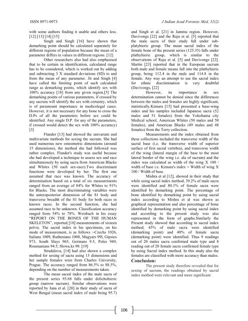
ISSN 0971-0973 J Indian Acad Forensic Med, <strong>32</strong>(2)with some authors finding it usable and others less.[12] [13] [14] [15]Singh and Singh [16] have shown thatdemarking point should be calculated separately fordifferent regions of population because the mean of aparameter differs in values in different regions. [12]Other researchers also had also emphasisedthat to be certain in identification, calculated rangehas to be considered, which is worked out by addingand subtracting 3 X standard deviations (SD) to andfrom the mean of any parameter. Jit and Singh [4]have called the limiting point of such calculatedrange as demarking points, which identify sex with100% accuracy [18] from any given region.[5] Thedemarking points of various parameters, if crossed byany sacrum will identify the sex with certainty, whichis of paramount importance in medicolegal cases.However, it is not necessary for any bone to cross theD.Ps of all the parameters before sex could beidentified. Any single D.P. for any of the parameters,if crossed would detect the sex with 100% accuracy)[3]Flander [13] had showed the univariate andmultivariate methods for sexing the sacrum. She hadused numerous new osteometric dimensions (around15 dimensions), the method she had followed wasrather complex. Flander's study was useful becauseshe had developed a technique to assess sex and racesimultaneously by using sacra from American Blacksand Whites (50 each sex-race).Two discriminantfunctions were developed by her. The first oneassumed that race was known. The accuracy ofdetermination based on a total of six measurementsranged from an average of 84% for Whites to 91%for Blacks. The most discriminating variables werethe anteroposterior dimension of the S1 body andtransverse breadth of the S1 body for both races inknown races. In the second function, she hadassumed race to be unknown. Classification accuracyranged from 54% to 78%. Weisbach in his essay“REPORT ON THE BONES OF THE HUMANSKELETON”, reported [18] measurements of sixteenpelvis. The sacral index in his specimens, on hismode of measurement, is as follows :-Czechs 1026,Italians 1009, Ruthenians 1008, Magyars 99l, Gipsies973, South Slays 965, Germans 9.5, Poles 949,Roumanians 94.5, Slowa.ks 90. [19]Stradalova, [14] had also shown a complexmethod for sexing of sacra using 15 dimensions andher sample females were from Charles University,Prague. The accuracy ranged from 86.5% to 88.5%,depending on the number of measurements taken.The mean sacral index of the male sacra ofthe present series 93.68 falls under dolichohiericgroup (narrow sacrum). Similar observations werereported by Jana et al. [20] in their study of sacra ofWest Bengal (mean sacral index of male being 95.7)and Singh et al. [21] in Jammu region. However,Davivongs [22] and the Raju et al. [5] reported thatthe male sacra of their study fall under subplatyhiericgroup. The mean sacral index of thefemale bone of the present series (125.35) falls underplathyhieric group, which is similar to theobservations of Raju et al. [5] and Davivongs [22].Martin [23] reported that in the European sacrumboth male and female means fall into the plathyhiericgroup, being 112.4 in the male and 114.8 in thefemale. Any way an attempt to use the sacral indexfor ethnic discrimination is very doubtful(Davivongs, [22]However, its importance in sexdetermination cannot be denied since the differencesbetween the males and females are highly significant,statistically.Kimura [15] had presented a base-wingindex and his samples included Japanese sacra (52males and 51 females) from the Yokohama cityMedical school, American Whites (50 males and 50females), and American Blacks (49 males and 48females) from the Terry collection.Measurements and the index obtained fromthese collections included the transverse width of thesacral base (i.e. the transverse width of superiorsurface of first sacral vertebra), and transverse widthof the wing (lateral margin of the base to the mostlateral border of the wing i.e. ala of sacrum) and theindex was calculated as width of the wing X 100 /width of base i.e. Kimura's index = Width of wing X100 / Width of base.Mishra et al [12], showed in their study thatwhile using sacral index method, 39.2% of male sacrawere identified and 80.1% of female sacra wereidentified by demarking point. The percentage ofbone identified by demarking point by using sacralindex according to Mishra et al was shown asgraphical representation and also percentage of boneidentified by demarking point by using sacral indexand according to the present study was alsorepresented in the form of graphs.Similarly thePresent study showed that according to sacral indexmethod; 45% of male sacra were identified(demarking point) and 40% of female sacra(demarking point) were identified. Thus 9 readingsout of 20 males sacra confirmed male type and 8reading out of 20 female sacra confirmed female typeby using Sacral index method. In this study also thefemales are classified with more accuracy than males.Conclusion:The present study therefore revealed that forsexing of sacrum, the readings obtained by sacralindex method were relevant and more significant.106
![syllabus in forensic medicine for m.b.b.s. students in india [pdf]](https://img.yumpu.com/48405011/1/190x245/syllabus-in-forensic-medicine-for-mbbs-students-in-india-pdf.jpg?quality=85)
![SPOTTING IN FORENSIC MEDICINE [pdf]](https://img.yumpu.com/45856557/1/190x245/spotting-in-forensic-medicine-pdf.jpg?quality=85)
![JAFM-33-2, April-June, 2011 [PDF] - forensic medicine](https://img.yumpu.com/43461356/1/190x245/jafm-33-2-april-june-2011-pdf-forensic-medicine.jpg?quality=85)
![JIAFM-33-4, October-December, 2011 [PDF] - forensic medicine](https://img.yumpu.com/31013278/1/190x245/jiafm-33-4-october-december-2011-pdf-forensic-medicine.jpg?quality=85)