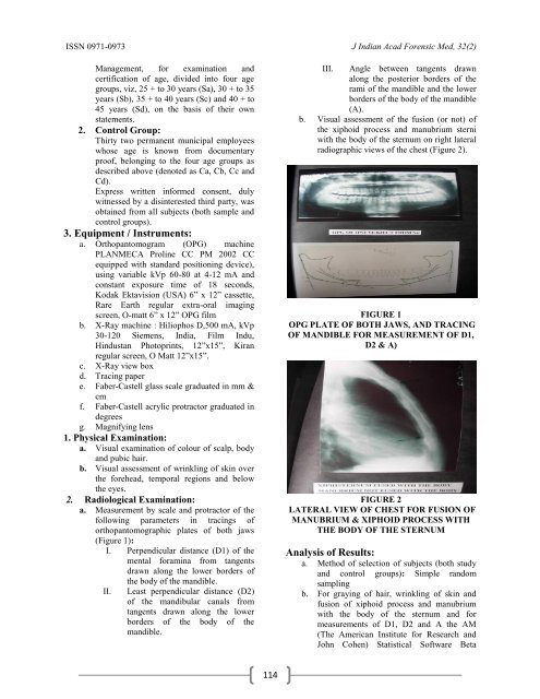
ISSN 0971-0973 J Indian Acad Forensic Med, <strong>32</strong>(2)Management, for examination andcertification of age, divided into four agegroups, viz, 25 + to 30 years (Sa), 30 + to 35years (Sb), 35 + to 40 years (Sc) and 40 + to45 years (Sd), on the basis of their ownstatements.2. Control Group:Thirty two permanent municipal employeeswhose age is known from documentaryproof, belonging to the four age groups asdescribed above (denoted as Ca, Cb, Cc andCd).Express written informed consent, dulywitnessed by a disinterested third party, wasobtained from all subjects (both sample andcontrol groups).3. Equipment / Instruments:a. Orthopantomogram (OPG) machinePLANMECA Proline CC PM 2002 CCequipped with standard positioning device),using variable kVp 60-80 at 4-12 mA andconstant exposure time of 18 seconds,Kodak Ektavision (USA) 6” x 12” cassette,Rare Earth regular extra-oral imagingscreen, O-matt 6” x 12” OPG filmb. X-Ray machine : Hiliophos D,500 mA, kVp30-120 Siemens, India, Film Indu,Hindustan Photoprints, 12”x15”, Kiranregular screen, O Matt 12”x15”.c. X-Ray view boxd. Tracing papere. Faber-Castell glass scale graduated in mm &cmf. Faber-Castell acrylic protractor graduated indegreesg. Magnifying lens1. Physical Examination:a. Visual examination of colour of scalp, bodyand pubic hair.b. Visual assessment of wrinkling of skin overthe forehead, temporal regions and belowthe eyes.2. Radiological Examination:a. Measurement by scale and protractor of thefollowing parameters in tracings oforthopantomographic plates of both jaws(Figure 1):I. Perpendicular distance (D1) of themental foramina from tangentsdrawn along the lower borders ofthe body of the mandible.II. Least perpendicular distance (D2)of the mandibular canals fromtangents drawn along the lowerborders of the body of themandible.III. Angle between tangents drawnalong the posterior borders of therami of the mandible and the lowerborders of the body of the mandible(A).b. Visual assessment of the fusion (or not) ofthe xiphoid process and manubrium sterniwith the body of the sternum on right lateralradiographic views of the chest (Figure 2).FIGURE 1OPG PLATE OF BOTH JAWS, AND TRACINGOF MANDIBLE FOR MEASUREMENT OF D1,D2 & A)FIGURE 2LATERAL VIEW OF CHEST FOR FUSION OFMANUBRIUM & XIPHOID PROCESS WITHTHE BODY OF THE STERNUMAnalysis of Results:a. Method of selection of subjects (both studyand control groups): Simple randomsamplingb. For graying of hair, wrinkling of skin andfusion of xiphoid process and manubriumwith the body of the sternum and formeasurements of D1, D2 and A the AM(The American Institute for Research andJohn Cohen) Statistical Software Beta114
J Indian Acad Forensic Med, <strong>32</strong>(2) ISSN 0971-0973Version – 0.06.02 was used for theexecution of the regression command andfor preparation of a prediction equation forthe age estimation.Observations and Results:The observations made in the <strong>32</strong> subjects ineach group were recorded in a master chart. Groupwiseobservations of each parameter are displayed inthe Tables.It was seen (Table No.1) that the scalp hairof 2 subjects in the control group of age group 25-30years (Ca) had grayed, while this was seen in only 1subject from this study group (Sa). None of thesubjects from either control or study groups hadgraying of hair over the body or pubic area at thisage.In the age group of 31-35 years, graying ofscalp hair was noticed in 2 subjects each in thecontrol group (Cb) and study group (Sb). Neithergroup had any subject with gray body or pubic hair.7 subjects in the control group in the age range of 36-40 years (Cc) had gray scalp hair and 2 showedgraying of body hair, but their pubic hair was stillblack in colour. In the study group (Sc), there were 7subjects with gray scalp hair, 3 with gray body hairand none with gray pubic hair.In the last age group (41-45 years) 8 subjectsin the control group (Cd) unmistakably had grayscalp hair and 6 of them had gray body hair.However, none of them had graying of pubic hair. Inthe study group (Sd), 8 subjects had evident grayingof scalp hair, 8 had gray body hair and only one ofthem showed graying of pubic hair.The first composite variable, i.e.,“Combined Hair Score” (CHS) was derived fromTable No. 2 by allocating a linear addition of 1 pointeach for the presence (0 for absence) of graying ofhair at the three different sites, viz., scalp, body andpubis. Thus the total score ranged from a minimum of0 to a maximum of 3.Table No. 2 shows the observations ofwrinkling of the skin. None of the subjects in thecontrol group (Ca) in the age group of 25-30 yearshad wrinkles on either the forehead or over thetemporal region, but only one had wrinkles below hiseyes. In the study group (Sa) none of the subjects hadwrinkles in any of these locations.In the second age group (31-35) years, noneof the subjects in the control group (Cb) had wrinklesover the forehead, 2 of them had overt wrinkles overthe temporal region and none of them had wrinklingof skin below the eyes. In the study group (Sb), 1subject was spotted with wrinkles over the forehead,two with wrinkles over the temporal region and onewith wrinkles below the eyes.In the age group of 36-40 years, fivesubjects in the control group (Cc), had wrinkles overthe forehead, seven wrinkles over the temporal regionand two wrinkles below the eyes. In the study group(Sc), wrinkles over forehead were observed in sixsubjects, wrinkles over the temporal region in seven,and wrinkles below the eyes in five.Eight subjects from the control group (Cd)in the age range of 41-45 years undeniably hadwrinkling of skin over the forehead, eight over thetemporal region and six below the eyes. In the studygroup (Sd), wrinkling of skin was noted in eightsubjects each at the three sites.A “Combined Skin Score” (CSS) wascalculated from this data, also by linear addition ofone point each for presence (0 for the absence) ofwrinkled skin at three different sites, viz., forehead,temporal region and below the eyes, with a total scorerange from 0 to 3.The combined skin score was found to be2.562, with a standard error of 0.613 and astatistically significant p > I t I value of 0.000. It was,therefore, used for the equation for estimation of agein a prediction model.It is evident from Table No. 3 that neitherthe xiphoid process nor the manubrium had fusedwith the body of sternum in either control group (Ca)or study group (Sa) in the age group of 25-30 years.Similarly, in the age group of 31-35 years, the studyof skiagrams did not display such fusion in any of thesubjects in the control group (Cb). In the study group(Sb), however, the xiphoid process of one subject hadfused with the body of the sternum. Fusion of themanubrium with the body of the sternum was notseen in any of them.Fusion of neither the xiphoid process normanubrium with the body of the sternum hadoccurred in any subject in the control group in the agegroup of 36-40 years (Cc). On the other hand, in thestudy group (Sc), the skiagrams of five subjectsmanifested fusion of xiphoid process with the body ofthe sternum, but manubrio-sternal fusion had nottaken place in even one of them.The xiphoid process had fused with the bodyof the sternum in eight subjects from the controlgroup (Cd) in the age group of 41-45 years; in noneof them had the manubrium fused with the body ofthe sternum. In the study group (Sd), eight subjectshad radiological evidence of xiphoid-sternal fusion,but in none of them had the manubrium fused withthe body of sternum.From this data, a Combined Bone FusionScore (CBFS) was calculated by linear addition of 1point each for the presence (0 point for the absence)of the fusion to the body of the sternum of its twoother components, viz., the manubrium and thexiphisternum. Thus the total score of the CBFS couldrange from a minimum of 0 to a maximum of 2.115


![syllabus in forensic medicine for m.b.b.s. students in india [pdf]](https://img.yumpu.com/48405011/1/190x245/syllabus-in-forensic-medicine-for-mbbs-students-in-india-pdf.jpg?quality=85)
![SPOTTING IN FORENSIC MEDICINE [pdf]](https://img.yumpu.com/45856557/1/190x245/spotting-in-forensic-medicine-pdf.jpg?quality=85)

![JAFM-33-2, April-June, 2011 [PDF] - forensic medicine](https://img.yumpu.com/43461356/1/190x245/jafm-33-2-april-june-2011-pdf-forensic-medicine.jpg?quality=85)
![JIAFM-33-4, October-December, 2011 [PDF] - forensic medicine](https://img.yumpu.com/31013278/1/190x245/jiafm-33-4-october-december-2011-pdf-forensic-medicine.jpg?quality=85)