Download - forensic medicine
Download - forensic medicine
Download - forensic medicine
Create successful ePaper yourself
Turn your PDF publications into a flip-book with our unique Google optimized e-Paper software.
88<br />
JIAFM, 2007 - 29(4); ISSN: 0971-0973<br />
nerve palsy had the presenting symptom and these signs get resolved spontaneously but child had died due to<br />
some other cause. The history or clinical evidence of trauma to jaw was specifically excluded in all cases in this<br />
study.<br />
Technique and Results:<br />
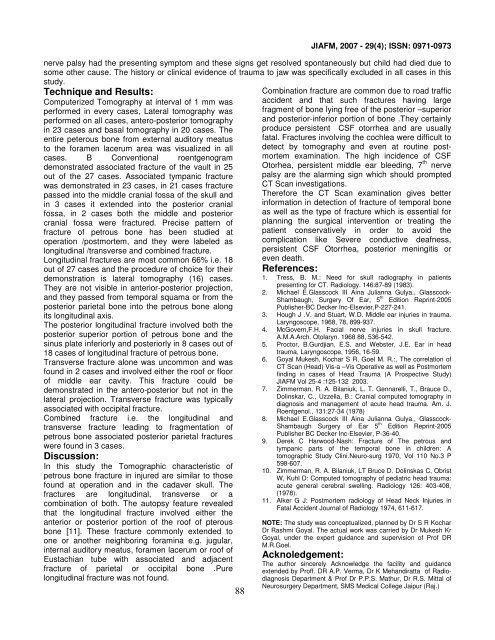
Computerized Tomography at interval of 1 mm was<br />
performed in every cases, Lateral tomography was<br />
performed on all cases, antero-posterior tomography<br />
in 23 cases and basal tomography in 20 cases. The<br />
entire peterous bone from external auditory meatus<br />
to the foramen lacerum area was visualized in all<br />
cases. B Conventional roentgenogram<br />
demonstrated associated fracture of the vault in 25<br />
out of the 27 cases. Associated tympanic fracture<br />
was demonstrated in 23 cases, in 21 cases fracture<br />
passed into the middle cranial fossa of the skull and<br />
in 3 cases it extended into the posterior cranial<br />
fossa, in 2 cases both the middle and posterior<br />
cranial fossa were fractured. Precise pattern of<br />
fracture of petrous bone has been studied at<br />
operation /postmortem, and they were labeled as<br />
longitudinal /transverse and combined fracture.<br />
Longitudinal fractures are most common 66% i.e. 18<br />
out of 27 cases and the procedure of choice for their<br />
demonstration is lateral tomography (16) cases.<br />
They are not visible in anterior-posterior projection,<br />
and they passed from temporal squama or from the<br />
posterior parietal bone into the petrous bone along<br />
its longitudinal axis.<br />
The posterior longitudinal fracture involved both the<br />
posterior superior portion of petrous bone and the<br />
sinus plate inferiorly and posteriorly in 8 cases out of<br />
18 cases of longitudinal fracture of petrous bone.<br />
Transverse fracture alone was uncommon and was<br />
found in 2 cases and involved either the roof or floor<br />
of middle ear cavity. This fracture could be<br />
demonstrated in the antero-posterior but not in the<br />
lateral projection. Transverse fracture was typically<br />
associated with occipital fracture.<br />
Combined fracture i.e. the longitudinal and<br />
transverse fracture leading to fragmentation of<br />
petrous bone associated posterior parietal fractures<br />
were found in 3 cases.<br />
Discussion:<br />
In this study the Tomographic characteristic of<br />
petrous bone fracture in injured are similar to those<br />
found at operation and in the cadaver skull. The<br />
fractures are longitudinal, transverse or a<br />
combination of both. The autopsy feature revealed<br />
that the longitudinal fracture involved either the<br />
anterior or posterior portion of the roof of pterous<br />
bone [11]. These fracture commonly extended to<br />
one or another neighboring foramina e.g. jugular,<br />
internal auditory meatus, foramen lacerum or roof of<br />
Eustachian tube with associated and adjacent<br />
fracture of parietal or occipital bone .Pure<br />
longitudinal fracture was not found.<br />
Combination fracture are common due to road traffic<br />
accident and that such fractures having large<br />
fragment of bone lying free of the posterior –superior<br />
and posterior-inferior portion of bone .They certainly<br />
produce persistent CSF otorrhea and are usually<br />
fatal. Fractures involving the cochlea were difficult to<br />
detect by tomography and even at routine postmortem<br />
examination. The high incidence of CSF<br />
Otorhea, persistent middle ear bleeding, 7 th nerve<br />
palsy are the alarming sign which should prompted<br />
CT Scan investigations.<br />
Therefore the CT Scan examination gives better<br />
information in detection of fracture of temporal bone<br />
as well as the type of fracture which is essential for<br />
planning the surgical intervention or treating the<br />
patient conservatively in order to avoid the<br />
complication like Severe conductive deafness,<br />
persistent CSF Otorrhea, posterior meningitis or<br />
even death.<br />
References:<br />
1. Tress, B. M.: Need for skull radiography in patients<br />
presenting for CT. Radiology. 146:87-89 (1983).<br />
2. Michael E.Glasscock III Aina Julianna Gulya., Glasscock-<br />
Shambaugh, Surgery Of Ear, 5 th Edition Reprint-2005<br />
Publisher-BC Decker Inc-Elsevier,P-227-241.<br />
3. Hough J .V. and Stuart, W.D. Middle ear injuries in trauma.<br />
Laryngoscope, 1968, 78, 899-937.<br />
4. McGovern,F.H. Facial nerve injuries in skull fracture.<br />
A.M.A.Arch. Otolaryn. 1968 88, 536-542.<br />
5. Proctor, B.Gurdjian, E.S. and Webster, J.E. Ear in head<br />
trauma, Laryngoscope, 1956, 16-59.<br />
6. Goyal Mukesh, Kochar S R, Goel M. R.:, The correlation of<br />
CT Scan (Head) Vis-a –Vis Operative as well as Postmortem<br />
finding in cases of Head Trauma (A Prospective Study)<br />
JIAFM Vol 25-4 :125-132 2003.<br />
7. Zimmerman, R. A. Bilaniuk, L. T. Gennarelli, T., Brauce D.,<br />
Dolinskar, C., Uzzella, B.: Cranial computed tomography in<br />
diagnosis and management of acute head trauma. Am. J.<br />
Roentgenol., 131:27-34 (1978)<br />
8. Michael E.Glasscock III Aina Julianna Gulya., Glasscock-<br />
Shambaugh Surgery of Ear 5 th Edition Reprint-2005<br />
Publisher BC Decker Inc-Elsevier, P-36-40.<br />
9. Derek C Harwood-Nash: Fracture of The petrous and<br />
tympanic parts of the temporal bone in children: A<br />
tomographic Study Clini.Neuro-surg 1970, Vol 110 No.3 P<br />
598-607.<br />
10. Zimmerman, R. A. Bilaniuk, LT Bruce D. Dolinskas C, Obrist<br />
W, Kuhl D: Computed tomography of pediatric head trauma:<br />
acute general cerebral swelling. Radiology 126: 403-408,<br />
(1978).<br />
11. Alker G J: Postmortem radiology of Head Neck Injuries in<br />
Fatal Accident Journal of Radiology 1974, 611-617.<br />
NOTE: The study was conceptualized, planned by Dr S R Kochar<br />
Dr Rashmi Goyal. The actual work was carried by Dr Mukesh Kr<br />
Goyal, under the expert guidance and supervision of Prof DR<br />
M.R.Goel.<br />
Acknoledgement:<br />
The author sincerely Acknowledge the facility and guidance<br />
extended by Proff. DR A.P. Verma, Dr K Mehandiratta of Radiodiagnosis<br />
Department & Prof Dr P.P.S. Mathur, Dr R.S. Mittal of<br />
Neurosurgery Department, SMS Medical College Jaipur (Raj.)



![syllabus in forensic medicine for m.b.b.s. students in india [pdf]](https://img.yumpu.com/48405011/1/190x245/syllabus-in-forensic-medicine-for-mbbs-students-in-india-pdf.jpg?quality=85)
![SPOTTING IN FORENSIC MEDICINE [pdf]](https://img.yumpu.com/45856557/1/190x245/spotting-in-forensic-medicine-pdf.jpg?quality=85)
![JAFM-33-2, April-June, 2011 [PDF] - forensic medicine](https://img.yumpu.com/43461356/1/190x245/jafm-33-2-april-june-2011-pdf-forensic-medicine.jpg?quality=85)
![JIAFM-33-4, October-December, 2011 [PDF] - forensic medicine](https://img.yumpu.com/31013278/1/190x245/jiafm-33-4-october-december-2011-pdf-forensic-medicine.jpg?quality=85)
