Gamma-ray Spectroscopy with LaBr3:Ce Scintillator ... - MPG HLL
Gamma-ray Spectroscopy with LaBr3:Ce Scintillator ... - MPG HLL
Gamma-ray Spectroscopy with LaBr3:Ce Scintillator ... - MPG HLL

You also want an ePaper? Increase the reach of your titles
YUMPU automatically turns print PDFs into web optimized ePapers that Google loves.
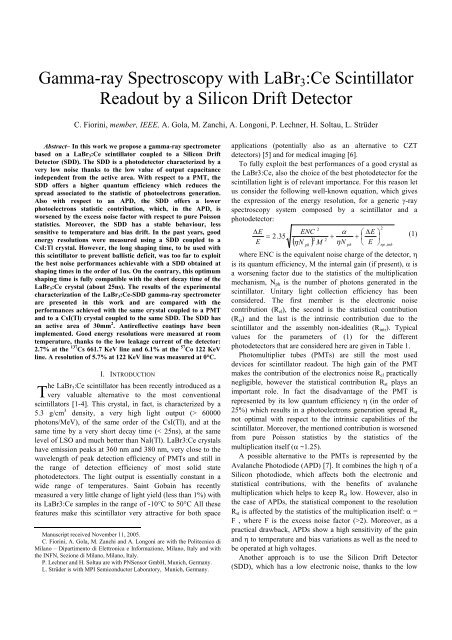
<strong>Gamma</strong>-<strong>ray</strong> <strong>Spectroscopy</strong> <strong>with</strong> LaBr 3 :<strong>Ce</strong> <strong>Scintillator</strong><br />
Readout by a Silicon Drift Detector<br />
C. Fiorini, member, IEEE, A. Gola, M. Zanchi, A. Longoni, P. Lechner, H. Soltau, L. Strüder<br />
Abstract– In this work we propose a gamma-<strong>ray</strong> spectrometer<br />
based on a LaBr 3 :<strong>Ce</strong> scintillator coupled to a Silicon Drift<br />
Detector (SDD). The SDD is a photodetector characterized by a<br />
very low noise thanks to the low value of output capacitance<br />
independent from the active area. With respect to a PMT, the<br />
SDD offers a higher quantum efficiency which reduces the<br />
spread associated to the statistic of photoelectrons generation.<br />
Also <strong>with</strong> respect to an APD, the SDD offers a lower<br />
photoelectrons statistic contribution, which, in the APD, is<br />
worsened by the excess noise factor <strong>with</strong> respect to pure Poisson<br />
statistics. Moreover, the SDD has a stable behaviour, less<br />
sensitive to temperature and bias drift. In the past years, good<br />
energy resolutions were measured using a SDD coupled to a<br />
CsI:Tl crystal. However, the long shaping time, to be used <strong>with</strong><br />
this scintillator to prevent ballistic deficit, was too far to exploit<br />
the best noise performances achievable <strong>with</strong> a SDD obtained at<br />
shaping times in the order of 1us. On the contrary, this optimum<br />
shaping time is fully compatible <strong>with</strong> the short decay time of the<br />
LaBr 3 :<strong>Ce</strong> crystal (about 25ns). The results of the experimental<br />
characterization of the LaBr 3 :<strong>Ce</strong>-SDD gamma-<strong>ray</strong> spectrometer<br />
are presented in this work and are compared <strong>with</strong> the<br />
performances achieved <strong>with</strong> the same crystal coupled to a PMT<br />
and to a CsI(Tl) crystal coupled to the same SDD. The SDD has<br />
an active area of 30mm 2 . Antireflective coatings have been<br />
implemented. Good energy resolutions were measured at room<br />
temperature, thanks to the low leakage current of the detector:<br />
2.7% at the 137 Cs 661.7 KeV line and 6.1% at the 57 Co 122 KeV<br />
line. A resolution of 5.7% at 122 KeV line was measured at 0°C.<br />
T<br />
I. INTRODUCTION<br />
he LaBr 3 :<strong>Ce</strong> scintillator has been recently introduced as a<br />
very valuable alternative to the most conventional<br />
scintillators [1-4]. This crystal, in fact, is characterized by a<br />
5.3 g/cm 3 density, a very high light output (> 60000<br />
photons/MeV), of the same order of the CsI(Tl), and at the<br />
same time by a very short decay time (< 25ns), at the same<br />
level of LSO and much better than NaI(Tl). <strong>LaBr3</strong>:<strong>Ce</strong> crystals<br />
have emission peaks at 360 nm and 380 nm, very close to the<br />
wavelength of peak detection efficiency of PMTs and still in<br />
the range of detection efficiency of most solid state<br />
photodetectors. The light output is essentially constant in a<br />
wide range of temperatures. Saint Gobain has recently<br />
measured a very little change of light yield (less than 1%) <strong>with</strong><br />
its <strong>LaBr3</strong>:<strong>Ce</strong> samples in the range of -10°C to 50°C All these<br />
features make this scintillator very attractive for both space<br />
Manuscript received November 11, 2005.<br />
C. Fiorini, A. Gola, M. Zanchi and A. Longoni are <strong>with</strong> the Politecnico di<br />
Milano – Dipartimento di Elettronica e Informazione, Milano, Italy and <strong>with</strong><br />
the INFN, Sezione di Milano, Milano, Italy.<br />
P. Lechner and H. Soltau are <strong>with</strong> PNSensor GmbH, Munich, Germany.<br />
L. Strüder is <strong>with</strong> MPI Semiconductor Laboratory, Munich, Germany.<br />
applications (potentially also as an alternative to CZT<br />
detectors) [5] and for medical imaging [6].<br />
To fully exploit the best performances of a good crystal as<br />
the <strong>LaBr3</strong>:<strong>Ce</strong>, also the choice of the best photodetector for the<br />
scintillation light is of relevant importance. For this reason let<br />
us consider the following well-known equation, which gives<br />
the expression of the energy resolution, for a generic γ-<strong>ray</strong><br />
spectroscopy system composed by a scintillator and a<br />
photodetector:<br />
ΔE<br />
E<br />
= 2.35<br />
2<br />
ENC<br />
2<br />
M<br />
( ηN<br />
)<br />
ph<br />
2<br />
α<br />
+<br />
ηN<br />
ph<br />
⎛ ΔE<br />
⎞<br />
+ ⎜ ⎟<br />
⎝ E ⎠<br />
2<br />
np , inh<br />
where ENC is the equivalent noise charge of the detector, η<br />
is its quantum efficiency, M the internal gain (if present), α is<br />
a worsening factor due to the statistics of the multiplication<br />
mechanism, N ph is the number of photons generated in the<br />
scintillator. Unitary light collection efficiency has been<br />
considered. The first member is the electronic noise<br />
contribution (R el ), the second is the statistical contribution<br />
(R st ) and the last is the intrinsic contribution due to the<br />
scintillator and the assembly non-idealities (R intr ). Typical<br />
values for the parameters of (1) for the different<br />
photodetectors that are considered here are given in Table 1.<br />
Photomultiplier tubes (PMTs) are still the most used<br />
devices for scintillator readout. The high gain of the PMT<br />
makes the contribution of the electronics noise R el practically<br />
negligible, however the statistical contribution R st plays an<br />
important role. In fact the disadvantage of the PMT is<br />
represented by its low quantum efficiency η (in the order of<br />
25%) which results in a photoelectrons generation spread R st<br />
not optimal <strong>with</strong> respect to the intrinsic capabilities of the<br />
scintillator. Moreover, the mentioned contribution is worsened<br />
from pure Poisson statistics by the statistics of the<br />
multiplication itself (α ~1.25).<br />
A possible alternative to the PMTs is represented by the<br />
Avalanche Photodiode (APD) [7]. It combines the high η of a<br />
Silicon photodiode, which affects both the electronic and<br />
statistical contributions, <strong>with</strong> the benefits of avalanche<br />
multiplication which helps to keep R el low. However, also in<br />
the case of APDs, the statistical component to the resolution<br />
R st is affected by the statistics of the multiplication itself: α =<br />
F , where F is the excess noise factor (>2). Moreover, as a<br />
practical drawback, APDs show a high sensitivity of the gain<br />
and η to temperature and bias variations as well as the need to<br />
be operated at high voltages.<br />
Another approach is to use the Silicon Drift Detector<br />
(SDD), which has a low electronic noise, thanks to the low<br />
(1)
value of output capacitance. In this case R el can be<br />
significantly low, <strong>with</strong>out using an internal multiplication<br />
mechanism. The statistical contribution is kept close to the<br />
pure Poisson limit thanks to both the high quantum efficiency<br />
of the photodetector, and by the absence of the multiplication,<br />
so that α = 1 (see Table 1).<br />
If we plot the energy resolution vs. γ-<strong>ray</strong> energy, as shown<br />
in Fig.1, there is a low-energy region where the electronic<br />
contribution is expected to be dominant, if present, like in<br />
APDs and SDDs; then we find a region dominated by the<br />
statistical contribution, and in this case the SDD is expected to<br />
have better energy resolution performances <strong>with</strong> respect to the<br />
other photodetectors. Finally there is a region where the<br />
intrinsic contribution of the scintillator is dominant. In this<br />
case, the choice of the photodetector has no significant effect.<br />
II. THE SDD DETECTOR<br />
The Silicon Drift Detector is a silicon detector,<br />
characterized by a particular charge collection mechanism,<br />
such as the electrons generated in the active area of the device<br />
drift towards the anode, which has a small size and a very<br />
small capacitance, independent from the total active area of<br />
the device. Equation 2 expresses the ENC of the detector, as a<br />
function of the shaping time used for signal processing.<br />
2 2<br />
⎛ A<br />
⎞<br />
1<br />
ENC = C<br />
T<br />
⎜ a + A2<br />
2 πa<br />
f<br />
+ A3τb<br />
τ<br />
⎟<br />
(2)<br />
⎝ s<br />
⎠<br />
In this equation C T is the value of the total anode<br />
capacitance (detector + preamplifier + parasitic), τ s is the<br />
shaping time, a is the white series noise, a f is the 1/f noise<br />
coefficient and b is the white parallel noise due to the leakage<br />
current, which can be reduced by cooling. From (2) it can be<br />
seen that a small value of C T reduces the electronic noise<br />
<strong>with</strong>out the need of a multiplication mechanism. Moreover, as<br />
the ENC is dependent on the shaping time, there is an<br />
optimum value for this parameter, which is usually of the<br />
order of 1 μs for the SDDs (this value is dependent on the<br />
operating temperature).<br />
SDDs used for scintillation readout have already<br />
demonstrated to achieve state-of-the-art energy resolution in<br />
γ-<strong>ray</strong> spectroscopy using a CsI:Tl scintillator [8] as well as<br />
sub-millimetre position resolution in γ-<strong>ray</strong> imaging [9].<br />
However, the use of a slow scintillator like CsI:Tl (decay time<br />
~1 μs) did not allow to fully exploit the best noise<br />
performances offered by the SDD, which are obtained using<br />
shaping times of the order of 1 μs. Longer shaping times were<br />
needed <strong>with</strong> CsI:Tl for not being excessively affected by<br />
ballistic deficit. In this case, the electronic noise of the SDD<br />
was dominated by the contribution of the leakage current<br />
(white parallel noise, see (2)) which had to be reduced by<br />
cooling.<br />
The recently introduced LaBr 3: <strong>Ce</strong> scintillator has a much<br />
shorter decay time, of the order of 25 ns, which does not<br />
generate ballistic deficit, even when used at shaping times of 1<br />
μs or less. This benefit has however to be considered together<br />
<strong>with</strong> the collection mechanism of the scintillation-generated<br />
charge inside the SDD. In fact, the drift time necessary for the<br />
charge to reach the anode of the photodetector prevents the<br />
use of a too short shaping time. For the device presented in the<br />
next section, the drift time from the outermost region of the<br />
active area is estimated to be about 0.7μs.<br />
III. DETECTOR CHARACTERIZATION<br />
The detector we have used for the measurements is a<br />
circular SDD, <strong>with</strong> an active area of 30mm 2 , produced at the<br />
Semiconductor Laboratory of Max Planck Institut in Munich.<br />
The light enters from the bottom side of the detector, which is<br />
made by an homogeneous entrance window. The input JFET<br />
of the readout electronics has been integrated in the detector<br />
itself, in order to further reduce the total anode capacitance,<br />
and consequently the electronic noise at short shaping times.<br />
Recent technological improvements in the fabrication process<br />
of the SDD include a reduced leakage current, of about<br />
200pA/cm 2 at 25°C, which makes the device very attractive<br />
for measurements near room temperature. Another feature is<br />
the presence of the bonding pads outside the active area; a<br />
layout of the chip is shown in Fig. 2.<br />
The principle schematic of the readout electronics used in<br />
the measurements <strong>with</strong> the SDD is shown in Fig.3. The JFET<br />
integrated on the detector is operated in a source follower<br />
configuration <strong>with</strong> an external current source. The voltage<br />
signal is obtained by the conversion of the charge signal into<br />
voltage on the total capacitance C d connected at the detector<br />
output. The signal is amplified by means of a voltage<br />
amplifier. This is realized by means of a coupling capacitor<br />
followed by a charge preamplifer. The voltage gain on the<br />
signal step is given by C K /C F while the decay time of the<br />
preamplifier waveform is equals to R F C F . The signal is further<br />
processed by a Tennelec TC244 shaping amplifier.<br />
The electronic noise of the device have been characterized<br />
by directly irradiating the device <strong>with</strong> an X-<strong>ray</strong> source of 55 Fe<br />
(5.9 KeV Mn Kα), at different temperatures, ranging from -<br />
10°C to 23°C. The optimum energy resolution achieved was<br />
145 eV FWHM (9.8 e - rms), obtained at -10°C and <strong>with</strong> a<br />
shaping time of 1 μs. At room temperature we obtained 237<br />
eV FWHM at 0.25 μs. The corresponding spectra are shown<br />
in Fig. 4.<br />
If we plot the measured electronics noise <strong>with</strong> respect to<br />
the shaping time we find a behavior, in accordance to (2),<br />
which is shown in Fig. 5. It is also worth mentioning that the<br />
shaping times compatible <strong>with</strong> LaBr 3: <strong>Ce</strong> correspond roughly<br />
to the minimum of the ENC curve, while the CsI:Tl lays in a<br />
region <strong>with</strong> higher noise, especially at room temperature.<br />
The entrance window of the detector was also provided<br />
<strong>with</strong> custom anti-reflective coatings (ARCs). The presence of<br />
these coatings is important, because they improve the<br />
Quantum Efficiency η at the wavelengths of interest,<br />
increasing the collected signal charge, and, from (1), this has<br />
positive effects both on the electronic contribution R el and on<br />
the statistical contribution R st . A plot of the measured η vs.
incident wavelength for these ARCs and for standard coatings<br />
is shown in Fig. 6. The ARC coatings were originally<br />
designed to have a maximum efficiency at λ = 565 nm<br />
(CsI:Tl), where we have measured η = 90%, but still have η =<br />
80% at λ = 400nm (the lower end of our QE measurement<br />
sensitivity).<br />
IV. γ-RAY SPECTROSCOPY SET-UP AND MEASUREMENTS<br />
For γ-<strong>ray</strong> measurements the SDD was optically coupled to a<br />
LaBr 3 :<strong>Ce</strong> Brillance 380 crystal module, produced by Saint<br />
Gobain. The crystal has a cylindrical shape <strong>with</strong> 5mm<br />
diameter and 5mm thickness. No specifications were available<br />
on the <strong>Ce</strong> doping of the crystal and on the light output of the<br />
assembled module. The crystal was already mounted <strong>with</strong> a<br />
light reflector and sealed in an aluminium housing. A picture<br />
of the scintillator coupled to the SDD photodetector is shown<br />
in Fig. 7.<br />
The back side of the detector was biased at -86 V, while the<br />
last ring, providing the charge drift mechanism, was biased at<br />
about -110V. The first measurement was done to evaluate the<br />
conversion gain between the energy of the γ-photon and the<br />
number of photoelectrons. Using the data from the X-<strong>ray</strong><br />
measurements, we obtained a conversion gain of about 28 e -<br />
/KeV. Assuming, from the data sheets, a light yield of the<br />
scintillator of 60 ph/KeV and extrapolating from the curve<br />
shown in Fig. 6 a value for η of about 70% at λ = 370 nm (the<br />
central wavelength of the emission spectrum of LaBr 3 :<strong>Ce</strong>), we<br />
can evaluate a light collection efficiency of about 70%. This<br />
means that there is still room for improvements, both in the<br />
assembly and in η, which should be tuned to match λ = 370<br />
nm.<br />
With the system described, we carried out a set of<br />
spectroscopic measurements at room temperature, <strong>with</strong><br />
different gamma sources. Fig. 8 shows the spectrum obtained<br />
irradiating the scintillator <strong>with</strong> a 137 Cs source. The energy<br />
resolution is 2.7% and the 662 keV escape peak, as shown in<br />
the expanded region plot on the right side, is clearly<br />
distinguishable. As far as the electronic noise contribution is<br />
already known from the X-<strong>ray</strong> spectroscopic measurements,<br />
and the statistical contribution can be obtained from the<br />
conversion gain, it is possible to evaluate from (1) the intrinsic<br />
contribution, due to the crystal and to the assembly, which is<br />
2% in this case. This value is quite high, if compared to the<br />
one measured by Shah and co-workers [7], who estimated the<br />
same contribution to be of 0.9%, using a LaBr 3 :<strong>Ce</strong> scintillator<br />
by RMD, coupled to a 8mm 2 APD cooled at 250K.<br />
It is worth mentioning that the same scintillator was used<br />
<strong>with</strong> a PMT by the manufacturer, and an energy resolution of<br />
3% have been measured. This is actually not among the best<br />
values measured <strong>with</strong> a LaBr 3 :<strong>Ce</strong> scintillator. The same<br />
manufacturer reports resolutions as good as 2.8% <strong>with</strong> other<br />
samples. We could estimate the potential improvement in<br />
resolution using the SDD photodetector <strong>with</strong> a LaBr 3 :<strong>Ce</strong><br />
scintillator characterized by a better intrinsic contribution. For<br />
instance, considering the intrinsic resolution of 0.9% quoted<br />
by RMD and using the R el and R st contributions for the SDD<br />
quoted in the first column of Table II, a resolution of 2.0% at<br />
662keV can be estimated.<br />
In Fig. 9, the spectrum of a 57 Co source measured at room<br />
temperature is shown. The energy resolution (FWHM) at the<br />
122keV peak is of 6.1%. In this spectrum, the peak at 14 keV<br />
is well distinguished from the noise threshold. The same<br />
measurement, carried out <strong>with</strong> the PMT by the manufacturer,<br />
has given a value of 6.5%.<br />
In this case the electronic noise is no longer negligible; to<br />
improve the results we repeated the measurements <strong>with</strong> a<br />
moderate cooling, operating the system at 0°C. The<br />
corresponding spectrum, reported in Fig. 10, shows an energy<br />
resolution of 5.7%.<br />
The energy resolutions measured <strong>with</strong> the SDD and PMT<br />
are listed in Table 2, together <strong>with</strong> the estimated contributions.<br />
Combining these measurements <strong>with</strong> others, made <strong>with</strong> a<br />
241 Am source, it is possible to draw a plot of the energy<br />
resolution vs. energy, shown in Fig. 11, which fits the<br />
experimental data.<br />
It is also interesting to compare the results obtained at 122<br />
keV <strong>with</strong> LaBr 3 :<strong>Ce</strong> <strong>with</strong> the ones obtained <strong>with</strong> a CsI:Tl<br />
crystal, using the same SDD as a photodetector. The<br />
comparison is shown in Fig. 12. The main difference between<br />
the two scintillators is that CsI:Tl has a much longer decay<br />
time which forces to use the SDD at longer shaping times and<br />
<strong>with</strong> a higher electronic noise, as anticipated in section II. The<br />
energy resolution of the LaBr 3 :<strong>Ce</strong> crystal readout by the PMT<br />
is also reported.<br />
V. CONCLUSIONS<br />
In conclusion the measurements have shown that the SDD<br />
coupled to a LaBr 3 :<strong>Ce</strong> scintillator is a good photodetector for<br />
γ-<strong>ray</strong> spectroscopy, and can obtain better results than a PMT.<br />
The performances achieved are due to the low electronic noise<br />
of the SDD and to an improved photodetection efficiency, but<br />
there is still room for improvements, especially in the<br />
optimization of the detection module. Applications in medical<br />
imaging and γ-<strong>ray</strong> spectrometry are foreseen in the near<br />
future.<br />
VI. ACKNOWLEDGMENTS<br />
The authors would like to thank Mike Mayhugh (Saint<br />
Gobain) for his support.<br />
REFERENCES<br />
[1] E.V.D.van Loef, P.Dorenbos, C.W.E. van Eijk, K.Krämer, H.U.Güdel,<br />
Appl.Phys.Lett., vol.79, p. 1573, 2001.<br />
[2] P. Dorenbos, Nucl. Instr. Meth., vol. A 486, pp. 208-213, 2002.<br />
[3] E. V. D. van Loef, et al., Nucl. Instr. Meth., vol. A 486, pp. 254-258,<br />
2002.<br />
[4] P. Dorenbos, J. T. M. de Haas, C. W. E. Van Eijk, IEEE Trans. Nucl.<br />
Sci., vol.51, n.3, p.1289, 2004.<br />
[5] M.L.McConnel, et al., SPIE 5488, 2004.<br />
[6] W.Moses, K.S.Shah, Nucl. Instr. Meth., vol. A 537, pp. 317-320, 2005.<br />
[7] K.S.Shah, et al., IEEE Trans. Nucl. Sci., vol.51, n.5, p.2395, 2004.
[8] C. Fiorini, et al., IEEE Trans. Nucl. Sci., vol. 44, no. 6, pp. 2553-2560,<br />
1997.<br />
[9] C.Fiorini, F.Perotti, Review of Scientific Instruments, 76, 044303,<br />
2005.
TABLE I<br />
PHOTODETECTOR TYPICAL PARAMETERS<br />
Parameter PMT APD SDD<br />
η ~25% >80% >80%<br />
α 1+υ (~1.25) F (>2) 1<br />
M ~ 10 6 ~ 10 3 1<br />
TABLE II<br />
DIFFERENT CONTRIBUTIONS TO THE ENERGY RESOLUTION<br />
SDD<br />
137 Cs<br />
T amb<br />
PMT<br />
137 Cs<br />
T amb<br />
SDD<br />
57 Co<br />
T amb<br />
PMT<br />
57 Co<br />
T amb<br />
SDD<br />
57 Co<br />
T = 0°C<br />
R el 0.5% ~0% 2.9% ~0% 1.9%<br />
R st 1.7% 2.2% 4.0% 5.4% 4.0%<br />
R intr 2% 2% 3.6% 3.6% 3.6%<br />
R tot 2.7% 3% 6.1% 6.5% 5.7%<br />
Energy resolution (%)<br />
10<br />
electronics<br />
noise<br />
total<br />
statistics<br />
scintillator<br />
non idealities<br />
1<br />
10 100 1000<br />
Energy (keV)<br />
Fig. 1. Plot of the energy resolution vs. incoming γ-<strong>ray</strong> energy, for a system composed of a scintillator crystal coupled to a photodetector. It is possible to<br />
distinguish three regions, each one dominated by a particular contribution to the resolution. In the region on the right side of the graph, above few hundreds<br />
of keV, scintillator non idealities dominates <strong>with</strong> respect to the other contributions.<br />
Fig. 2. Layout of the Silicon Drift Detector used for the γ-<strong>ray</strong> measurement. On the left side of the figure one can see the bonding pads, which are external to<br />
the active area and are connected to the central region of the device.
Fig. 3. Principle schematic of the readout electronics used in the measurements.<br />
4000<br />
5.9 keV<br />
5000<br />
55 Fe spectrum<br />
5.9 keV<br />
Counts<br />
3000<br />
2000<br />
55 Fe spectrum 4000 5000 6000 7000<br />
237eV<br />
FWHM<br />
Counts<br />
4000<br />
3000<br />
2000<br />
145eV<br />
FWHM<br />
1000<br />
1000<br />
4000 5000 6000 7000<br />
Energy [eV]<br />
Energy [eV]<br />
Fig. 4. Energy spectra of a 55 Fe source, measured at room temperature (left) <strong>with</strong> 0.25μs shaping time and at -10°C (right) <strong>with</strong> 1μs shaping time. The longer<br />
shaping time used at -10°C is used thanks to the reduced leakage current (see also Fig. 4).
Fig. 5. ENC curve of the 30 mm 2 SDD measured at different temperatures. In this figure the shaping time range to be used <strong>with</strong> two different scintillators, due<br />
to their decay times, are also shown.<br />
Fig. 6. Graph of the Quantum Efficiency η measured <strong>with</strong> the 30 mm 2 SDD detector, <strong>with</strong> custom and standard anti-reflection coatings.<br />
Fig. 7. Picture of the circular SDD coupled to the LaBr 3:<strong>Ce</strong> module. The detector is placed on a ceramic substrate for biasing and signal extraction.<br />
2x10 6 2x10 4<br />
137<br />
Cs spectrum<br />
661.7 keV<br />
6x10 4<br />
661.7 keV<br />
1x10 6<br />
4x10 4<br />
2.7%<br />
FWHM<br />
escape<br />
5x10 5<br />
32 keV<br />
Ba X-<strong>ray</strong>s<br />
0 200 400 600<br />
E [k V]<br />
600 650 700<br />
Energy [keV]
Fig. 8. Spectrum of a 137 Cs source, measured at room temperature (23°C). In the left side of the figure, the same spectrum is shown in two different vertical<br />
scales to better show both 32keV X-<strong>ray</strong> lines and 662keV line. The escape peak of the 661.7 KeV line (shown in the expanded region on the right side of the<br />
figure) is clearly distinguishable and the noise threshold is well below the 32 KeV X-<strong>ray</strong> line.<br />
2000<br />
57<br />
Co spectrum<br />
122 keV<br />
Counts<br />
1500<br />
1000<br />
6.1%<br />
FWHM<br />
500<br />
0<br />
14 keV<br />
Escape<br />
136.4 keV<br />
0 40 80 120 160<br />
Energy [keV]<br />
Fig. 9. Spectrum of a 57 Co source, measured at room temperature (23°C). The 14 KeV line is clearly distinguished from noise. Laescape<br />
peak is visible in figure.<br />
2000<br />
57<br />
Co spectrum<br />
122 keV<br />
Counts<br />
1500<br />
1000<br />
5.7%<br />
FWHM<br />
500<br />
0<br />
14 keV<br />
Escape<br />
136.4 keV<br />
0 40 80 120 160<br />
Energy [keV]<br />
Fig. 10. Same measurements of Fig. 8, but made at a lower temperature, in order to reduce the electronic noise contribution. The energy<br />
resolution has improved to 5.7%.
Fig. 11. Plot of the energy resolution vs. energy for the 30 mm 2 SDD used for γ-<strong>ray</strong> measurements.<br />
Fig. 12. Comparison between the energy resolution obtained at different shaping times for LaBr 3 :<strong>Ce</strong> and CsI:Tl. Also the measurement <strong>with</strong> the<br />
LaBr 3:<strong>Ce</strong> crystal using a PMT is reported.