Interesting Case Series Bilateral Macrostomia - ePlasty
Interesting Case Series Bilateral Macrostomia - ePlasty
Interesting Case Series Bilateral Macrostomia - ePlasty

You also want an ePaper? Increase the reach of your titles
YUMPU automatically turns print PDFs into web optimized ePapers that Google loves.
<strong>Interesting</strong> <strong>Case</strong> <strong>Series</strong><br />
<strong>Bilateral</strong> <strong>Macrostomia</strong><br />
DarleneM.Guse,BS,BA, a and Vincent A. Naman, MD b<br />
a Mayo Clinic—Mayo Medical School, Rochester, Minn; and b Chattahoochee Plastic Surgery,<br />
Columbus, Ga<br />
Correspondence: dguse55@hotmail.com<br />
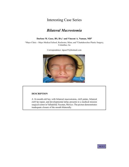
DESCRIPTION<br />
A 16-month-old boy with bilateral macrostomia, cleft palate, bilateral<br />
cleft lip repair, and developmental delay presents to a medical mission<br />
surgical center in Valladolid, Yucatan, Mexico. The picture demonstrates<br />
inadequate closure of the mouth bilaterally.<br />
NEXT
QUESTIONS<br />
1. What is the epidemiology of this condition?<br />
2. What other symptoms may be present in a patient presenting with macrostomia?<br />
3. What surgical techniques are used for reconstruction of this defect?<br />
BACK<br />
NEXT
DISCUSSION<br />
<strong>Macrostomia</strong> is most often associated with a transverse facial cleft, classified<br />
by Tessier as a number 7 cleft. It is found in 1 out of 80,000 births and<br />
occurs more frequently in males. Most cases are unilateral, but bilateral<br />
involvement can be seen in 10% to 20% of cases. More than 50% of bilateral<br />
cases are isolated without additional ear or skin deformities. However,<br />
the defect is often larger than in unilateral macrostomia.<br />
The severity of the cleft can vary, and thus, additional symptoms may or<br />
may not be present. Examples include preauricular tags; microtia; zygomatic<br />
arch deficiency; deformities of the mandibular ramus, condyle, and<br />
coronoid process; and other craniofacial clefts. The cleft can also be part of<br />
an underlying syndrome and present with an absent orbital rim and eyelash<br />
hypoplasia in Treacher Collins syndrome, facial asymmetry in hemifacial<br />
microsomia, and vertebral anomalies in Goldenhar syndrome.<br />
Surgical repair is more complicated in bilateral macrostomia than unilateral,<br />
because the surgeon lacks a comparative normal side and must<br />
define the location of the commissure without oral anatomical aide. Fortunately,<br />
the vermilion often thins at the approximate location of the natural<br />
commissure and can align with a line from the pupils to the infraorbital<br />
and submental foramina.<br />
Three layers must be considered for repair: mucosal, muscular, and skin<br />
layers. The mucosal layer is closed with simple interrupted absorbable<br />
sutures, with the commissural sutures attaching the mucosal layer to either<br />
the overlying muscle or skin. Function for speaking, eating, and blowing<br />
is dependent upon correction of the muscular layer. Involved muscles consist<br />
of the orbicularis, buccinator, and zygomatic muscles. The superior<br />
orbicularis muscle is sutured at a 90-degree angle over the inferior orbicularis<br />
muscle. The remaining muscles are similarly overlapped and sutured<br />
together.<br />
The skin layer is the final layer to consider and many techniques have<br />
been described in the literature, including simple linear closure, Z-plasty,<br />
and W-plasty for the lateral portion of the cleft. The simple line closure can<br />
result in a noticeable long, linear scar that can contract or become hypertrophic.<br />
The Z-plasty, positioned to lie within the nasolabial fold, and the<br />
W-plasty minimize this and prevent lateralization of the commissure. Final<br />
consideration must be given to the vermilion. Although direct suturing of<br />
mucosa to skin is most common, various flaps have been described which<br />
result in superior repositioning to further minimize scar visibility.<br />
BACK<br />
NEXT
REFERENCES<br />
1. Gleizal A, Wan DC, Picard A, et al. <strong>Bilateral</strong> macrostomia as an isolated pathology. Cleft Palate Craniofac<br />
J. 2007;44(1):58-61.<br />
2. Fadeyibi IO, Ugburo AO, Ogunbanjo CV, et al. The surgical repair of macrostomia. Cleft Palate Craniofac<br />
J. 2009;46(6):642-7.<br />
3. Jaworski S. <strong>Macrostomia</strong>. A modified technique of surgical repair. Acta Chir Plast 1976;(18):117-21.<br />
4. Tessier, P. Anatomical classification of facial, cranio-facial and latero-facial clefts. J. Maxillofac Surg.<br />
1976;(4):69.<br />
5. Woods RH, Varma S, David DJ. Tessier no. 7 cleft: a new subclassification and management protocol.<br />
Plast Reconstr Surg. 2008;122(3):898-905.<br />
Darlene M. Guse. <strong>Bilateral</strong> <strong>Macrostomia</strong>. www.<strong>ePlasty</strong>.com, <strong>Interesting</strong> <strong>Case</strong>, April 1, 2010<br />
BACK