Living Images: Fluorescence microscopy Camera Lens ... - Carl Zeiss
Living Images: Fluorescence microscopy Camera Lens ... - Carl Zeiss
Living Images: Fluorescence microscopy Camera Lens ... - Carl Zeiss

Create successful ePaper yourself
Turn your PDF publications into a flip-book with our unique Google optimized e-Paper software.
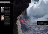
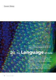
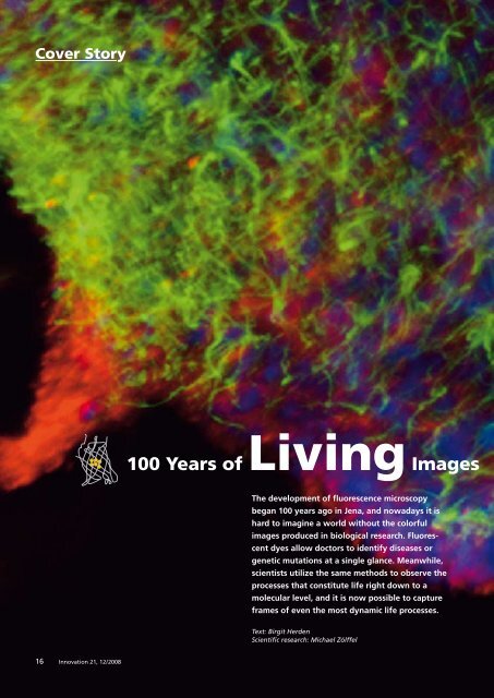
Cover Story<br />
100 Years of <strong>Living</strong> <strong>Images</strong><br />
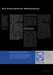
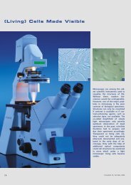
The development of fluorescence <strong>microscopy</strong><br />
began 100 years ago in Jena, and nowadays it is<br />
hard to imagine a world without the colorful<br />
images produced in biological research. Fluorescent<br />
dyes allow doctors to identify diseases or<br />
genetic mutations at a single glance. Meanwhile,<br />
scientists utilize the same methods to observe the<br />
processes that constitute life right down to a<br />
molecular level, and it is now possible to capture<br />
frames of even the most dynamic life processes.<br />
Text: Birgit Herden<br />
Scientific research: Michael Zölffel<br />
16<br />
Innovation 21, 12 / 2008