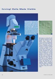
Living Images: Fluorescence microscopy Camera Lens ... - Carl Zeiss
Living Images: Fluorescence microscopy Camera Lens ... - Carl Zeiss
Living Images: Fluorescence microscopy Camera Lens ... - Carl Zeiss

Create successful ePaper yourself
Turn your PDF publications into a flip-book with our unique Google optimized e-Paper software.
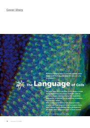
Cover story: <strong>Living</strong> images<br />
jellyfish has been modified and is<br />
now seeing the highest level of application.<br />
Perhaps the potentially<br />
most useful fluorescent proteins belong<br />
to a new class termed “optical<br />
highlighters.” These are proving to<br />
be very efficacious in tracking dynamic<br />
events in living cells. In general,<br />
fluorescent proteins are non-toxic<br />
and feature excellent photostability,<br />
although their brightness falls far<br />
short of that exhibited by synthetic<br />
dyes and quantum dots.<br />
In the future, we can expect to see<br />
significant advances in the realm of<br />
fluorophore technologies. Improvements<br />
in synthetics will continue as<br />
manufacturers strive to compete<br />
with fluorescent proteins. Furthermore,<br />
quantum dots will become<br />
very useful. Hybrid systems are also<br />
receiving a considerable amount of<br />
attention and many new strategies<br />
are emerging. Finally, there appears<br />
to be no end to the new and promising<br />
fluorophore candidates emerging<br />
from fluorescent proteins. Dynamic<br />
biosensors that monitor a<br />
variety of cellular processes, including<br />
pH, voltage and sugar metabolism<br />
are being introduced in laboratories<br />
around the world.<br />
Littered with acronyms such as PAL-<br />
M, STED, 4Pi, STORM, SIM, and<br />
RESOLFT, the field of superresolution<br />
<strong>microscopy</strong> is currently the most rapidly<br />
evolving frontier in fluorescence<br />
imaging. Almost every month heralds<br />
the introduction of a new technique<br />
that promises to stretch or<br />
break the diffraction barrier. Although<br />
seemingly unrelated in many<br />
respects, the diverse technologies<br />
that comprise the foundation of superresolution<br />
<strong>microscopy</strong> all have<br />
the common goal of imaging biological<br />
specimens on a molecular level.<br />
Among the important questions for<br />
the future are exactly where are the<br />
resolution limits and how quickly<br />
and effectively can we bridge the<br />
gap (if at all) between optical and<br />
electron <strong>microscopy</strong>? More importantly,<br />
can all of this be done with<br />
living cells and, at some point, even<br />
live animals?<br />
Several manufacturers are now starting<br />
to offer solutions or are are<br />
working on developments that<br />
promise to spread this technology to<br />
the mainstream. Still, the complex<br />
fluorophore technology for superresolution<br />
<strong>microscopy</strong> is highly demanding<br />
and most methods have<br />
not yet been extrapolated to live-cell<br />
imaging. Hopefully this technology<br />
will be developed to the point of<br />
having auxiliary devices that attach<br />
to widefield or confocal microscopes<br />
with plug-in software modules for<br />
gathering images.<br />
The complete article can be found<br />
at www.zeiss.com/innovation<br />
The details<br />
Virtual Campus<br />
Together with biophysicist Michael<br />
W. Davidson from Florida<br />
State University, <strong>Carl</strong> <strong>Zeiss</strong> has<br />
created an education and science<br />
platform on the Internet<br />
regarding the topic of <strong>microscopy</strong><br />
and digital images. The<br />
virtual “ZEISS Campus” is a<br />
knowledge source that presents<br />
theory and technology<br />
based on applications. On this<br />
website, methods and techniques<br />
of fluorescence <strong>microscopy</strong><br />
are described with the aid<br />
of detailed descriptions, interactive<br />
animations, application<br />
examples and picture galleries.<br />
All materials were developed<br />
on microscope systems from<br />
<strong>Carl</strong> <strong>Zeiss</strong>.<br />
The websites not only provide<br />
information about current scientific<br />
topics regarding fluorescence<br />
<strong>microscopy</strong> – users also<br />
have the opportunity to publish<br />
their applications. With this<br />
website, <strong>Carl</strong> <strong>Zeiss</strong> is addressing<br />
all “microscopers,” particularly<br />
young scientists who can<br />
expand their knowledge at the<br />
“ZEISS Campus.”<br />
Further information<br />
is available at<br />
www.zeiss.com/campus<br />
Innovation 21, 12 / 2008<br />
31