Living Images: Fluorescence microscopy Camera Lens ... - Carl Zeiss
Living Images: Fluorescence microscopy Camera Lens ... - Carl Zeiss
Living Images: Fluorescence microscopy Camera Lens ... - Carl Zeiss

You also want an ePaper? Increase the reach of your titles
YUMPU automatically turns print PDFs into web optimized ePapers that Google loves.
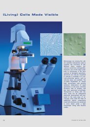
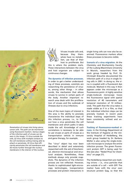
Penetration of viruses into living uterus<br />
cancer cells. The path can be monitored<br />
using fluorescent markers. Various stages<br />
of the infection path of viruses can be<br />
seen in the image. (1) Virus outside the<br />
cells, (2) Virus with multiple contacts to<br />
the cell membrane without being able to<br />
attach or penetrate, (3) Virus that efficiently<br />
penetrates the cell membrane and<br />
(4) moves further through the nucleus<br />
membrane in the interior of the nucleus.<br />
Viruses invade cells and,<br />
because they themselves<br />
have no metabolism,<br />
use that of their<br />
host to proliferate. But<br />
this is where the problem starts.<br />
These interactions between the virus<br />
and host organism are subject to<br />
continuous changes.<br />
The dynamics of infection processes.<br />
In order to get a better understanding<br />
of these processes, scientists are<br />
researching the persistence of viruses,<br />
among other things – in other<br />
words, the mechanism that allows<br />
viruses to survive in certain parts of<br />
the body. Another important research<br />
area deals with the proliferation<br />
of viruses and the outbreak of<br />
illnesses due to virus infections.<br />
One of the main topics of interest in<br />
this area is the ability to precisely<br />
characterize the individual steps of<br />
this infection process, i.e., to find<br />
out how a virus penetrates the cell<br />
membrane and which paths it takes<br />
inside the cell. Knowledge of such<br />
correlations is necessary to be able<br />
to use viruses or parts of viruses as a<br />
transportation vehicle in immune<br />
and gene therapy, for example.<br />
The “virus” object has now been<br />
described in detail and extensively<br />
researched with the aid of biochemical,<br />
electron-microscopical and structural<br />
analyses. Nonetheless, these<br />
methods always only provide snapshots.<br />
The dynamics of the infection<br />
process are not reflected. However,<br />
thanks to sophisticated light-<strong>microscopy</strong><br />
technologies, transportation<br />
processes and protein interactions in<br />
single living cells can now also be examined.<br />
<strong>Fluorescence</strong> markers allow<br />
these processes to become visible.<br />
Scenario of a virus migration. At the<br />
Chemistry and Biochemistry Faculty<br />
of the Ludwig-Maximilians University<br />
in Munich, researchers from the<br />
work group headed by Prof. Dr.<br />
Christoph Bräuchle documented the<br />
infection path of a virus in single living<br />
cells in 2001. In doing so, the virus<br />
is coupled with a fluorescent dye<br />
molecule. Marked in this way it then<br />
appears under the microscope as a<br />
fluorescence point. A highly sensitive<br />
single-molecule microscope traces<br />
the fluorescence signal with spatial<br />
resolution of 40 nanometers and<br />
temporal resolution of 10 milliseconds.<br />
The path that the virus takes is<br />
made visible as if in a film, so that<br />
the individual infection steps can be<br />
precisely tracked. In recent years<br />
these tracking experiments have<br />
been consistently refined and enhanced.<br />
Tracking of fluorescence-marked viruses.<br />
In the Virology Department at<br />
the Institute of Hygiene at the University<br />
Hospital of Heidelberg, the<br />
work group headed by Dr. Barbara<br />
Müller is also using the single-molecule<br />
microscope to analyze the entire<br />
infection process. The green fluorescent<br />
protein GFP is being used for<br />
this (see also: “Nobel Prize for Chemistry”<br />
on page 21).<br />
The Heidelberg researchers are marking<br />
virions – i.e., virus particles that<br />
are outside of a cell – with GFP. The<br />
marker fuses with the virus' own<br />
structure protein Gag, so that the<br />
28 Innovation 21, 12 / 2008