Machine Learning Methods for Protein Structure ... - ResearchGate
Machine Learning Methods for Protein Structure ... - ResearchGate
Machine Learning Methods for Protein Structure ... - ResearchGate

Create successful ePaper yourself
Turn your PDF publications into a flip-book with our unique Google optimized e-Paper software.
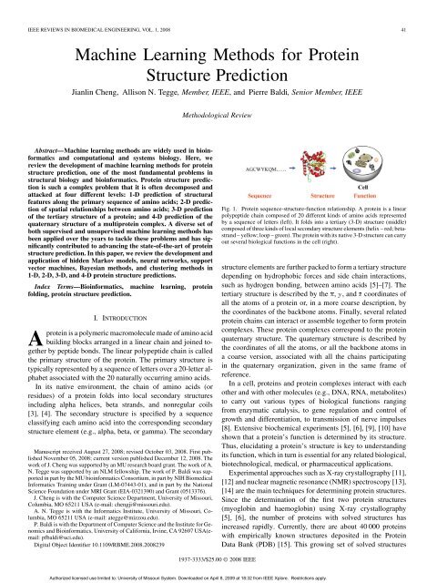
IEEE REVIEWS IN BIOMEDICAL ENGINEERING, VOL. 1, 2008 41<br />
<strong>Machine</strong> <strong>Learning</strong> <strong>Methods</strong> <strong>for</strong> <strong>Protein</strong><br />
<strong>Structure</strong> Prediction<br />
Jianlin Cheng, Allison N. Tegge, Member, IEEE, and Pierre Baldi, Senior Member, IEEE<br />
Methodological Review<br />
Abstract—<strong>Machine</strong> learning methods are widely used in bioin<strong>for</strong>matics<br />
and computational and systems biology. Here, we<br />
review the development of machine learning methods <strong>for</strong> protein<br />
structure prediction, one of the most fundamental problems in<br />
structural biology and bioin<strong>for</strong>matics. <strong>Protein</strong> structure prediction<br />
is such a complex problem that it is often decomposed and<br />
attacked at four different levels: 1-D prediction of structural<br />
features along the primary sequence of amino acids; 2-D prediction<br />
of spatial relationships between amino acids; 3-D prediction<br />
of the tertiary structure of a protein; and 4-D prediction of the<br />
quaternary structure of a multiprotein complex. A diverse set of<br />
both supervised and unsupervised machine learning methods has<br />
been applied over the years to tackle these problems and has significantly<br />
contributed to advancing the state-of-the-art of protein<br />
structure prediction. In this paper, we review the development and<br />
application of hidden Markov models, neural networks, support<br />
vector machines, Bayesian methods, and clustering methods in<br />
1-D, 2-D, 3-D, and 4-D protein structure predictions.<br />
Index Terms—Bioin<strong>for</strong>matics, machine learning, protein<br />
folding, protein structure prediction.<br />
I. INTRODUCTION<br />
Aprotein is a polymeric macromolecule made of amino acid<br />
building blocks arranged in a linear chain and joined together<br />
by peptide bonds. The linear polypeptide chain is called<br />
the primary structure of the protein. The primary structure is<br />
typically represented by a sequence of letters over a 20-letter alphabet<br />
associated with the 20 naturally occurring amino acids.<br />
In its native environment, the chain of amino acids (or<br />
residues) of a protein folds into local secondary structures<br />
including alpha helices, beta strands, and nonregular coils<br />
[3], [4]. The secondary structure is specified by a sequence<br />
classifying each amino acid into the corresponding secondary<br />
structure element (e.g., alpha, beta, or gamma). The secondary<br />
Manuscript received August 27, 2008; revised October 03, 2008. First published<br />
November 05, 2008; current version published December 12, 2008. The<br />
work of J. Cheng was supported by an MU research board grant. The work of A.<br />
N. Tegge was supported by an NLM fellowship. The work of P. Baldi was supported<br />
in part by the MU bioin<strong>for</strong>matics Consortium, in part by NIH Biomedical<br />
In<strong>for</strong>matics Training under Grant (LM-07443-01), and in part by the National<br />
Science Foundation under MRI Grant (EIA-0321390) and Grant (0513376).<br />
J. Cheng is with the Computer Science Department, University of Missouri,<br />
Columbia, MO 65211 USA (e-mail: chengji@missouri.edu).<br />
A. N. Tegge is with the In<strong>for</strong>matics Institute, University of Missouri, Columbia,<br />
MO 65211 USA (e-mail: ategge@mizzou.edu).<br />
P. Baldi is with the Department of Computer Science and the Institute <strong>for</strong> Genomics<br />
and Bioin<strong>for</strong>matics, University of Cali<strong>for</strong>nia, Irvine, CA 92697 USA(email:<br />
pfbaldi@uci.edu).<br />
Digital Object Identifier 10.1109/RBME.2008.2008239<br />
Fig. 1. <strong>Protein</strong> sequence-structure-function relationship. A protein is a linear<br />
polypeptide chain composed of 20 different kinds of amino acids represented<br />
by a sequence of letters (left). It folds into a tertiary (3-D) structure (middle)<br />
composed of three kinds of local secondary structure elements (helix – red; betastrand<br />
– yellow; loop – green). The protein with its native 3-D structure can carry<br />
out several biological functions in the cell (right).<br />
structure elements are further packed to <strong>for</strong>m a tertiary structure<br />
depending on hydrophobic <strong>for</strong>ces and side chain interactions,<br />
such as hydrogen bonding, between amino acids [5]–[7]. The<br />
tertiary structure is described by the , , and coordinates of<br />
all the atoms of a protein or, in a more coarse description, by<br />
the coordinates of the backbone atoms. Finally, several related<br />
protein chains can interact or assemble together to <strong>for</strong>m protein<br />
complexes. These protein complexes correspond to the protein<br />
quaternary structure. The quaternary structure is described by<br />
the coordinates of all the atoms, or all the backbone atoms in<br />
a coarse version, associated with all the chains participating<br />
in the quaternary organization, given in the same frame of<br />
reference.<br />
In a cell, proteins and protein complexes interact with each<br />
other and with other molecules (e.g., DNA, RNA, metabolites)<br />
to carry out various types of biological functions ranging<br />
from enzymatic catalysis, to gene regulation and control of<br />
growth and differentiation, to transmission of nerve impulses<br />
[8]. Extensive biochemical experiments [5], [6], [9], [10] have<br />
shown that a protein’s function is determined by its structure.<br />
Thus, elucidating a protein’s structure is key to understanding<br />
its function, which in turn is essential <strong>for</strong> any related biological,<br />
biotechnological, medical, or pharmaceutical applications.<br />
Experimental approaches such as X-ray crystallography [11],<br />
[12] and nuclear magnetic resonance (NMR) spectroscopy [13],<br />
[14] are the main techniques <strong>for</strong> determining protein structures.<br />
Since the determination of the first two protein structures<br />
(myoglobin and haemoglobin) using X-ray crystallography<br />
[5], [6], the number of proteins with solved structures has<br />
increased rapidly. Currently, there are about 40 000 proteins<br />
with empirically known structures deposited in the <strong>Protein</strong><br />
Data Bank (PDB) [15]. This growing set of solved structures<br />
1937-3333/$25.00 © 2008 IEEE<br />
Authorized licensed use limited to: University of Missouri System. Downloaded on April 8, 2009 at 18:32 from IEEE Xplore. Restrictions apply.
42 IEEE REVIEWS IN BIOMEDICAL ENGINEERING, VOL. 1, 2008<br />
Fig. 2. One-dimensional protein structure prediction. Three-dimensional example of 1-D protein structure prediction where the input primary sequence of amino<br />
acid is “translated” into an output sequence of secondary structure assignments <strong>for</strong> each amino acid (C =coil; H=helix; E=beta-strand[extended sheet)].<br />
provides invaluable in<strong>for</strong>mation to help further understand how<br />
a protein chain folds into its unique 3-D structure, how chains<br />
interact in quaternary complexes, and how to predict structures<br />
from primary sequences [16].<br />
Since the pioneering experiments [1], [2], [5], [6], [17]<br />
showing that a protein’s structure is dictated by its sequence,<br />
predicting protein structure from its sequence has become one<br />
of the most fundamental problems in structural biology (Fig. 1).<br />
This is not only a fundamental theoretical challenge but also<br />
a practical one due to the discrepancy between the number<br />
of protein sequences and solved structures. In the genomic<br />
era, with the application of high-throughput DNA and protein<br />
sequencing technologies, the number of protein sequences<br />
has increased exponentially, at a pace that exceeds the pace at<br />
which protein structures are solved experimentally. Currently,<br />
only about 1.5% of protein sequences (about 40 000 out of<br />
2.5 million known sequences available) have solved structures<br />
and the gap between proteins with known structures and with<br />
unknown structures is still increasing.<br />
In spite of progress in robotics and other areas, experimental<br />
determination of a protein structure can still be expensive, labor<br />
intensive, time consuming, and not always possible. Some of the<br />
hardest challenges involve large quaternary complexes or particular<br />
classes of proteins, such as membrane proteins which are<br />
associated with a complex lipid bilayer environment. These proteins<br />
are particularly difficult to crystallize. Although membrane<br />
proteins are extremely important <strong>for</strong> biology and medicine, only<br />
a few dozen membrane protein structures are available in the<br />
PDB. Thus, in the remainder of this paper we focus almost exclusively<br />
on globular, nonmembrane proteins that are typically<br />
found in the cytoplasm or the nucleus of the cell, or that are secreted<br />
by the cell.<br />
<strong>Protein</strong> structure prediction software is becoming an important<br />
proteomic tool <strong>for</strong> understanding phenomena in modern<br />
molecular and cell biology [18] and has important applications<br />
in biotechnology and medicine [19]. Here, we look at protein<br />
structure prediction at multiple levels, from 1-D to 4-D [20]<br />
and focus on the contributions made by machine learning approaches<br />
[21]. The 1-D prediction focuses on predicting structural<br />
features such as secondary structure [22]–[25] and relative<br />
solvent accessibility [26], [27] of each residue along the<br />
primary 1-D protein sequence (Fig. 2). The 2-D prediction focuses<br />
on predicting the spatial relationship between residues,<br />
such as distance and contact map prediction [28], [29] and disulfide<br />
bond prediction [30]–[33] (Fig. 3). One essential characteristic<br />
of these 2-D representations is that they are independent of<br />
any rotations and translations of the protein, there<strong>for</strong>e independent<br />
of any frame of coordinates, which appear only in the 3-D<br />
level. The 3-D prediction focuses on predicting the coordinates<br />
<strong>for</strong> all the residues or atoms of a protein in a 3-D space. Although<br />
the ultimate goal is to predict 3-D structure, 1-D and 2-D predictions<br />
are often used as input <strong>for</strong> 3-D coordinate predictors; furthermore,<br />
1-D and 2-D predictions are also of intrinsic interest<br />
(Fig. 4). Finally, 4-D prediction focuses on the prediction of the<br />
structure of protein complexes comprised of several folded protein<br />
chains (Fig. 5).<br />
The 1-D, 2-D, and 3-D protein structure prediction methods<br />
are routinely evaluated in the Critical Assessment of Techniques<br />
<strong>for</strong> the <strong>Protein</strong> <strong>Structure</strong> Prediction (CASP) [34] experiment—a<br />
community-wide experiment <strong>for</strong> blind protein structure prediction<br />
held every two years since 1994. The 4-D prediction<br />
methods are currently evaluated in the Critical Assessment of<br />
Techniques <strong>for</strong> <strong>Protein</strong> Interaction (CAPRI) [35]—a community-wide<br />
experiment <strong>for</strong> protein interaction. The assessment<br />
results are published in the supplemental issues of the journal<br />
<strong>Protein</strong>s.<br />
To date, the most successful structure prediction methods<br />
have been knowledge based. Knowledge-based methods involve<br />
learning or extracting knowledge from existing solved<br />
protein structures and generalizing the gained knowledge to<br />
new proteins whose structures are unknown. <strong>Machine</strong> learning<br />
methods [21] that can automatically extract knowledge from<br />
the PDB are an important class of tools and have been widely<br />
used in all aspects of protein structure prediction. Here, we<br />
review the development and application of machine learning<br />
methods in 1-D, 2-D, 3-D, and 4-D structure prediction.<br />
We focus primarily on unsupervised clustering methods and<br />
three supervised machine learning methods including hidden<br />
Markov models (HMMs) [21], [36], [37], neural networks [21],<br />
[38], and support vector machines [39] <strong>for</strong> 1-D, 2-D, 3-D, and<br />
4-D structure prediction problems. We emphasize their applications<br />
to the problem of predicting the structure of globular proteins,<br />
which are the most abundant proteins—roughly 75% of<br />
a typical proteome—and <strong>for</strong> which several prediction methods<br />
have been developed. We also briefly review some applications<br />
of these methods to the prediction of the structure of membrane<br />
proteins, although far less training data is available <strong>for</strong> this class<br />
of proteins.<br />
II. MACHINE LEARNING METHODS FOR 1-D<br />
STRUCTURE PREDICTION<br />
Many protein structural feature predictions are 1-D prediction<br />
problems, including, <strong>for</strong> example, secondary structure prediction,<br />
solvent accessibility prediction, disordered region predic-<br />
Authorized licensed use limited to: University of Missouri System. Downloaded on April 8, 2009 at 18:32 from IEEE Xplore. Restrictions apply.
CHENG et al.: MACHINE LEARNING METHODS FOR PROTEIN STRUCTURE PREDICTION 43<br />
Fig. 3. Two-dimensional protein structure prediction. Example depicts a predicted 2-D contact map with an 8 Angstrom cutoff. The protein sequence is aligned<br />
along the sides of the contact map both horizontally and vertically. Each dot represents a predicted contact, i.e., a residue pair whose spatial distance is below 8<br />
Angstroms. For instance, the red dotted lines mark a predicted contact associated with the pair (D, T).<br />
Fig. 4. Three-dimensional protein structure prediction. Three-dimensional<br />
structure predictors often combine in<strong>for</strong>mation from the primary sequence and<br />
the predicted 1-D and 2-D structures to produce 3-D structure predictions.<br />
tion, binding site prediction, functional site prediction, protein<br />
domain boundary prediction, and transmembrane helix prediction<br />
[22], [23], [33], [40]–[45], [96].<br />
The input <strong>for</strong> 1-D prediction problems is a protein primary<br />
sequence and the output is a sequence of predicted features <strong>for</strong><br />
each amino acid in the sequence. The learning goal is to map<br />
the input sequence of amino acids to the output sequence of<br />
features. The 1-D structure prediction problem is often viewed<br />
as a classification problem <strong>for</strong> each individual amino acid in<br />
the protein sequence. Historically, protein secondary structure<br />
prediction has been the most studied 1-D problem and has had<br />
a fundamental impact on the development of protein structure<br />
prediction methods [22], [23], [47]–[49]. Here, we will mainly<br />
focus on machine learning methods <strong>for</strong> secondary structure prediction<br />
of globular proteins. Similar techniques have also been<br />
applied to other 1-D prediction problems.<br />
Early secondary structure prediction methods [47] were<br />
based on extracting the statistical correlations between a<br />
window of consecutive amino acids in a protein sequence and<br />
the secondary structure classification of the amino acid in the<br />
center of the window. Simple correlation methods capture a<br />
certain amount of in<strong>for</strong>mation and can reach an accuracy of<br />
about 50%, well above chance levels. With the development<br />
Authorized licensed use limited to: University of Missouri System. Downloaded on April 8, 2009 at 18:32 from IEEE Xplore. Restrictions apply.
44 IEEE REVIEWS IN BIOMEDICAL ENGINEERING, VOL. 1, 2008<br />
Fig. 5. Four-dimensional protein structure prediction. Four-dimensional prediction<br />
derived by docking individual protein chains to create a protein complex.<br />
of more powerful pattern recognition and nonlinear function<br />
fitting methods, new approaches have been used to predict<br />
protein secondary structures. In the 1980s, feed<strong>for</strong>ward neural<br />
networks were first applied to secondary structure prediction<br />
and significantly improved prediction accuracy to a level in<br />
the 60% to 70% range [48]. This was probably the first time a<br />
large-scale machine learning method was successfully applied<br />
to a difficult problem in bioin<strong>for</strong>matics. A third important<br />
breakthrough occurred with the realization that higher accuracy<br />
could be achieved by using a richer input derived from a multiple<br />
alignment of a sequence to its homologs. This is due to<br />
the fact that protein secondary structure is more conserved than<br />
protein primary sequence—i.e., protein sequences in the same<br />
protein family evolving from the same ancestor have different<br />
amino acid sequences but often maintain the same secondary<br />
structure [50], [51]. Rost and Sander [22], [23] were the first to<br />
combine neural networks with multiple sequence alignments<br />
to improve secondary structure prediction accuracy to about<br />
70%–74%. In this approach, instead of encoding each amino<br />
acid with a sparse binary vector of length 20 containing a single<br />
1-bit located at a different position <strong>for</strong> each different amino<br />
acid, the empirical probabilities (i.e., normalized frequencies)<br />
of the 20 amino acids appearing in the corresponding column<br />
of the multiple sequence alignment are used. The positional<br />
frequency vector, called the profile of the family at the corresponding<br />
position, captures evolutionary in<strong>for</strong>mation related<br />
to the structural properties of the protein family. Profiles are<br />
relatively easy to create and allow one to leverage in<strong>for</strong>mation<br />
contained in the sequence databases (e.g., SWISSPROT [53])<br />
that are much larger than the PDB. Profiles are now used<br />
in virtually all knowledge-based protein structure prediction<br />
methods and have been further refined. For instance, PSI-PRED<br />
[24] uses PSI-BLAST [54] to derive new profiles based on<br />
position specific scoring matrices to further improve secondary<br />
structure prediction.<br />
New algorithmic developments [49], [27] inspired by the<br />
theory of probabilistic graphical models [21] have led to more<br />
sophisticated recursive neural network architectures to try to<br />
improve prediction accuracy by incorporating in<strong>for</strong>mation<br />
that extends beyond the fixed-size window input of traditional<br />
feed<strong>for</strong>ward neural networks. Large ensembles of hundreds of<br />
neural networks have also been used [55]. The new technologies<br />
available along with the increase of protein sequence databases<br />
used to build profiles have improved secondary structure prediction<br />
accuracy to about 78%–80%. Moreover, hybrid methods<br />
[45], [57] that combine neural network approaches with homology<br />
searches have been developed to improve secondary<br />
structure prediction. Homologous proteins are proteins that are<br />
derived from the same evolutionary ancestor and there<strong>for</strong>e tend<br />
to share structural and functional characteristics. A protein that<br />
is strongly homologous to another protein with known structures<br />
in the PDB [15] will likely share a similar structure. In<br />
addition to neural networks, support vector machines (SVMs)<br />
are also another set of statistical machine learning methods<br />
used to predict protein secondary structures and other 1-D<br />
features of globular proteins with good accuracy [58].<br />
<strong>Machine</strong> learning methods are also frequently used to predict<br />
1-D feature of membrane proteins. For instance, neural networks<br />
as well as HMMs have been used to identify membrane<br />
proteins and predict their topology, which include predicting the<br />
location of their alpha-helical or beta-strand regions and the intracellular<br />
or extracellular localization of the loop regions [59],<br />
[96].<br />
While 1-D prediction methods have made good progress<br />
over the past three decades, there is still room <strong>for</strong> some improvement<br />
in both the accuracy and scope of these methods.<br />
For instance, secondary structure prediction accuracy is still at<br />
least 8% below the predicted limit of 88% [60]. The prediction<br />
of protein domain boundaries [33], [40]–[42] and disordered<br />
regions [43]–[45] are still at an early stage of development,<br />
while already showing promising results. Some improvements<br />
may come from algorithmic improvements, <strong>for</strong> instance using<br />
ensemble and meta-learning techniques such as bagging and<br />
boosting [62] to combine classifiers to improve accuracy. Other<br />
improvements may require exploiting new sources of biological<br />
in<strong>for</strong>mation. For instance, gene structure in<strong>for</strong>mation, such<br />
as alternative splicing sites, may be used to improve domain<br />
boundary prediction [42].<br />
III. MACHINE LEARNING METHODS FOR 2-D<br />
STRUCTURE PREDICTION<br />
The classic 2-D structure prediction problem is the prediction<br />
of protein contact maps [28], [63], [64]. A protein contact map<br />
is a matrix , where is either one or zero, depending on<br />
whether the Euclidean distance between the two amino acids at<br />
linear positions and is above a specified distance threshold<br />
(e.g., 8 Angstroms) or not. Distances can be measured, <strong>for</strong><br />
instance, between corresponding backbone carbon atoms.<br />
A coarser contact map can be derived in a similar way by<br />
considering secondary structure elements. Finer contact maps<br />
Authorized licensed use limited to: University of Missouri System. Downloaded on April 8, 2009 at 18:32 from IEEE Xplore. Restrictions apply.
CHENG et al.: MACHINE LEARNING METHODS FOR PROTEIN STRUCTURE PREDICTION 45<br />
can be derived by considering all the atoms of each amino<br />
acid. As previously mentioned, contact map representations<br />
are particularly interesting due to their invariance with respect<br />
to rotations and translations. Given an accurate contact map,<br />
several algorithms can be used to reconstruct the corresponding<br />
protein 3-D structure [65]–[67]. Since a contact map is essentially<br />
another representation of a protein 3-D structure, the<br />
difficulty of predicting a contact map is more or less equivalent<br />
to the difficulty of predicting the corresponding 3-D structure.<br />
Contact maps can also be used to try to infer protein folding<br />
rates [68], [69].<br />
Several machine learning methods, including neural networks<br />
[28], [70]–[72], self-organizing maps [73], and support vector<br />
machines [74] have been applied to contact map prediction.<br />
Standard feed<strong>for</strong>ward neural networks and support vector machines<br />
approaches use two windows around two target amino<br />
acids, and , to predict if they are in contact or not. This can<br />
be viewed as a binary classification problem. Each position in<br />
a window is usually a vector consisting of 20 numbers corresponding<br />
to the 20 profile probabilities, as in the 1-D prediction<br />
problem. Additional useful 1-D in<strong>for</strong>mation that can be<br />
leveraged includes the predicted secondary structure or relative<br />
solvent accessibility of each amino acid. As in 1-D prediction,<br />
methods based on local windows approaches cannot take into<br />
account the effect of amino acids outside of the window. To<br />
overcome this problem, a 2-D-recursive neural network architecture<br />
[29] that in principle can use the entire sequence to derive<br />
each prediction was designed to improve contact map prediction.<br />
In the latest Critical Assessment of Techniques <strong>for</strong> protein<br />
structure prediction (CASP) [34], three methods using standard<br />
neural networks [72], 2-D recursive neural networks [45], and<br />
support vector machines [74] achieved the best results [75].<br />
Despite progress made in the last several years, contact map<br />
prediction remains largely an unsolved problem. The current<br />
precision and recall of medium and long-range contact predictions<br />
is around 28% [74]. Although this number is quite low,<br />
its accuracy is better than the accuracy of contacts generated<br />
by other ab initio 3-D structure prediction methods. Predicted<br />
contact maps are likely to provide some help in 3-D structure<br />
prediction because even a small fraction of correctly predicted<br />
long-range contacts can effectively help build a protein topology<br />
[76].<br />
In addition to the prediction of general residue-residue contact<br />
maps, special attention has been paid to more specific contact<br />
predictions: beta-strand pairing prediction [77] and disulfide<br />
bond prediction [33], [78], [79]. Disulfide bonds are covalent<br />
bonds that can <strong>for</strong>m between cysteine residues. These disulfide<br />
bonds play a crucial role in stabilizing proteins, particularly<br />
small proteins. Disulfide bond prediction involves predicting if<br />
a disulfide bond exists between any two cysteine residues in<br />
a protein. Both neural networks and support vector machines<br />
have been used to predict disulfide bonds. The average precision<br />
and recall per<strong>for</strong>mance measures are slightly above 50%.<br />
Likewise, one can try to predict if two amino acids in two different<br />
beta-strands are paired or not in the same beta sheet. Usually,<br />
two paired beta-residues <strong>for</strong>m hydrogen bonds with each<br />
other or their neighbors and contribute to the stabilization of the<br />
corresponding beta-sheet. In part because of the requirements<br />
imposed by the hydrogen bonding constraints, the accuracy of<br />
amino acid pairing in beta-sheets is above 41%, higher than the<br />
accuracy <strong>for</strong> generic contacts in contact maps. As with other<br />
2-D prediction problems, feed<strong>for</strong>ward and recursive neural networks<br />
have been used to predict beta-sheet pairings. Currently,<br />
the most successful method is a 2-D recursive neural network<br />
approach which takes a grid of beta-residues as inputs [77] and,<br />
together with graph matching algorithms, predicts pairings at<br />
the residue, strand, and sheet levels.<br />
In addition to 2-D prediction <strong>for</strong> globular proteins, these techniques<br />
have recently been used to predict contacts in transmembrane<br />
beta-barrel proteins. Prediction of transmembrane betabarrel<br />
proteins have been used to reconstruct 3-D structures with<br />
reasonable accuracy [59].<br />
To use 2-D prediction more effectively as input features <strong>for</strong><br />
3-D structure prediction, one important task is to further improve<br />
2-D prediction accuracy. As <strong>for</strong> 1-D predictions, progress<br />
may come from improvements in machine learning methods or,<br />
perhaps more effectively, from incorporating more in<strong>for</strong>mative<br />
features in the inputs. For instance, recently mutual in<strong>for</strong>mation<br />
has been shown to be a useful feature <strong>for</strong> 2-D prediction<br />
[72], [74]. On the reconstruction side, several optimization algorithms<br />
exist to try to reconstruct 3-D structures from contact<br />
maps by using Monte Carlo methods [66], [82] and incorporating<br />
experimentally determined contacts or contacts extracted<br />
from template structures into protein structure prediction<br />
[83]–[85] or protein structure determination by NMR methods.<br />
However, these methods cannot reliably reconstruct 3-D structures<br />
from very noisy contact maps that were predicted from<br />
primary sequence in<strong>for</strong>mation alone [66], [82]. Thus, of parallel<br />
importance is the development of more robust 3-D reconstruction<br />
algorithms that can tolerate the noise contained in predicted<br />
contact maps.<br />
IV. MACHINE LEARNING METHODS FOR 3-D<br />
STRUCTURE PREDICTION<br />
<strong>Machine</strong> learning methods have been used in several aspects<br />
of protein 3-D structure prediction such as fold recognition [33],<br />
[86], [87], model generation [89], and model evaluation [90],<br />
[91].<br />
Fold recognition aims to identify a protein, with known structure,<br />
that is presumably similar to the unknown structure of a<br />
query protein. Identification of structural homologs is an essential<br />
step <strong>for</strong> the most successful template-based 3-D structure<br />
prediction approaches. Neural networks were first used <strong>for</strong> this<br />
task in combination with threading [86]. More recently, a general<br />
machine learning framework has been proposed to improve<br />
both the sensitivity and specificity of fold recognition based on<br />
pairwise similarity features between query and template proteins<br />
[88]. Although the current implementation of the framework<br />
uses support vector machines to identify folds, it can be<br />
extended to any other supervised learning method.<br />
In addition to classification methods, HMMs are among the<br />
most important techniques <strong>for</strong> protein fold recognition. Earlier<br />
HMM approaches, such as SAM [92] and HMMer [93], built an<br />
HMM <strong>for</strong> a query with its homologous sequences and then used<br />
this HMM to score sequences with known structures in the PDB<br />
Authorized licensed use limited to: University of Missouri System. Downloaded on April 8, 2009 at 18:32 from IEEE Xplore. Restrictions apply.
46 IEEE REVIEWS IN BIOMEDICAL ENGINEERING, VOL. 1, 2008<br />
using the Viterbi algorithm, an instance of dynamic programming<br />
methods. This can be viewed as a <strong>for</strong>m of profile-sequence<br />
alignment. More recently, profile–profile methods have been<br />
shown to significantly improve the sensitivity of fold recognition<br />
over profile–sequence, or sequence–sequence, methods<br />
[94]. In the HMM version of profile–profile methods, the HMM<br />
<strong>for</strong> the query is aligned with the prebuilt HMMs of the template<br />
library. This <strong>for</strong>m of profile–profile alignment is also computed<br />
using standard dynamic programming methods.<br />
Optimization techniques, such as conjugate gradient descent<br />
and Monte Carlo methods (e.g., simulated annealing) that are<br />
widely used in statistical machine learning methods are also<br />
essential techniques <strong>for</strong> 3-D protein structure generation and<br />
sampling. Conjugate gradient descent (a technique also used in<br />
neural network learning) is used to generate structures in the<br />
most widely used comparative modeling tool Modeller [95].<br />
Lattice Monte Carlo sampling is used in both template-based<br />
and ab initio structure modeling [85] and the most widely used<br />
ab initio fragment assembly tool, Rosetta, uses simulated annealing<br />
sampling techniques [89].<br />
In addition to model generation, machine learning methods<br />
are also widely used to evaluate and select protein models. Most<br />
ab initio structure prediction methods use clustering techniques<br />
to select models [96]. These methods first generate a large population<br />
of candidate models and then cluster them into several<br />
clusters based on the structure similarity between the models,<br />
using -means clustering or some other similar clustering algorithm.<br />
Representative elements from each cluster, such as the<br />
centroids, are then proposed as possible 3-D structures. Usually,<br />
the centroid of the largest cluster is used as the most confident<br />
prediction, although occasionally the centroid of a different<br />
cluster can be even closer to the native structure. In addition<br />
to clustering, supervised learning techniques have been<br />
used to directly assess the quality of a protein model. Neural networks<br />
have been used to estimate the root mean square distance<br />
(RMSD) between a model and the native structure [90]. Support<br />
vector machines have been used to rank protein models [91].<br />
One main challenge of model selection is that current methods<br />
cannot consistently select the best model with lowest RMSD.<br />
For model quality evaluation, the correlation between predicted<br />
scores and real quality scores <strong>for</strong> hard targets (poor models) is<br />
still low [97], i.e., some poor models may receive good predicted<br />
scores. In addition, a statistical confidence score should be assigned<br />
to the predicted quality scores <strong>for</strong> better model usage<br />
and interpretation. It is likely that additional machine learning<br />
methods will have to be developed to better deal with these problems.<br />
V. MACHINE LEARNING METHODS FOR 4-D<br />
STRUCTURE PREDICTION<br />
The aim of 4-D structure prediction is to predict the structure<br />
of a protein complex consisting of two or more protein<br />
chains, also known as protein docking [98]–[106]. Like 3-D<br />
structure prediction, 4-D structure prediction is often reduced<br />
to a problem of con<strong>for</strong>mation sampling with the use of energy<br />
functions [107]–[110].<br />
Assuming the 3-D structures of each protein subunit are<br />
known, some docking methods use 3-D grid Fourier trans<strong>for</strong>mation<br />
methods [111] to dock protein subunits together.<br />
More recently, RosettaDock uses the same simulated annealing<br />
technique as Rosetta <strong>for</strong> 3-D, with some adjustments to the 4-D<br />
problem [106]. More broadly, several ongoing ef<strong>for</strong>ts aim to<br />
adapt 3-D methods to 4-D problems. For instance, clustering<br />
methods have been adapted to cluster docking con<strong>for</strong>mations<br />
and to select centroids of clusters to generate final predictions<br />
[112].<br />
Four-dimensional prediction is closely related to 1-D, 2-D,<br />
and 3-D prediction. For instance, if the protein interaction interfaces<br />
(sites) can be accurately predicted by 1-D predictors [113],<br />
the con<strong>for</strong>mation search space <strong>for</strong> the protein docking phase can<br />
be drastically reduced. Since one of the major bottlenecks of<br />
4-D prediction is the size of the con<strong>for</strong>mation space to be sampled,<br />
which is even larger than in the 3-D case, improving interface<br />
prediction is an essential step to address this bottleneck.<br />
Currently, neural networks, HMMs and support vector machine<br />
methods have been used to predict interface sites [114]. Most of<br />
these methods use some features extracted from the 3-D structures<br />
of the protein subunits. Since in most practical cases the<br />
3-D structures themselves are currently not available, it may be<br />
worthwhile to further develop methods to predict interactions<br />
from protein sequences alone.<br />
The other major bottleneck in protein docking comes from<br />
induced con<strong>for</strong>mational changes, which introduce an additional<br />
layer of complexity that is not well handled by current methods<br />
[107]. Most current docking methods assume that the structures<br />
of the subunits are subjected to little or no changes during<br />
docking. However, upon protein binding, individual proteins<br />
may undergo substantial or even large-scale con<strong>for</strong>mational<br />
changes, which cannot be handled by current docking methods.<br />
Developing machine learning methods to identify regions, such<br />
as flexible hinges, that facilitate large-scale movement may be<br />
of some help in predicting the overall structure of these protein<br />
complexes, although the amount of available training data <strong>for</strong><br />
this problem may not be as abundant as one would like.<br />
Finally, as in the case of 3-D structure prediction, machine<br />
learning methods may help in developing better methods <strong>for</strong><br />
assessing the quality of 4-D models and predict their quality and<br />
confidence levels.<br />
VI. CONCLUSION<br />
<strong>Machine</strong> learning methods have played, and continue to play,<br />
an important role in 1-D-4-D protein structure predictions, as<br />
well as in many other related problems. For example, machine<br />
learning methods are being used to predict protein solubility<br />
[115], protein stability [116], protein signal peptides [117],<br />
[118], protein cellular localization [117], protein post-translation<br />
modification sites, such as phosphorilation sites [119], and<br />
protein epitopes [120]–[123]. Here, we have tried to give a selected<br />
and nonexhaustive overview of some of the applications<br />
of machine learning methods to protein structure prediction<br />
problems.<br />
A common question often asked by students is which machine<br />
learning method is “better” or more suitable <strong>for</strong> a given<br />
problem? In short, should I use a neural network, an HMM, an<br />
SVM, or something else? In our opinion, it turns out that this<br />
question is not as fundamental as it may seem. While a given<br />
machine learning approach may be easier to implement <strong>for</strong> a<br />
given problem, or more suited to a particular data <strong>for</strong>mat, to<br />
Authorized licensed use limited to: University of Missouri System. Downloaded on April 8, 2009 at 18:32 from IEEE Xplore. Restrictions apply.
CHENG et al.: MACHINE LEARNING METHODS FOR PROTEIN STRUCTURE PREDICTION 47<br />
tackle difficult problems what matters in the end is the expertise<br />
a scientist has in a particular machine learning technology.<br />
What can be obtained with a general-purpose machine learning<br />
method can be achieved using another general-purpose machine<br />
learning method, provided the learning architecture and algorithms<br />
are properly crafted.<br />
In the <strong>for</strong>eseeable future, machine learning methods will continue<br />
to play a role in protein structure prediction and its multiple<br />
facets. The growth in the size of the available training<br />
sets coupled with the gap between the number of sequences<br />
and the number of solved structures remain powerful motivators<br />
<strong>for</strong> further developments. Furthermore, in many cases machine<br />
learning methods are relatively fast compared to other<br />
methods. <strong>Machine</strong> learning methods spend most of their time in<br />
the learning phase, which can be done offline. In “production”<br />
mode, a pretrained feed<strong>for</strong>ward neural network, <strong>for</strong> instance,<br />
can produce predictions rather fast. Both accuracy and speed<br />
considerations are likely to remain important as genomic, proteomic,<br />
and protein engineering projects continue to generate<br />
great challenges and opportunities in this area.<br />
REFERENCES<br />
[1] F. Sanger and E. O. Thompson, “The amino-acid sequence in the glycyl<br />
chain of insulin.1. The identification of lower peptides from partial hydrolysates,”<br />
J. Biochem., vol. 53, no. 3, pp. 353–366, 1953a.<br />
[2] F. Sanger and E. O. Thompson, “The amino-acid sequence in the glycyl<br />
chain of insulin. II. The investigation of peptides from enzymic hydrolysates,”<br />
J. Biochem., vol. 53, no. 3, pp. 366–374, 1953b.<br />
[3] L. Pauling and R. B. Corey, “The pleated sheet, a new layer configuration<br />
of the polypeptide chain,” Proc. Nat. Acad. Sci., vol. 37, pp.<br />
251–256, 1951.<br />
[4] L. Pauling, R. B. Corey, and H. R. Branson, “The structure of proteins:<br />
Two hydrogenbonded helical configurations of the polypeptide chain,”<br />
Proc. Nat. Acad. Sci., vol. 37, pp. 205–211, 1951.<br />
[5] J. C. Kendrew, R. E. Dickerson, B. E. Strandberg, R. J. Hart, D. R.<br />
Davies, D. C. Phillips, and V. C. Shore, “<strong>Structure</strong> of myoglobin: A<br />
three-dimensional Fourier synthesis at 2_a resolution,” Nature, vol.<br />
185, pp. 422–427, 1960.<br />
[6] M. F. Perutz, M. G. Rossmann, A. F. Cullis, G. Muirhead, G. Will, and<br />
A. T. North, “<strong>Structure</strong> of haemoglobin: A three-dimensional Fourier<br />
synthesis at 5.5 Angstrom resolution, obtained by x-ray analysis,” Nature,<br />
vol. 185, pp. 416–422, 1960.<br />
[7] K. A. Dill, “Dominant <strong>for</strong>ces in protein folding,” Biochemistry, vol. 31,<br />
pp. 7134–7155, 1990.<br />
[8] R. A. Laskowski, J. D. Watson, and J. M. Thornton, “From protein<br />
structure to biochemical function?,” J. Struct. Funct. Genomics, vol. 4,<br />
pp. 167–177, 2003.<br />
[9] A. Travers, “DNA con<strong>for</strong>mation and protein binding,” Ann. Rev.<br />
Biochem., vol. 58, pp. 427–452, 1989.<br />
[10] P. J. Bjorkman and P. Parham, “<strong>Structure</strong>, function and diversity<br />
of class I major histocompatibility complex molecules,” Ann. Rev.<br />
Biochem., vol. 59, pp. 253–288, 1990.<br />
[11] L. Bragg, The Development of X-Ray Analysis. London, U.K.: G.<br />
Bell, 1975.<br />
[12] T. L. Blundell and L. H. Johnson, <strong>Protein</strong> Crystallography. New<br />
York: Academic, 1976.<br />
[13] K. Wuthrich, NMR of <strong>Protein</strong>s and Nucleic Acids. New York: Wiley,<br />
1986.<br />
[14] E. N. Baldwin, I. T. Weber, R. S. Charles, J. Xuan, E. Appella, M.<br />
Yamada, K. Matsushima, B. F. P. Edwards, G. M. Clore, A. M. Gronenborn,<br />
and A. Wlodawar, “Crystal structure of interleukin 8: Symbiosis<br />
of NMR and crystallography,” Proc. Nat. Acad. Sci., vol. 88, pp.<br />
502–506, 1991.<br />
[15] H. M. Berman, J. Westbrook, Z. Feng, G. Gilliland, T. N. Bhat, H.<br />
Weissig, I. N. Shindyalov, and P. E. Bourne, “The protein data bank,”<br />
Nucl. Acids Res., vol. 28, pp. 235–242, 2000.<br />
[16] J. M. Chandonia and S. E. Brenner, “The impact of structural genomics:<br />
Expectations and outcomes,” Science, vol. 311, pp. 347–351, 2006.<br />
[17] C. B. Anfinsen, “Principles that govern the folding of protein chains,”<br />
Science, vol. 181, pp. 223–230, 1973.<br />
[18] D. Petrey and B. Honig, “<strong>Protein</strong> structure prediction: Inroads to biology,”<br />
Mol. Cell., vol. 20, pp. 811–819, 2005.<br />
[19] M. Jacobson and A. Sali, “Comparative protein structure modeling<br />
and its applications to drug discovery,” in Annual Reports in Medical<br />
Chemistry, J. Overington, Ed. London, U.K.: Academic, 2004, pp.<br />
259–276.<br />
[20] B. Rost, J. Liu, D. Przybylski, R. Nair, K. O. Wrzeszczynski, H.<br />
Bigelow, and Y. Ofran, “Prediction of protein structure through<br />
evolution,” in Handbook of Chemoin<strong>for</strong>matics – From Data to Knowledge,<br />
J. Gasteiger and T. Engel, Eds. New York: Wiley, 2003, pp.<br />
1789–1811.<br />
[21] P. Baldi and S. Brunak, Bioin<strong>for</strong>matics: The <strong>Machine</strong> <strong>Learning</strong> Approach,<br />
2nd ed. Cambridge, MA: MIT Press, 2001.<br />
[22] B. Rost and C. Sander, “Improved prediction of protein secondary<br />
structure by use of sequence profiles and neural networks,” Proc. Nat.<br />
Acad. Sci., vol. 90, no. 16, pp. 7558–7562, 1993a.<br />
[23] B. Rost and C. Sander, “Prediction of protein secondary structure at<br />
better than 70% accuracy,” J. Mol. Bio., vol. 232, no. 2, pp. 584–599,<br />
1993b.<br />
[24] D. T. Jones, “<strong>Protein</strong> secondary structure prediction based on positionspecific<br />
scoring matrices,” J. Mol. Bio., vol. 292, pp. 195–202, 1999b.<br />
[25] G. Pollastri, D. Przybylski, B. Rost, and P. Baldi, “Improving the prediction<br />
of protein secondary structure in three and eight classes using<br />
recurrent neural networks and profiles,” <strong>Protein</strong>s, vol. 47, pp. 228–235,<br />
2002a.<br />
[26] B. Rost and C. Sander, “Conservation and prediction of solvent accessibility<br />
in protein families,” <strong>Protein</strong>s, vol. 20, no. 3, pp. 216–226, 1994.<br />
[27] G. Pollastri, P. Baldi, P. Fariselli, and R. Casadio, “Prediction of coordination<br />
number and relative solvent accessibility in proteins,” <strong>Protein</strong>s,<br />
vol. 47, pp. 142–153, 2002b.<br />
[28] P. Fariselli, O. Olmea, A. Valencia, and R. Casadio, “Prediction of contact<br />
maps with neural networks and correlated mutations,” Prot. Eng.,<br />
vol. 13, pp. 835–843, 2001.<br />
[29] G. Pollastri and P. Baldi, “Prediction of contact maps by GIOHMMs<br />
and recurrent neural networks using lateral propagation from all four<br />
cardinal corners,” Bioinfo., vol. 18, no. Suppl 1, pp. S62–S70, 2002.<br />
[30] P. Fariselli and R. Casadio, “Prediction of disulfide connectivity in proteins,”<br />
Bioinfo., vol. 17, pp. 957–964, 2004.<br />
[31] A. Vullo and P. Frasconi, “A recursive connectionist approach <strong>for</strong> predicting<br />
disulfide connectivity in proteins,” in Proc. 18th Annu. ACM<br />
Symp. Applied Computing, 2003, pp. 67–71.<br />
[32] P. Baldi, J. Cheng, and A. Vullo, “Large-scale prediction of disulphide<br />
bond connectivity,” in Advances in Neural In<strong>for</strong>mation Processing Systems,<br />
L. Bottou, L. Saul, and Y. Weiss, Eds. Cambridge, MA: MIT<br />
Press, 2005, vol. 17, NIPS04 Conf., pp. 97–104.<br />
[33] J. Cheng, H. Saigo, and P. Baldi, “Large-scale prediction of disulphide<br />
bridges using kernel methods, two-dimensional recursive neural networks,<br />
and weighted graph matching,” <strong>Protein</strong>s: <strong>Structure</strong>, Function,<br />
Bioin<strong>for</strong>matics, vol. 62, no. 3, pp. 617–629, 2006b.<br />
[34] J. Moult, K. Fidelis, A. Kryshtafovych, B. Rost, T. Hubbard, and A.<br />
Tramontano, “Critical assessment of methods of protein structure prediction-Round<br />
VII,” <strong>Protein</strong>s, vol. 29, pp. 179–187, 2007.<br />
[35] S. J. Wodak, “From the Mediterranean coast to the shores of Lake Ontario:<br />
CAPRI’s premiere on the American continent,” <strong>Protein</strong>s, vol. 69,<br />
pp. 687–698, 2007.<br />
[36] P. Baldi, Y. Chauvin, T. Hunkapillar, and M. McClure, “Hidden<br />
Markov models of biological primary sequence in<strong>for</strong>mation,” Proc.<br />
Nat. Acad. Sci., vol. 91, no. 3, pp. 1059–1063, 1994.<br />
[37] A. Krogh, M. Brown, I. S. Mian, K. Sjolander, and D. Haussler,<br />
“Hidden Markov models in computational biology: Applications to<br />
protein modeling,” J. Mol. Biol., vol. 235, pp. 1501–1531, 1994.<br />
[38] D. E. Rumelhart, G. E. Hinton, and R. J. Williams, “<strong>Learning</strong> representations<br />
by back-propagating error,” Nature, vol. 323, pp. 533–536,<br />
1986.<br />
[39] V. Vapnik, The Nature of Statistical <strong>Learning</strong> Theory. Berlin, Germany:<br />
Springer-Verlag, 1995.<br />
[40] K. Bryson, D. Cozzetto, and D. T. Jones, “Computer-assisted protein<br />
domain boundary prediction using the DomPred server,” Curr <strong>Protein</strong><br />
Pept Sci., vol. 8, pp. 181–188, 2007.<br />
[41] M. Tress, J. Cheng, P. Baldi, K. Joo, J. Lee, J. H. Seo, J. Lee, D.<br />
Baker, D. Chivian, D. Kim, A. Valencia, and I. Ezkurdia, “Assessment<br />
of predictions submitted <strong>for</strong> the CASP7 domain prediction category,”<br />
<strong>Protein</strong>s: <strong>Structure</strong>, Function and Bioin<strong>for</strong>matics, vol. 68, no. S8, pp.<br />
137–151, 2007.<br />
[42] J. Cheng, “DOMAC: An accurate, hybrid protein domain prediction<br />
server,” Nucleic Acids Res., vol. 35, pp. w354–w356, 2007.<br />
[43] Z. Obradovic, K. Peng, S. Vucetic, P. Radivojac, and A. K. Dunker,<br />
“Exploiting heterogeneous sequence properties improves prediction of<br />
protein disorder,” <strong>Protein</strong>s, vol. 61, no. Suppl 1, pp. 176–182, 2005.<br />
[44] J. J. Ward, L. J. McGuffin, K. Bryson, B. F. Buxton, and D. T.<br />
Jones, “The DISOPRED server <strong>for</strong> the prediction of protein disorder,”<br />
Bioinfo., vol. 20, pp. 2138–2139, 2004.<br />
[45] J. Cheng, M. J. Sweredoski, and P. Baldi, “Accurate prediction of protein<br />
disordered regions by mining protein structure data,” Data Mining<br />
Knowledge Discovery, vol. 11, pp. 213–222, 2005.<br />
Authorized licensed use limited to: University of Missouri System. Downloaded on April 8, 2009 at 18:32 from IEEE Xplore. Restrictions apply.
48 IEEE REVIEWS IN BIOMEDICAL ENGINEERING, VOL. 1, 2008<br />
[46] A. Krogh, B. Larsson, G. von Heijne, and E. L. L. Sonnhammer, “Predicting<br />
transmembrane protein topology with a hidden Markov model:<br />
Application to complete genomes,” J. Mol. Biol., vol. 305, no. 3, pp.<br />
567–580, 2001.<br />
[47] P. Y. Chou and G. D. Fasman, “Prediction of the secondary structure<br />
of proteins from their amino acid sequence,” Adv. Enzymol., vol. 47,<br />
pp. 45–148, 1978.<br />
[48] N. Qian and T. J. Sejnowski, “Predicting the secondary structure of<br />
globular proteins using neural network models,” J. Mol. Biol., vol. 202,<br />
pp. 265–884, 1988.<br />
[49] P. Baldi, S. Brunak, P. Frasconi, G. Soda, and G. Pollastri, “Exploiting<br />
the past and the future in protein secondary structure prediction,” Bioin<strong>for</strong>matics,<br />
vol. 15, no. 11, pp. 937–946, 1999.<br />
[50] I. P. Craw<strong>for</strong>d, T. Niermann, and K. Kirchner, “Prediction of secondary<br />
structure by evolutionary comparison: Application to the a subunit of<br />
tryptophan synthase,” <strong>Protein</strong>s, vol. 2, pp. 118–129, 1987.<br />
[51] G. J. Barton, R. H. Newman, P. S. Freemont, and M. J. Crumpton,<br />
“Amino acid sequence analysis of the annexin supergene family of proteins,”<br />
Eur. J. Biochem., vol. 198, pp. 749–760, 1991.<br />
[52] B. Rost and C. Sander, “Improved prediction of protein secondary<br />
structure by use of sequence profiles and neural networks,” Proc. Nat.<br />
Acad. Sci., vol. 90, pp. 7558–7562, 1993.<br />
[53] A. Bairoch, R. Apweiler, C. H. Wu, W. C. Barker, B. Boeckmann, S.<br />
Ferro, E. Gasteiger, H. Huang, R. Lopez, M. Magrane, M. J. Martin, D.<br />
A. Natale, C. O’Donovan, N. Redaschi, and L. S. Yeh, “The universal<br />
protein resource (UniProt),” Nucleic Acids Res., vol. 33, pp. D154–159,<br />
2005.<br />
[54] S. F. Altschul, T. L. Madden, A. A. Scha_er, J. Zhang, Z. Zhang, W.<br />
Miller, and D. J. Lipman, “Gapped BLAST and PSI-BLAST: A new<br />
generation of protein database search programs,” Nuc. As. Res., vol.<br />
25, no. 17, pp. 3389–3402, 1997.<br />
[55] G. Pollastri and A. McLysaght, “Porter: A new, accurate server <strong>for</strong><br />
protein secondary structure prediction,” Bioinfo., vol. 21, no. 8, pp.<br />
1719–20, 2005.<br />
[56] J. Cheng, A. Z. Randall, M. J. Sweredoski, and P. Baldi, “SCRATCH:<br />
A protein structure and structural feature prediction server,” Nuc. As.<br />
Res., vol. 33, pp. 72–76, 2005.<br />
[57] R. Bondugula and D. Xu, “MUPRED: A tool <strong>for</strong> bridging the gap between<br />
template based methods and sequence profile based methods <strong>for</strong><br />
protein secondary structure prediction,” <strong>Protein</strong>s, vol. 66, no. 3, pp.<br />
664–670, 2007.<br />
[58] J. J. Ward, L. J. McGuffin, B. F. Buxton, and D. T. Jones, “Secondary<br />
structure prediction using support vector machines,” Bioinfo., vol. 19,<br />
pp. 1650–1655, 2003.<br />
[59] Randall, J. Cheng, M. Sweredoski, and P. Baldi, “TMBpro: Secondary<br />
structure, beta-contact, and tertiary structure prediction of transmembrane<br />
beta-barrel proteins,” Bioinfo., vol. 24, pp. 513–520, 2008.<br />
[60] B. Rost, “Rising accuracy of protein secondary structure prediction,”<br />
in <strong>Protein</strong> <strong>Structure</strong> Determination, Analysis, and Modeling <strong>for</strong> Drug<br />
Discovery, D. Chasman, Ed. New York: Marcel Dekker, 2003, pp.<br />
207–249.<br />
[61] J. Cheng, M. Sweredoski, and P. Baldi, “DOMpro: <strong>Protein</strong> domain<br />
prediction using profiles, secondary structure, relative solvent accessibility,<br />
and recursive neural networks,” Data Mining Knowledge Discovery,<br />
vol. 13, pp. 1–10, 2006.<br />
[62] Y. Freund, “Boosting a weak learning algorithm by majority,” in Proc.<br />
Third Annu. Workshop Computational <strong>Learning</strong> Theory, 1990.<br />
[63] O. Olmea and A. Valencia, “Improving contact predictions by the combination<br />
of correlated mutations and other sources of sequence in<strong>for</strong>mation,”<br />
Fold Des, vol. 2, pp. s25–s32, 1997.<br />
[64] P. Baldi and G. Pollastri, “A machine learning strategy <strong>for</strong> protein analysis,”<br />
IEEE Intelligent Systems, Special Issue Intelligent Systems in Biology,<br />
vol. 17, no. 2, pp. 28–35, Feb. 2002.<br />
[65] A. Aszodi, M. Gradwell, and W. Taylor, “Global fold determination<br />
from a small number of distance restraints,” J. Mol. Biol., vol. 251, pp.<br />
308–326, 1995.<br />
[66] M. Vendruscolo, E. Kussell, and E. Domany, “Recovery of protein<br />
structure from contact maps,” Folding Design, vol. 2, pp. 295–306,<br />
1997.<br />
[67] J. Skolnick, A. Kolinski, and A. Ortiz, “MONSSTER: A method <strong>for</strong><br />
folding globular proteins with a small number of distance restraints,” J<br />
Mol. Biol., vol. 265, pp. 217–241, 1997.<br />
[68] K. Plaxco, K. Simons, and D. Baker, “Contact order, transition state<br />
placement and the refolding rates of single domain proteins,” J. Mol.<br />
Biol., vol. 277, pp. 985–994, 1998.<br />
[69] M. Punta and B. Rost, “<strong>Protein</strong> folding rates estimated from contact<br />
predictions,” J. Mol. Biol., pp. 507–512, 2005a.<br />
[70] P. Baldi and G. Pollastri, “The principled design of large-scale recursive<br />
neural network architectures—DAG-RNNs and the protein<br />
structure prediction problem,” J. <strong>Machine</strong> <strong>Learning</strong> Res., vol. 4, pp.<br />
575–602, 2003.<br />
[71] M. Punta and B. Rost, “PROFcon: Novel prediction of long-range contacts,”<br />
Bioinfo., vol. 21, pp. 2960–2968, 2005b.<br />
[72] G. Shackel<strong>for</strong>d and K. Karplus, “Contact prediction using mutual in<strong>for</strong>mation<br />
and neural nets,” <strong>Protein</strong>s, vol. 69, pp. 159–164, 2007.<br />
[73] R. MacCallum, “Striped sheets and protein contact prediction,”<br />
Bioinfo., vol. 20, no. Supplement 1, pp. i224–i231, 2004.<br />
[74] J. Cheng and P. Baldi, “Improved residue contact prediction using support<br />
vector machines and a large feature set,” BMC Bioin<strong>for</strong>matics, vol.<br />
8, p. 113, 2007.<br />
[75] J. M. G. Izarzugaza, O. Graña, M. L. Tress, A. Valencia, and N. D.<br />
Clarke, “Assessment of intramolecular contact predictions <strong>for</strong> CASP7,”<br />
<strong>Protein</strong>s, vol. 69, pp. 152–158.<br />
[76] S. Wu and Y. Zhang, “A comprehensive assessment of sequence-based<br />
and template-based methods <strong>for</strong> protein contact prediction,” Bioinfo.,<br />
2008, to be published.<br />
[77] J. Cheng and P. Baldi, “Three-stage prediction of protein beta-sheets<br />
by neural networks, alignments, and graph algorithms,” Bioinfo., vol.<br />
21, pp. i75–i84, 2005.<br />
[78] P. Fariselli, P. Riccobelli, and R. Casadio, “Role of evolutionary in<strong>for</strong>mation<br />
in predicting the disulfide-bonding state of cysteine in proteins,”<br />
<strong>Protein</strong>s, vol. 36, pp. 340–346, 1999.<br />
[79] A. Vullo and P. Frasconi, “Disulfide connectivity prediction using recursive<br />
neural networks and evolutionary in<strong>for</strong>mation,” Bioinfo., vol.<br />
20, pp. 653–659, 2004.<br />
[80] J. Cheng, H. Saigo, and P. Baldi, “Large-scale prediction of disulphide<br />
bridges using kernel methods, two-dimensional recursive neural networks,<br />
and weighted graph matching,” <strong>Protein</strong>s: <strong>Structure</strong>, Function,<br />
Bioin<strong>for</strong>matics, vol. 62, no. 3, pp. 617–629, 2006b.<br />
[81] A. Z. Randall, J. Cheng, M. Sweredoski, and P. Baldi, “TMBpro: Secondary<br />
structure, beta-contact, and tertiary structure prediction of transmembrane<br />
beta-barrel proteins,” Bioinfo., vol. 24, pp. 513–520, 2008.<br />
[82] M. Vassura, L. Margara, P. Di Lena, F. Medri, P. Fariselli, and R.<br />
Casadio, “FT-COMAR: Fault tolerant three-dimensional structure<br />
reconstruction from protein contact maps,” Bioinfo., vol. 24, pp.<br />
1313–1315, 2008.<br />
[83] C. A. Rohl and D. Baker, “De novo determination of protein backbone<br />
structure from residual dipolar couplings using Rosetta,” J. Amer.<br />
Chemical Soc., vol. 124, pp. 2723–2729, 2004.<br />
[84] P. M. Bowers, C. E. Strauss, and D. Baker, “De novo protein structure<br />
determination using sparse NMR data,” J. Biomol. NMR, vol. 18, no.<br />
4, pp. 311–318, 2000.<br />
[85] Y. Zhang and J. Skolnick, “Automated structure prediction of weakly<br />
homologous proteins on a genomic scale,” Proc Nat. Acad. Sci., vol.<br />
101, no. 20, pp. 7594–7599, 2004a.<br />
[86] D. T. Jones, “GenTHREADER: An efficient and reliable protein fold<br />
recognition method <strong>for</strong> genomic sequences,” J. Mol. Biol., vol. 287, pp.<br />
797–815, 1999a.<br />
[87] D. Kim, D. Xu, J. Guo, K. Ellrott, and Y. Xu, “PROSPECT II: <strong>Protein</strong><br />
structure prediction method <strong>for</strong> genome-scale applications,” <strong>Protein</strong><br />
Eng., vol. 16, no. 9, pp. 641–650, 2003.<br />
[88] J. Cheng and P. Baldi, “A machine learning in<strong>for</strong>mation retrieval<br />
approach to protein fold recognition,” Bioinfo., vol. 22, no. 12, pp.<br />
1456–1463, 2006.<br />
[89] K. T. Simons, C. Kooperberg, E. Huang, and D. Baker, “Assembly of<br />
protein tertiary structures from fragments with similar local sequences<br />
using simulated annealing and Bayesian scoring functions,” J. Mol.<br />
Biol., vol. 268, pp. 209–225, 1997.<br />
[90] B. Wallner and A. Elofsson, “Prediction of global and local model<br />
quality in CASP7 using Pcons and ProQ,” <strong>Protein</strong>s, vol. 69, pp.<br />
184–193, 2007.<br />
[91] J. Qiu, W. Sheffler, D. Baker, and W. S. Noble, “Ranking predicted<br />
protein structures with support vector regression,” <strong>Protein</strong>s, vol. 71,<br />
pp. 1175–1182, 2007.<br />
[92] K. Karplus, C. Barrett, and R. Hughey, “Hidden Markov models <strong>for</strong><br />
detecting remote protein homologies,” Bioinfo., vol. 14, no. 10, pp.<br />
846–856, 1998.<br />
[93] S. R. Eddy, “Profile hidden Markov models,” Bioinfo., vol. 14, pp.<br />
755–763, 1998.<br />
[94] J. Soeding, “<strong>Protein</strong> homology detection by HMM-HMM comparison,”<br />
Bioinfo., vol. 21, pp. 951–960, 2005.<br />
[95] A. Sali and T. L. Blundell, “Comparative protein modelling by satisfaction<br />
of spatial restraints,” J. Mol. Biol., vol. 234, pp. 779–815, 1993.<br />
[96] Y. Zhang and J. Skolnick, “SPICKER: A clustering approach to identify<br />
near-native protein folds,” J. Comp. Chem., vol. 25, pp. 865–871,<br />
2004b.<br />
[97] D. Cozzetto, A. Kryshtafovych, M. Ceriani, and A. Tramontano, “Assessment<br />
of predictions in the model quality assessment category,” <strong>Protein</strong>s,<br />
vol. 69, no. S8, pp. 175–183, 2007.<br />
[98] P. Aloy, G. Moont, H. A. Gabb, E. Querol, F. X. Aviles, and M. J.<br />
E. Sternberg, “Modelling protein docking using shape complementarity,<br />
electrostatics and biochemical in<strong>for</strong>mation,” <strong>Protein</strong>s, vol. 33,<br />
pp. 535–549, 1998.<br />
[99] A. J. Bordner and A. A. Gorin, “<strong>Protein</strong> docking using surface<br />
matching and supervised machine learning,” <strong>Protein</strong>s, vol. 68, pp.<br />
488–502, 2007.<br />
Authorized licensed use limited to: University of Missouri System. Downloaded on April 8, 2009 at 18:32 from IEEE Xplore. Restrictions apply.
CHENG et al.: MACHINE LEARNING METHODS FOR PROTEIN STRUCTURE PREDICTION 49<br />
[100] V. Chelliah, T. L. Blundell, and J. Fernandez-Recio, “Efficient restraints<br />
<strong>for</strong> protein-protein docking by comparison of observed amino<br />
acid substitution patterns with those predicted from local environment,”<br />
J. Mol. Biol., vol. 357, pp. 1669–1682, 2006.<br />
[101] R. Chen, L. Li, and Z. Weng, “ZDOCK: An initial-stage protein<br />
docking algorithm,” <strong>Protein</strong>s, vol. 52, pp. 80–87, 2003.<br />
[102] S. R. Comeau, D. W. Gatchell, S. Vajda, and C. J. Camacho, “ClusPro:<br />
An automated docking and discrimination method <strong>for</strong> the prediction of<br />
protein complexes,” Bioinfo., vol. 20, pp. 45–50, 2004.<br />
[103] M. D. Daily, D. Masica, A. Sivasubramanian, S. Somarouthu, and J.<br />
J. Gray, “CAPRI rounds 3–5 reveal promising successes and future<br />
challenges <strong>for</strong> RosettaDock,” <strong>Protein</strong>s, vol. 60, pp. 181–186, 2005.<br />
[104] C. Dominguez, R. Boelens, and A. Bonvin, “HADDOCK: A proteinprotein<br />
docking approach based on biochemical or biophysical in<strong>for</strong>mation,”<br />
J. Amer. Chem. Soc., vol. 125, pp. 1731–1737, 2003.<br />
[105] H. A. Gabb, R. M. Jackson, and M. J. E. Sternberg, “Modelling protein<br />
docking using shape complementarity, electrostatics, and biochemical<br />
in<strong>for</strong>mation,” J. Mol. Biol., vol. 272, pp. 106–120, 1997.<br />
[106] J. J. Gray, S. E. Moughan, C. Wang, O. Schueler-Furman, B. Kuhlman,<br />
C. A. Rohl, and D. Baker, “<strong>Protein</strong>-protein docking with simultaneous<br />
optimization of rigid body displacement and side chain con<strong>for</strong>mations,”<br />
J. Mol. Biol., vol. 331, pp. 281–299, 2003.<br />
[107] S. J. Wodak and R. Mendez, “Prediction of protein-protein interactions:<br />
The CAPRI experiment, its evaluation and implications,” Curr. Opin.<br />
Struct. Biol., vol. 14, pp. 242–249, 2004.<br />
[108] H. Lu, L. Lu, and J. Skolnick, “Development of unified statistical potentials<br />
describing protein-protein interactions,” Biophysical J., vol. 84,<br />
pp. 1895–1901, 2003.<br />
[109] J. Mintseris, B. Pierce, K. Wiehe, R. Anderson, R. Chen, and Z. Weng,<br />
“Integrating statistical pair potentials into protein complex prediction,”<br />
<strong>Protein</strong>s, vol. 69, pp. 511–520, 2007.<br />
[110] G. Moont, H. A. Gabb, and M. J. Sternberg, “Use of pair potentials<br />
across protein interfaces in screening predicted docked complexes,”<br />
<strong>Protein</strong>s, vol. 35, pp. 364–373, 1999.<br />
[111] E. Katchalski-Katzir, I. Shariv, M. Eisenstein, A. A. Friesem, C. Aflalo,<br />
and I. A. Vakser, “Molecular surface recognition: Determination of<br />
geometric fit between proteins and their ligands by correlation techniques,”<br />
Proc. Nat. Acad. Sci., vol. 89, pp. 2195–2199, 1992.<br />
[112] S. Lorenzen and Y. Zhang, “Identification of near-native structures<br />
by clustering protein docking con<strong>for</strong>mations,” <strong>Protein</strong>s, vol. 68, pp.<br />
187–194, 2007.<br />
[113] H. X. Zhou and S. Qin, “Interaction-site prediction <strong>for</strong> protein complexes:<br />
A critical assessment,” Bioinfo., vol. 23, no. 17, pp. 2203–2209,<br />
2007.<br />
[114] H. X. Zhou and Y. Shan, “Prediction of protein interaction sites from<br />
sequence profile and residue neighbor list,” <strong>Protein</strong>s, vol. 44, pp.<br />
336–343, 2001.<br />
[115] P. Smialowski, A. J. Martin-Galiano, A. Mikolajka, T. Girschick,<br />
T. A. Holak, and D. Frishman, “<strong>Protein</strong> solubility: Sequence based<br />
prediction and experimental verification,” Bioin<strong>for</strong>matics, vol. 23, pp.<br />
2536–2542, 2007.<br />
[116] J. Cheng, A. Randall, and P. Baldi, “Prediction of protein stability<br />
changes <strong>for</strong> single site mutations using support vector machines,” <strong>Protein</strong>s,<br />
vol. 62, no. 4, pp. 1125–1132, 2006c.<br />
[117] O. Emanuelsson, S. Brunak, G. V. Heijne, and H. Nielsen, “Locating<br />
proteins in the cell using TargetP, SignalP, and related tools,” Nature<br />
Protocols, vol. 2, pp. 953–971, 2007.<br />
[118] J. D. Bendtsen, H. Nielsen, G. V. Heijne, and S. Brunak, “Improved<br />
prediction of signal peptides: SignalP 3.0,” J. Mol. Biol., vol. 340, pp.<br />
783–795, 2004.<br />
[119] N. Blom, S. Gammeltoft, and S. Brunak, “Sequence- and structurebased<br />
prediction of eukaryotic protein phosphorylation sites,” J. Molecular<br />
Biol., vol. 294, pp. 1351–1362, 1999.<br />
[120] P. H. Andersen, M. Nielsen, and O. Lund, “Prediction of residues in<br />
discontinuous B-cell epitopes using protein 3D structures,” <strong>Protein</strong><br />
Sci., vol. 15, pp. 2558–2567, 2006.<br />
[121] J. Larsen, O. Lund, and M. Nielsen, “Improved method <strong>for</strong> predicting<br />
linear B-cell epitopes,” Immunome Res., vol. 2, p. 2, 2006.<br />
[122] J. Sweredoski and P. Baldi, “PEPITO: Improved discontinuous B-cell<br />
epitope prediction using multiple distance thresholds and half-sphere<br />
exposure,” Bioin<strong>for</strong>matics, vol. 24, pp. 1459–1460, 2008a.<br />
[123] J. Sweredoski and P. Baldi, COBEpro: A Novel System <strong>for</strong> Predicting<br />
Continuous B-Cell Epitopes, 2008, submitted <strong>for</strong> publication.<br />
Jianlin Cheng received the Ph.D. degree from the<br />
University of Cali<strong>for</strong>nia, Irvine, 2006.<br />
He is an Assistant Professor of bioin<strong>for</strong>matics in<br />
the Computer Science Department, University of<br />
Missouri, Columbia (MU). He is affiliated with the<br />
MU In<strong>for</strong>matics Institute, the MU Interdisciplinary<br />
Plant Group, and the National Center <strong>for</strong> Soybean<br />
Biotechnology. His research is focused on bioin<strong>for</strong>matics,<br />
systems biology, and machine learning.<br />
Allison N. Tegge (M’08) received the B.Sc. degree<br />
in animal science and the M.Sc. degree in bioin<strong>for</strong>matics,<br />
both from the University of Illinois, Urbana-<br />
Champaign. She is working toward the Ph.D. degree<br />
in bioin<strong>for</strong>matics at the University of Missouri, Columbia<br />
(MU).<br />
She is a National Library of Medicine (NLM)<br />
Fellow. Her research interests include protein structure<br />
prediction and systems biology.<br />
Pierre Baldi (M’88–SM’01) received the Ph.D. degree<br />
from the Cali<strong>for</strong>nia Institute of Technology, in<br />
1986.<br />
He is the Chancellor’s Professor in the School of<br />
In<strong>for</strong>mation and Computer Sciences and the Department<br />
of Biological Chemistry and the Director of the<br />
UCI Institute <strong>for</strong> Genomics and Bioin<strong>for</strong>matics at the<br />
University of Cali<strong>for</strong>nia, Irvine. From 1986 to 1988,<br />
he was a Postdoctoral Fellow at the University of Cali<strong>for</strong>nia,<br />
San Diego. From 1988 to 1995, he held faculty<br />
and member of the technical staff positions at the<br />
Cali<strong>for</strong>nia Institute of Technology and at the Jet Propulsion Laboratory. He was<br />
CEO of a startup company from 1995 to 1999 and joined UCI in 1999. His research<br />
work is at the intersection of the computational and life sciences, in particular<br />
the application of AI/statistical/machine learning methods to problems<br />
in bio- and chemical in<strong>for</strong>matics. He has published over 200 peer-reviewed research<br />
articles and four books: Modeling the Internet and the We–Probabilistic<br />
<strong>Methods</strong> and Algorithms (Wiley, 2003); DNA Microarrays and Gene Regulation–From<br />
Experiments to Data Analysis and Modeling (Cambridge University<br />
Press, 2002); The Shattered Self–The End of Evolution, (MIT Press, 2001); and<br />
Bioin<strong>for</strong>matics: the <strong>Machine</strong> <strong>Learning</strong> Approach (MIT Press, Second Edition,<br />
2001).<br />
Dr. Baldi is the recipient of a 1993 Lew Allen Award, a 1999 Laurel<br />
Wilkening Faculty Innovation Award, and a 2006 Microsoft Research Award<br />
and was elected an AAAI Fellow in 2007.<br />
Authorized licensed use limited to: University of Missouri System. Downloaded on April 8, 2009 at 18:32 from IEEE Xplore. Restrictions apply.