Transfer Membranes
Transfer Membranes
Transfer Membranes
You also want an ePaper? Increase the reach of your titles
YUMPU automatically turns print PDFs into web optimized ePapers that Google loves.
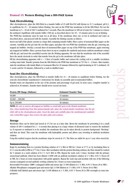
Protocol 15: Western Blotting from a SDS-PAGE System<br />
Tank Electroblotting<br />
After electrophoresis, place the SDS-PAGel in a transfer buffer of 25 mM Tris/192 mM Glycine in 15 % methanol, pH 8.2,<br />
to equilibrate for 15 – 20 minutes before blotting. Size and cut the PVDF-Star membrane to fit the SDS-PAGel. Pre-wet the<br />
membrane in 5 – 10 ml of 100 % methanol for 5 seconds. Place the membrane in 500 ml of water for 5 minutes to remove<br />
the methanol. Equilibrate with transfer buffer (500 ml, as described above) for 10 – 15 minutes prior to use in blotting.<br />
The PVDF-Star membrane must be kept wet at all times. If the membrane dries out, re-wet in methanol and water as<br />
described above, and proceed with the transfer. Assemble the blotting cassette as follows:<br />
Insert one half of the plastic cassette in a large dish containing transfer buffer. Place a piece of moistened filter paper on the<br />
cassette. Carefully put the gel onto the wet filter paper, and place the wet PVDF-Star membrane onto the gel, removing any<br />
trapped air bubbles. Overlay a second sheet of moistened filter paper on top of the PVDF-Star membrane, again removing<br />
any trapped air bubbles. Place a porous foam sheet on top to complete the sandwich and ensure uniform membrane contact<br />
with the gel, and insert the assembled cassette into the blotting apparatus. Be sure that the membrane side of the assembly<br />
faces the anode electrode in order that transfer from the gel to the membrane will take place.<br />
Fill the electroblotting apparatus with 4 – 5 liters of transfer buffer and connect the cooling coil to a suitable circulation<br />
cooling water bath. <strong>Transfer</strong> proteins from the SDS-PAGel to the PVDF-Star membrane at 70 V for 1 – 2 hours. After transfer,<br />
stain the membrane with Amido Black or Coomassie Blue for 10 minutes followed by rapid destaining in 50 % methanol, 10<br />
% acetic acid for 10 minutes, followed by a wash in distilled water.<br />
Semi-Dry Electroblotting<br />
After electrophoresis, place the SDS-PAGel in transfer buffer for 15 – 20 minutes to equilibrate before blotting. See the<br />
semi-dry electroblotter manufacturer's instructions for details on assembly and recommended buffers.<br />
<strong>Transfer</strong> times are dependent on the size of the proteins and percentage of gel used. In most cases, complete transfer is<br />
achieved in 30 minutes. <strong>Transfer</strong> times should never exceed one hour.<br />
Protein MW Range (Daltons)<br />
Estimated <strong>Transfer</strong> Time<br />
50,000 5 minutes<br />
50,000 – 200,000 30 minutes<br />
Up to 250,000<br />
40 minutes<br />
Note: Be sure to remove all trapped air bubbles to avoid bald spots in the blotted membrane.<br />
Also, when transferring from thin polyacrylamide gels, place the wetted transfer membrane onto the gel<br />
while still on the electrophoresis plate. Invert the plate and carefully place the membrane/gel sandwich<br />
onto wetted filter paper, then remove the glass plate and continue.<br />
Storage<br />
Membrane blots can be dried and stored at 4°C for use at a later date. Rewet the membrane by prewetting it in a small<br />
volume of 100 % methanol for 2 – 4 seconds then placing it in a large volume of deionized water to remove the methanol<br />
or, if exposure to methanol is to be avoided, the membrane blot can be taken directly to protein background “blocking”<br />
and then air dried. This coats the membrane with hydrophilic protein and allows easy rewetting in antibody incubation<br />
solution.<br />
Using this method requires that the membrane strips be stored at 4°C. The blots are stable for up to 1 year when stored dry.<br />
15<br />
protocol<br />
protocols<br />
Immunostaining<br />
Begin by incubating blots in a protein blocking solution of 5 % BSA in TBS for 1 hour at 37°C or, by incubating blots in<br />
0.5 % non-fat milk in PBS at 37°C for 1 hour. After incubation with the protein blocking solution, the blots should be washed<br />
for 5 minutes in a wash solution of 0.1 % (w/v) BSA in TBS. Repeat the wash cycle 3 times. Incubate with the primary<br />
monoclonal antibody, which has been diluted 1:1000 with an antibody incubation solution of 1 % BSA, 0.05 % Tween-20<br />
in TBS, for 2 hours at room temperature with gentle agitation. Repeat the wash step and incubate with one of the following<br />
enzyme-conjugated second antibody working solutions for 2 hours at room temperature.<br />
Horseradish Peroxidase (HRP) conjugated rabbit anti-mouse IgG (1:500 dilution in 1 % BSA, 0.05 % Tween-20 in TBS).<br />
Alkaline Phosphatase conjugated goat anti-mouse IgG (1:500 dilution in 1 % BSA, 0.05 % Tween-20 in TBS).<br />
Colloidal Gold labeled goat anti-mouse IgG (1:100 dilution in 1 % BSA, 0.05 % Tween-20 in TBS overnight at room temperature).<br />
© 2008 AppliChem • <strong>Transfer</strong> <strong>Membranes</strong> 37


