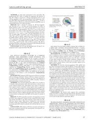
UROLOGY either placebo, vardenafil (10 mg), sildenafil (50 mg) or tadalafil (20 mg), after 3 days of sexual abst<strong>in</strong>ence. Despite the absence of any sexual stimulation, 10%, 41%, 26% and 55% of men <strong>in</strong> the placebo, sildenafil, tadalafil and vardenafil groups, respectively, were able to achieve ≥60% base or tip rigidities (P
ather <strong>in</strong>formation on the 95 th percentile of semen analysis data. As such, they <strong>in</strong>form the reader that 95% of men who achieve pregnancy with<strong>in</strong> 12 months of try<strong>in</strong>g will have sperm concentrations of >15 million cells/ml, >40% of observed sperm demonstrat<strong>in</strong>g good movement, and >4% of sperm with normal morphology. Unfortunately, these criteria do not predict the likelihood of achiev<strong>in</strong>g a pregnancy <strong>in</strong> the follow<strong>in</strong>g 12 months, which is typically when urologists first see these patients <strong>in</strong> consultation. Furthermore, the reference range must be viewed as a cont<strong>in</strong>uum, given that many patients at the low end of the range might still achieve pregnancy, and men at the high end might not. The nondef<strong>in</strong>itive nature of these guidel<strong>in</strong>es confirms the need for further sophisticated test<strong>in</strong>g—such as DNA fragmentation analysis, oxidative stress analysis and sperm evaluation for genomic, proteomic and metabolic factors—<strong>in</strong> certa<strong>in</strong> cases of male factor or unexpla<strong>in</strong>ed <strong>in</strong>fertility. For example, a patient whose semen parameters fall with<strong>in</strong> the new reference ranges but has not achieved pregnancy might benefit from DNA fragmentation test<strong>in</strong>g to identify subtle sperm abnormalities. Esteves et al. 2 have analyzed the new reference values and highlight further issues that may face the urologist, <strong>in</strong>clud<strong>in</strong>g whether the referral of male partners will decrease, whether we were previously overtreat<strong>in</strong>g our male patients and how to better <strong>in</strong>terpret these reference values by focus<strong>in</strong>g on the 50 th percentile of data. Over the past decade, progress <strong>in</strong> the field of assisted reproduction has led to a change <strong>in</strong> the management of severe male factor <strong>in</strong>fertility not amenable to medical or surgical correction. Currently, <strong>in</strong>tracytoplasmic sperm <strong>in</strong>jection (ICSI) is the treatment of choice for patients who suffer from either severe oligospermia or non obstructive azoospermia (NOA). Historically, any motile sperm present <strong>in</strong> the ejaculate would be preferentially utilized for ICSI. Alternatively, if no sperm were found <strong>in</strong> the ejaculate, sperm surgically extracted from the testis were used <strong>in</strong>stead. When very low numbers of sperm were present <strong>in</strong> the ejaculate and <strong>in</strong>itial ICSI results with motile ejaculated sperm were poor, then testis sperm was considered. Evidence aga<strong>in</strong>st this ejaculate-first approach was recently reported by Hauser et al., 3 who found that fertilization rates <strong>in</strong> patients with relative or virtual azoospermia were higher when fresh or frozen-thawed testicular sperm cells were used than when ejaculated sperm cells were used. This f<strong>in</strong>d<strong>in</strong>g is particularly <strong>in</strong>terest<strong>in</strong>g consider<strong>in</strong>g that although more motile sperm cells were found <strong>in</strong> the ejaculated specimens than <strong>in</strong> the testicular samples, the quality of embryos from testicular sperm (fresh and frozen) was significantly higher than of those from ejaculated sperm. This observa tion led the authors to conclude that it is the source of sperm cells, and not their motility, that plays a crucial role <strong>in</strong> fertility outcome. This pilot study suggests a possible role for testicular sperm extraction (TESE) coupled with ICSI <strong>in</strong> patients with severe oligo asthenospermia or relative or virtual azoo spermia. If testicular sperm leads to better fertility outcomes, does it matter if fresh or frozen-thawed testicular spermatozoa are retrieved? Accord<strong>in</strong>g to Hauser et al., 3 the answer is yes. While frozen-thawed spermatozoa may be more conveniently obta<strong>in</strong>ed, the researchers found that for patients with virtual or relative azoospermia, fresh testis sperm yielded better implantation rates than frozen testicular sperm. Although these results support the use of fresh testicular sperm for patients with relative or virtual azoospermia, there is still no consensus on the best approach for retriev<strong>in</strong>g testis sperm from men with pure NOA. This year, the value of diagnostic testis biopsy <strong>in</strong> the era of ICSI was addressed by Kalsi et al., 4 who provide evidence that microsurgical TESE (m-TESE)— <strong>in</strong>troduced by Schlegel and Li 5 <strong>in</strong> 1998—is the optimum sperm retrieval method <strong>in</strong> patients with NOA, preferential to f<strong>in</strong>e-needle aspira tion and traditional TESE. Researchers were able to successfully retrieve spermatozoa from 50 of 100 men with NOA who underwent m-TESE at their center, which <strong>in</strong>cludes a success rate of 57% <strong>in</strong> men with previously failed attempts at sperm retrieval. The only significant positive predictor of a successful retrieval was a previous histological diagnosis of hypospermatogenesis, and therefore the authors recommend aga<strong>in</strong>st the common practice of perform<strong>in</strong>g isolated diagnostic testicular biopsies on men with NOA, and suggest <strong>in</strong>stead that biopsy should always be comb<strong>in</strong>ed with a TESE procedure. Therefore, the take home message for any urologist treat<strong>in</strong>g a patient with NOA is to either proceed with a comb<strong>in</strong>ed diagnostic testis biopsy and send tissue to an andrology laboratory for process<strong>in</strong>g and cryo preservation, or to refer the patient to a reproductive <strong>Key</strong> advances UROLOGY ■ The updated 5 th edition of the WHO semen guidel<strong>in</strong>es <strong>in</strong>cludes significant changes from prior versions and is the first edition to <strong>in</strong>clude evidence-based data 1 ■ In certa<strong>in</strong> cases, testicular sperm may be used for <strong>in</strong>tracytoplasmic sperm <strong>in</strong>jection, <strong>in</strong> preference to ejaculated sperm, for patients with relative or virtual azoospermia 3 ■ Diagnostic testis biopsy alone (without tissue process<strong>in</strong>g) has limited value <strong>in</strong> the management of nonobstructive azoospermia 4 ■ The underly<strong>in</strong>g cause of hematospermia can be evaluated us<strong>in</strong>g transrectal ultrasonography 6 ■ F<strong>in</strong>asteride is a feasible treatment option for men with recurrent idiopathic hematospermia 7 urologist who has this capability. Our current approach is to beg<strong>in</strong> with a standard TESE and if no sperm are observed to proceed with an immediate m-TESE. Abnormal f<strong>in</strong>d<strong>in</strong>gs <strong>in</strong> ejaculate are not always related to male factor <strong>in</strong>fertility. In fact, one of the most frequently encountered problems <strong>in</strong> general urology is hematospermia, which can be a cause of great concern and anxiety for affected men. Until recently, hematospermia was assumed to be idiopathic and patients were reassured that their condition was benign. A recent study by Zhao et al., 6 however, may alter the urologist’s approach to hematospermia. In their study, researchers performed transrectal ultrasonography on 270 men with hematospermia, and found abnormalities <strong>in</strong> 95% of the cohort. These abnormalities were universally benign <strong>in</strong> patients under 40 years of age, <strong>in</strong>clud<strong>in</strong>g prostatic calcifications, ejaculatory duct calculi, and benign prostatic hyperplasia. Patients over the age of 40 years, however, were significantly more likely to have a malignant disease; 8 Courtesy of A. Kilchevsky, Yale–New Haven Hospital, USA KEY ADVANCES IN MEDICINE JANUARY 2012 | S83