Subcellular Colocalization of Hypericin with ... - Anticancer Research
Subcellular Colocalization of Hypericin with ... - Anticancer Research
Subcellular Colocalization of Hypericin with ... - Anticancer Research

You also want an ePaper? Increase the reach of your titles
YUMPU automatically turns print PDFs into web optimized ePapers that Google loves.
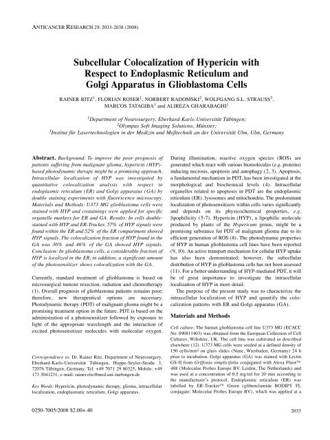
ANTICANCER RESEARCH 28: 2033-2038 (2008)<br />
<strong>Subcellular</strong> <strong>Colocalization</strong> <strong>of</strong> <strong>Hypericin</strong> <strong>with</strong><br />
Respect to Endoplasmic Reticulum and<br />
Golgi Apparatus in Glioblastoma Cells<br />
RAINER RITZ 1 , FLORIAN ROSER 1 , NORBERT RADOMSKI 2 , WOLFGANG S.L. STRAUSS 3 ,<br />
MARCOS TATAGIBA 1 and ALIREZA GHARABAGHI 1<br />
1 Department <strong>of</strong> Neurosurgery, Eberhard-Karls-Universität Tübingen;<br />
2 Olympus S<strong>of</strong>t Imaging Solutions, Münster;<br />
3 Institut für Lasertechnologien in der Medizin und Meßtechnik an der Universität Ulm, Ulm, Germany<br />
Abstract. Background: To improve the poor prognosis <strong>of</strong><br />
patients suffering from malignant glioma, hypericin (HYP)based<br />
photodynamic therapy might be a promising approach.<br />
Intracellular localization <strong>of</strong> HYP was investigated by<br />
quantitative colocalization analysis <strong>with</strong> respect to<br />
endoplasmic reticulum (ER) and Golgi apparatus (GA) by<br />
double staining experiments <strong>with</strong> fluorescence microscopy.<br />
Materials and Methods: U373 MG glioblastoma cells were<br />
stained <strong>with</strong> HYP and costainings were applied for specific<br />
organelle markers for ER and GA. Results: In cells doublestained<br />
<strong>with</strong> HYP and ER-Tracker, 57% <strong>of</strong> HYP signals were<br />
found <strong>with</strong>in the ER and 52% <strong>of</strong> the ER compartment showed<br />
HYP signals. The colocalization fraction <strong>of</strong> HYP found in the<br />
GA was 36% and 46% <strong>of</strong> the GA showed HYP signals.<br />
Conclusion: In glioblastoma cells, a considerable fraction <strong>of</strong><br />
HYP is localized in the ER; in addition, a significant amount<br />
<strong>of</strong> the photosensitizer shows colocalization <strong>with</strong> the GA.<br />
Currently, standard treatment <strong>of</strong> glioblastoma is based on<br />
microsurgical tumour resection, radiation and chemotherapy<br />
(1). Overall prognosis <strong>of</strong> glioblastoma patients remains poor;<br />
therefore, new therapeutical options are necessary.<br />
Photodynamic therapy (PDT) <strong>of</strong> malignant glioma might be a<br />
promising treatment option in the future. PDT is based on the<br />
administration <strong>of</strong> a photosensitizer followed by exposure to<br />
light <strong>of</strong> the appropriate wavelength and the interaction <strong>of</strong><br />
excited photosensitizer molecules <strong>with</strong> molecular oxygen.<br />
Correspondence to: Dr. Rainer Ritz, Department <strong>of</strong> Neurosurgery,<br />
Eberhard-Karls-Universität Tübingen, Hoppe-Seyler-Straße 3,<br />
72076 Tübingen, Germany. Tel: +49 7071 29 80325, Mobile: +49<br />
173 3041231, e-mail: rainer.ritz@med.uni-tuebingen.de<br />
Key Words: <strong>Hypericin</strong>, photodynamic therapy, glioma, intracellular<br />
localization, endoplasmatic reticulum, Golgi apparatus.<br />
0250-7005/2008 $2.00+.40<br />
During illumination, reactive oxygen species (ROS) are<br />
generated which react <strong>with</strong> various biomolecules (e.g. proteins)<br />
inducing necrosis, apoptosis and autophagy (2, 3). Apoptosis,<br />
a fundamental mechanism in PDT, has been investigated at the<br />
morphological and biochemical levels (4). Intracellular<br />
organelles related to apoptosis in PDT are the endoplasmic<br />
reticulum (ER), lysosomes and mitochondria. The predominant<br />
localization <strong>of</strong> photosensitizers <strong>with</strong>in cells varies significantly<br />
and depends on its physicochemical properties, e.g.<br />
lipophilicity (5-7). <strong>Hypericin</strong> (HYP), a lipophilic molecule<br />
produced by plants <strong>of</strong> the Hypericum genus, might be a<br />
promising substance for PDT <strong>of</strong> malignant glioma due to its<br />
efficient generation <strong>of</strong> ROS (8). The photodynamic properties<br />
<strong>of</strong> HYP in human glioblastoma cell lines have been reported<br />
(9, 10). An active transport mechanism for cellular HYP uptake<br />
has also been demonstrated; however, the subcellular<br />
distribution <strong>of</strong> HYP in glioblastoma cells has not been assessed<br />
(11). For a better understanding <strong>of</strong> HYP-mediated PDT, it will<br />
be <strong>of</strong> great importance to investigate the intracellular<br />
localization <strong>of</strong> HYP in more detail.<br />
The purpose <strong>of</strong> the present study was to characterize the<br />
intracellular localization <strong>of</strong> HYP and quantify the colocalization<br />
patterns <strong>with</strong> ER and Golgi apparatus (GA).<br />
Materials and Methods<br />
Cell culture. The human glioblastoma cell line U373 MG (ECACC<br />
No. 890811403) was obtained from the European Collection <strong>of</strong> Cell<br />
Cultures, Wiltshire, UK. The cell line was cultivated as described<br />
elsewhere (12). U373 MG cells were seeded at a defined density <strong>of</strong><br />
150 cells/mm 2 on glass slides (Nunc, Wiesbaden, Germany) 24 h<br />
prior to incubation. Golgi apparatus (GA) was stained <strong>with</strong> Lectin<br />
GS-II from Griffonia simplicifolia conjugated <strong>with</strong> Alexa Fluor<br />
488 (Molecular Probes Europe BV, Leiden, The Netherlands) and<br />
was used at a concentration <strong>of</strong> 0.5 mg/ml for 20 min according to<br />
the manufacturer’s protocol. Endoplasmic reticulum (ER) was<br />
labelled by ER-Tracker Green (glibenclamide BODIPY FL<br />
conjugate; Molecular Probes Europe BV), which was applied at a<br />
2033
concentration <strong>of</strong> 2 μM for 20 min. After staining <strong>with</strong> one <strong>of</strong> the<br />
organelle markers, slides were washed twice <strong>with</strong> phosphate-buffered<br />
saline (PBS). Thereafter, samples were incubated <strong>with</strong> 1 μM HYP<br />
for 2 h. After washing again <strong>with</strong> PBS, cells were fixed by 3.7%<br />
paraformaldehyde for 10 min. Double stained ER-HYP and GA-<br />
HYP slides were covered <strong>with</strong> DAKO Cytomation fluorescence<br />
mounting medium (Dako, Hamburg, Germany). In all cases, at least<br />
three independent experiments were performed. Cultures incubated<br />
solely <strong>with</strong> one <strong>of</strong> the organelle markers served as controls.<br />
Fluorescence microscopy. Fluorescence microscopy was carried out<br />
using an Olympus BX61 microscope (Olympus-Europa GmbH,<br />
Hamburg, Germany) equipped <strong>with</strong> a PLAPO60X (N.A. 1.4) oil<br />
immersion objective and filter sets for the detection <strong>of</strong> green<br />
ANTICANCER RESEARCH 28: 2033-2038 (2008)<br />
Figure 1. Fluorescence microscopic images <strong>of</strong> U373 MG glioblastoma cells, co-stained <strong>with</strong> ER-Tracker (2 μM/20 min) and HYP (1 μM/2 h). The<br />
same cells visualized by U-MWBA3 filter (band-pass excitation filter 460-495 nm, band-pass emission filter 510-550 nm) for green fluorescence (ER)<br />
(A). U-MWG2 filter (band pass excitation filter 510-550 nm, long-pass emission filter 590 nm) for red fluorescence (HYP) (B). Superimposition and<br />
quantitative colocalization analysis is shown for a region <strong>of</strong> interest in (C), (C-I) original image, (C-II) after image processing. Magnification x60,<br />
Olympus BX61 microscope, Hamburg, Germany. (D) <strong>Colocalization</strong> analysis ER-HYP: 57% ±21% (median±MAD) <strong>of</strong> HYP signals were found<br />
<strong>with</strong>in the ER and 52% ±15% <strong>of</strong> the ER compartment showed a HYP signal; n=77.<br />
2034<br />
fluorescence (U-MWBA3; excitation: 460-495 nm, emission: 510-<br />
550 nm) and red fluorescence (U-MWG2; excitation: 510-550 nm,<br />
emission: >590 nm). Images were obtained using an F-View II<br />
charge-coupled device camera (Olympus S<strong>of</strong>t Imaging Solutions<br />
GmbH, Münster, Germany). Images <strong>of</strong> single stained cells were<br />
acquired <strong>with</strong> both filters to determine possible cross-talks.<br />
Additionally, intensity measurements to determine the specific to<br />
non-specific ratio for the green fluorescence <strong>of</strong> both organelle<br />
markers and the red fluorescence <strong>of</strong> HYP were performed. Images<br />
<strong>of</strong> co-stained cells were analyzed <strong>with</strong> Cell^ P s<strong>of</strong>tware (Olympus<br />
S<strong>of</strong>t Imaging Solutions GmbH). <strong>Colocalization</strong> was determined<br />
applying a logical AND operation on two binary images derived<br />
from the original images <strong>of</strong> the green (organelle marker) and red<br />
channel (HYP) by appointing a specific intensity threshold in each
case. The quantitative analysis was calculated from n=77<br />
fluorescence images for HYP-ER and n=68 fluorescence images<br />
for HYP-GA.<br />
Results<br />
Intracellular localization <strong>of</strong> HYP was investigated in U373<br />
MG glioblastoma for a noncytotoxic incubation<br />
concentration <strong>of</strong> 1 μM (incubation time 2 h). At the<br />
beginning <strong>of</strong> three independent experiments, single stained<br />
cells were acquired <strong>with</strong> both filters to monitor possible<br />
cross-talks. No significant visible cross-talk was observed.<br />
Intensity measurements revealed a specific to non-specific<br />
Ritz et al: <strong>Subcellular</strong> Localization <strong>of</strong> <strong>Hypericin</strong><br />
Figure 2. Fluorescence microscopic images <strong>of</strong> U373 MG glioblastoma cells, co-stained <strong>with</strong> Golgi (0.5 mg/ml/20 min) and HYP (1 μM/2 h). Golgi<br />
apparatus was visualized by U-MWBA3 filter (band-pass excitation filter 460-495 nm, band-pass emission filter 510-550 nm) (A), HYP was<br />
visualized by U-MWG2 filter (band-pass excitation filter 510-550 nm, long-pass emission filter 590 nm) (B). Superimposition and quantitative<br />
colocalization analysis performed by s<strong>of</strong>tware Cell^ P (C); (C-I) original image, (C-II) after image processing. Magnification x60, Olympus BX61<br />
microscope, Hamburg, Germany. (D) <strong>Colocalization</strong> analysis Golgi-HYP: 36% ±19% (median±MAD) <strong>of</strong> HYP signals were found <strong>with</strong>in the GA<br />
and 46% ±23% <strong>of</strong> the GA compartment showed a HYP signal; n=68.<br />
ratio <strong>of</strong> 9.8 and 7.3 for green (organelle markers) and red<br />
fluorescence (HYP), respectively. Thus, fluorescence signals<br />
could be determined quite selectively <strong>with</strong> only minor crosstalk<br />
<strong>of</strong> below 15% in each case.<br />
Micrographs <strong>of</strong> the U373 MG glioblastoma cells doublestained<br />
<strong>with</strong> HYP and ER Tracker Green are presented in<br />
Figure 1; cells double-stained <strong>with</strong> HYP and the GA marker<br />
are shown in Figure 2. HYP was mainly localized in the<br />
perinuclear region, irrespective <strong>of</strong> the applied organelle<br />
marker (Figure 1B / Figure 2B). Only weak fluorescence<br />
intensity was found in the nucleus.<br />
The endoplasmic reticulum was predominantly found in the<br />
perinuclear region and exhibited a broad plane character<br />
2035
(Figure 1A). In contrast, the Golgi apparatus was found in a<br />
more granular pattern, even more distant from the nucleus<br />
(Figure 2A). Glioblastoma cells double-stained by HYP and<br />
ER-Tracker are given in Figure 1C as a superinposition <strong>of</strong><br />
Figures 1A and 1B. Yellow areas correspond to areas <strong>of</strong><br />
colocalization <strong>of</strong> HYP and ER-Tracker. Only weak<br />
fluorescence <strong>of</strong> HYP was seen in the nucleus. <strong>Colocalization</strong><br />
in the cytoplasm <strong>with</strong> enrichment <strong>of</strong> both compounds in the<br />
perinuclear region was demonstrated quite nicely, as<br />
predictable from the staining patterns <strong>of</strong> each compound<br />
(Figure 1A and 1B).<br />
Figure 2C demonstrates the glioblastoma cells doublestained<br />
by HYP and the GA marker. Again, yellow areas<br />
correspond to the colocalization fraction <strong>of</strong> HYP and the<br />
GA. The Golgi apparatus exhibited a granular fluorescence<br />
pattern mainly at the same site <strong>of</strong> the nucleus where HYP<br />
accumulation occured. For both colocalization experiments,<br />
regions <strong>of</strong> interest <strong>of</strong> the fluorescence images used for<br />
quantitative analysis are exemplarily shown prior to<br />
(Figures 1C-I and 2C-I) and after image processing<br />
(Figures1C-II and 2C-II).<br />
In order to increase the significance <strong>of</strong> the co-staining<br />
experiments, fluorescence images were analyzed quantitatively<br />
and results are depicted in Figures 1D and 2D. In cells doublestained<br />
<strong>with</strong> HYP and ER marker, a median <strong>of</strong> 57% (±21% ,<br />
median absolute deviation (MAD)) <strong>of</strong> HYP signals was found<br />
<strong>with</strong>in the ER and 52% ±15% (median±MAD) <strong>of</strong> the ER<br />
compartment showed HYP signals (Figure 1D). In the<br />
colocalization experiment, the colocalization fraction <strong>of</strong> HYP<br />
found in the GA was 36% (MAD 19% ), whereas 46%<br />
(MAD 23% ) <strong>of</strong> the GA coincided <strong>with</strong> the HYP fluorescence<br />
(Figure 2D).<br />
Discussion<br />
This study aimed at analysing the subcellular distribution <strong>of</strong><br />
the lipophilic molecule HYP, a natural compound originating<br />
from plants <strong>of</strong> the genus Hypericum, in more detail.<br />
Commonly, HYP is found in the plant Hypericum perforatum,<br />
better known as St. John’s wort. HYP, <strong>with</strong> its high triplet<br />
quantum yield, is a highly effective photosensitizer in<br />
malignant glioma (11). PDT is based on the reaction <strong>of</strong><br />
singlet-oxygen <strong>with</strong> biomolecules. Due to their very short<br />
life-span, the cytotoxic species are only able to diffuse over<br />
short distances (10-20 nm). Considering cell sizes <strong>of</strong><br />
approximately 20 μm, it is obvious that the subcellular<br />
localization <strong>of</strong> the photosensitizer becomes essential as the<br />
site <strong>of</strong> activity (13). As previously reported by several<br />
authors, HYP is enriched mainly in the perinuclear region,<br />
suggested as the ER (10, 12). To investigate HYP-mediated<br />
PDT in more detail, the intracellular distribution <strong>of</strong> these<br />
compounds <strong>with</strong> respect to the ER and GA was elucidated in<br />
a quantitative manner.<br />
2036<br />
ANTICANCER RESEARCH 28: 2033-2038 (2008)<br />
The ER consists <strong>of</strong> membrane-surrounded tubuli and<br />
cisterns extending from the nucleus into the cytoplasm.<br />
One main function <strong>of</strong> the ER is the processing and sorting<br />
<strong>of</strong> proteins. This is done in the so-called rough ER, which<br />
is enriched by ribosomes on the outer membrane, mainly<br />
producing proteins. From the rough ER, the smooth ER is<br />
distinguished. The smooth ER is the major site at which<br />
membrane lipids are synthesized. The GA, consisting <strong>of</strong><br />
cisternae and associated vesicules, is in close vicinity to<br />
the ER.<br />
The proteins received from the ER are processed and<br />
sorted in the GA for their further application. Additionally,<br />
the GA has also important functions in lipid metabolism. In<br />
particular, glycolipids and sphingomyelins are synthesized in<br />
this cell organelle. GA and ER are in a close morphological<br />
and dynamic functional relationship, while there is a<br />
continuous cycling between the two cell organelles (14-16).<br />
PDT requires a photosensitizer, light <strong>of</strong> appropriate<br />
wavelength and molecular oxygen to generate ROS. Oxidative<br />
damage <strong>of</strong> lipids, proteins and nucleic acids leads to cell death<br />
by several pathways. Apoptosis is a fundamental mechanism<br />
<strong>of</strong> cell death. Intracellular organelles, e.g. mitochondria,<br />
lysosomes and the ER, are involved in apoptosis. Ca 2+<br />
homeostasis and shifts in Ca 2+ compartmentalization play an<br />
important role in triggering apoptosis (17). In the case <strong>of</strong> the<br />
photosenitizer vertepr<strong>of</strong>in, a derivative <strong>of</strong> benzoporphyrine,<br />
PDT results in the release <strong>of</strong> Ca 2+ from the ER and<br />
mitochondria, which plays a major role in apoptosis (18).<br />
Recently, deregulated ER-Ca 2+ homeostasis after HYP-PDT<br />
was reported (19). Based on these data, the present data on<br />
the subcellular distribution <strong>of</strong> HYP may contribute to a better<br />
understanding <strong>of</strong> HYP-mediated PDT.<br />
Golgi apparatus has important functions in lipid<br />
metabolism. Therefore, a high affinity <strong>of</strong> HYP, a lipophilic<br />
compound, to the GA was supposed and investigated. To date,<br />
only minor endeavour has been made to investigate the<br />
relation between damage <strong>of</strong> the GA and PDT, particularly in<br />
the case <strong>of</strong> HYP-mediated PDT. <strong>Subcellular</strong> localization as<br />
well as mechanisms <strong>of</strong> PDT varies for different<br />
photosensitizers. HPD (Phot<strong>of</strong>rin), a first-generation<br />
photosensitizer composed <strong>of</strong> a complex mixture <strong>of</strong><br />
porphyrins, was first applied by Lipson and Schwarz in 1960.<br />
Mitochondrial localization was demonstrated in radiationinduced<br />
double-labelled fibrosarcoma tumor cells by confocal<br />
fluorescence microscopy (20). The data concerning the<br />
subcellular localization <strong>of</strong> Phot<strong>of</strong>rin differ. Hsieh et al.<br />
demonstrated a very high colocalization <strong>of</strong> Phot<strong>of</strong>rin and<br />
GA complex by selective fluorescence staining <strong>with</strong><br />
BODOPY FL C5-ceramide for long incubation times and<br />
plasma membranes after short incubation times (6).<br />
Second-generation sensitizers such as phthalocyanines and<br />
chlorines are typically located in the cytoplasm. More<br />
detailed information about intracellular location exists for
Foscan (meso-tetra(m-hydroxyphenyl)chlorin, m-THPC),<br />
a second-generation photosensitizer. It was localized in ER<br />
and GA as demonstrated by Teiten et al. and Marchal et al.<br />
(21, 22). Teiten et al. evaluated enzymatic activities after<br />
photosensitizing MCF-7 cells (human breast cancer cells)<br />
<strong>with</strong> Foscan and demonstrated that uridine 5’-diphosphate<br />
galactosyl transferase, an enzyme located in the GA, was<br />
inactivated after PDT (21). Marchal et al. demonstrated good<br />
colocalization <strong>of</strong> m-THPC <strong>with</strong> the GA after an incubation<br />
time <strong>of</strong> 3 hours. The authors found only weak correlation to<br />
a mitochondrial marker. In contrast, Yow et al. demonstrated<br />
in the nasopharyngeal carcinoma cell line NPS/HK1 that<br />
most mitochondria were targeted by m-THPC (7). These<br />
authors found a similar intracellular localization pattern <strong>with</strong><br />
respect to mitochondria for HPD and m-THPC.<br />
Chiu et al. showed a high release <strong>of</strong> cytochrome c from<br />
mitochondria by PDT <strong>with</strong> phthalocyanine 4, suggesting that<br />
the PDT-induced cell death by apoptosis mainly correlates<br />
<strong>with</strong> the release <strong>of</strong> cytochrome c from mitochondria (5).<br />
Chen et al. demonstrated the mitochondrion as an important<br />
cell organelle in PDT-mediated apoptosis by merocyanine<br />
540 in murine myeloid leukaemia (JCS) cells by confocal<br />
laser scanning microscopy (23). Lysosomes have been<br />
shown as a potential site <strong>of</strong> hydrophilic sensitizers in PDT,<br />
e.g. tetraphenylporphyrins, or Nile blue derivatives (24-27).<br />
A modern fluorescence marker (and photosensitizer), well<br />
established in neurosurgery for visualisation <strong>of</strong> malignant<br />
glioma, is protoporphyrin IX (PP IX). Accumulation <strong>of</strong> PP<br />
IX in malignant cells occurs after administration <strong>of</strong> the prodrug<br />
5-aminolevulinic acid (5-ALA) (28), possibly based on<br />
a diminished activity <strong>of</strong> ferrochelatase, one <strong>of</strong> the key<br />
enzymes <strong>of</strong> heme-biosynthesis. Recently, in a fluorescence<br />
microscopic study, Ji et al. revealed that 5-ALA-induced PP<br />
IX was mainly located in mitochondria (29). Similar results<br />
were obtained by investigating subcellular colocalization<br />
analysis <strong>of</strong> Phot<strong>of</strong>rin and PP IX <strong>with</strong> mitochondriaspecific<br />
markers by confocal fluorescence microscopy (20).<br />
The photosensitizer used in the present study was HYP, a<br />
strongly hydrophobic compound. As reported previously by<br />
our group, HYP showed a quite different fluorescence<br />
pattern as compared to lysosomes and mitochondria, stained<br />
by acridine orange and rhodamine 123, respectively (11).<br />
The present study reveals both descriptively and<br />
quantitatively the subcellular distribution <strong>of</strong> HYP in human<br />
glioma cells by fluorescence microscopy. In order to gain<br />
reliable results, the selection <strong>of</strong> the appropriate filter sets is<br />
quite crucial. Due to the broad fluorescence emission above<br />
600 nm, the green fluorescence <strong>of</strong> the organelle marker was<br />
detected <strong>with</strong> a band-pass emission filter for 510-550 nm.<br />
Thus, a significant reduction <strong>of</strong> fluorescence cross-talk was<br />
found as compared to a standard filter set (long-pass<br />
emission filter), which improved our results considerably<br />
(data not shown).<br />
Ritz et al: <strong>Subcellular</strong> Localization <strong>of</strong> <strong>Hypericin</strong><br />
In conclusion, the present data suggest that the ER seems<br />
to be an important target <strong>of</strong> HYP-mediated PDT. In addition,<br />
damage to the GA might also be involved in PDT-induced<br />
cell death; however the colocalization data indicate that its<br />
contribution seems to be <strong>of</strong> minor importance as compared<br />
to ER damage. These data may lead to a better appreciation<br />
<strong>of</strong> new approaches <strong>of</strong> PDT in the therapy <strong>of</strong> malignant<br />
glioma. For the future it will be <strong>of</strong> great interest to find<br />
concepts for improving efficacy <strong>of</strong> HYP-PDT, e.g. by<br />
increasing the photosensitization <strong>of</strong> malignant cells.<br />
Acknowledgements<br />
The authors appreciate the technical assistance by A. Gruber,<br />
Department <strong>of</strong> Neurosurgery, University Hospital Tübingen, Germany.<br />
References<br />
1 Stupp R, Mason WP, van den Bent MJ, Weller M, Fisher B,<br />
Taphoorn MJ, Belanger K, Brandes AA, Marosi C, Bogdahn U,<br />
Curschmann J, Janzer RC, Ludwin SK, Gorlia T, Allgeier A,<br />
Lacombe D, Cairncross JG, Eisenhauer E and Miriman<strong>of</strong>f RO:<br />
Radiotherapy plus concomitant and adjuvant temozolomide for<br />
glioblastoma. N Engl J Med 352: 987-996, 2005.<br />
2 Dougherty TJ, Gomer CJ, Henderson BW, Jori G, Kessel D,<br />
Korbelik M, Moan J and Peng Q: Photodynamic therapy. J Natl<br />
Cancer Inst 90: 889-905, 1998.<br />
3 Vrouenraets MB, Visser GW, Snow GB and van Dongen GA:<br />
Basic principles, applications in oncology and improved selectivity<br />
<strong>of</strong> photodynamic therapy. <strong>Anticancer</strong> Res 23: 505-522, 2003.<br />
4 Kessel D, Vicente MG and Reiners JJ Jr: Initiation <strong>of</strong> apoptosis<br />
and autophagy by photodynamic therapy. Lasers Surg Med 38:<br />
482-488, 2006.<br />
5 Chiu S, Evans HH, Lam M, Nieminen A and Oleinick NL:<br />
Phthalocyanine 4 photodynamic therapy-induced apoptosis <strong>of</strong><br />
mouse L5178Y-R cells results from a delayed but extensive release<br />
<strong>of</strong> cytochrome c from mitochondria. Cancer Lett 165: 51-58, 2001.<br />
6 Hsieh YJ, Wu CC, Chang CJ and Yu JS: <strong>Subcellular</strong> localization<br />
<strong>of</strong> Phot<strong>of</strong>rin determines the death phenotype <strong>of</strong> human<br />
epidermoid carcinoma A431 cells triggered by photodynamic<br />
therapy when plasma membranes are the main targets. J Cell<br />
Physiol 194: 363-375, 2003.<br />
7 Yow CM, Chen JY, Mak NK, Cheung NH and Leung AW:<br />
Cellular uptake, subcellular localization and photodamaging<br />
effect <strong>of</strong> temoporfin (mTHPC) in nasopharyngeal carcinoma<br />
cells: comparison <strong>with</strong> hematoporphyrin derivative. Cancer Lett<br />
157: 123-131, 2000.<br />
8 Diwu Z and Lown JW: Photosensitization <strong>with</strong> anticancer<br />
agents. 17. EPR studies <strong>of</strong> photodynamic action <strong>of</strong> hypericin:<br />
formation <strong>of</strong> semiquinone radical and activated oxygen species<br />
on illumination. Free Radic Biol Med 14: 209-215, 1993.<br />
9 Couldwell WT, Gopalakrishna R, Hinton DR, He S, Weiss MH,<br />
Law RE, Apuzzo ML and Law RE: <strong>Hypericin</strong>: a potential<br />
antiglioma therapy. Neurosurgery 35: 705-709, 1994.<br />
10 Uzdensky AB, Ma LW, Iani V, Hjortland GO, Steen HB and<br />
Moan J: Intracellular localisation <strong>of</strong> hypericin in human<br />
glioblastoma and carcinoma cell lines. Lasers Med Sci 16: 276-<br />
283, 2001.<br />
2037
11 Ritz R, Wein HT, Dietz K, Schenk M, Roser F, Tatagiba M and<br />
Strauss WS: Photodynamic therapy <strong>of</strong> malignant glioma <strong>with</strong><br />
hypericin: comprehensive in vitro study in human glioblastoma<br />
cell lines. Int J Oncol 30: 659-667, 2007.<br />
12 Ritz R, Muller M, Weller M, Dietz K, Kuci S, Roser F and<br />
Tatagiba M: <strong>Hypericin</strong>: a promising fluorescence marker for<br />
differentiating between glioblastoma and neurons in vitro. Int J<br />
Oncol 27: 1543-1549, 2005.<br />
13 Moan J and Berg K: The photodegradation <strong>of</strong> porphyrins in cells<br />
can be used to estimate the lifetime <strong>of</strong> singlet oxygen.<br />
Photochem Photobiol 53: 549-553, 1991.<br />
14 Rhee SW, Starr T, Forsten-Williams K and Storrie B: The<br />
steady-state distribution <strong>of</strong> glycosyltransferases between the<br />
Golgi apparatus and the endoplasmic reticulum is approximately<br />
90:10. Traffic 6: 978-990, 2005.<br />
15 Storrie B: Maintenance <strong>of</strong> Golgi apparatus structure in the face<br />
<strong>of</strong> continuous protein recycling to the endoplasmic reticulum:<br />
making ends meet. Int Rev Cytol 244: 69-94, 2005.<br />
16 Cooper G: Cell structure and function. In: The Cell. A Molecular<br />
Approach. Cooper GM (ed.). www.ncbi.nlm.nih.gov/books/<br />
bv.fcgi?rid=cooper. 2000.<br />
17 Orrenius S, Zhivotovsky B and Nicotera P: Regulation <strong>of</strong> cell<br />
death: the calcium-apoptosis link. Nat Rev Mol Cell Biol 4: 552-<br />
565, 2003.<br />
18 Granville DJ, Ruehlmann DO, Choy JC, Cassidy BA, Hunt DW,<br />
van Breemen C and McManus BM: Bcl-2 increases emptying <strong>of</strong><br />
endoplasmic reticulum Ca 2+ stores during photodynamic<br />
therapy-induced apoptosis. Cell Calcium 30: 343-350, 2001.<br />
19 Buytaert E, Callewaert G, Hendrickx N, Scorrano L, Hartmann<br />
D, Missiaen L, Vandenheede JR, Heirman I, Grooten J and<br />
Agostinis P: Role <strong>of</strong> endoplasmic reticulum depletion and<br />
multidomain proapoptotic BAX and BAK proteins in shaping<br />
cell death after hypericin-mediated photodynamic therapy.<br />
FASEB J 20: 756-758, 2006.<br />
20 Wilson BC, Olivo M and Singh G: <strong>Subcellular</strong> localization <strong>of</strong><br />
Phot<strong>of</strong>rin and aminolevulinic acid and photodynamic crossresistance<br />
in vitro in radiation-induced fibrosarcoma cells<br />
sensitive or resistant to Phot<strong>of</strong>rin-mediated photodynamic<br />
therapy. Photochem Photobiol 65: 166-176, 1997.<br />
21 Teiten MH, Marchal S, D’Hallewin MA, Guillemin F and<br />
Bezdetnaya L: Primary photodamage sites and mitochondrial<br />
events after Foscan photosensitization <strong>of</strong> MCF-7 human breast<br />
cancer cells. Photochem Photobiol 78: 9-14, 2003.<br />
2038<br />
ANTICANCER RESEARCH 28: 2033-2038 (2008)<br />
22 Marchal S, Francois A, Dumas D, Guillemin F and Bezdetnaya<br />
L: Relationship between subcellular localisation <strong>of</strong> Foscan and<br />
caspase activation in photosensitised MCF-7 cells. Br J Cancer<br />
96: 944-951, 2007.<br />
23 Chen JY, Cheung NH, Fung MC, Wen JM, Leung WN and Mak<br />
NK: <strong>Subcellular</strong> localization <strong>of</strong> merocyanine 540 (MC540) and<br />
induction <strong>of</strong> apoptosis in murine myeloid leukemia cells.<br />
Photochem Photobiol 72: 114-120, 2000.<br />
24 Berg K and Moan J: Lysosomes and microtubules as targets for<br />
photochemotherapy <strong>of</strong> cancer. Photochem Photobiol 65: 403-<br />
409, 1997.<br />
25 Geze M, Morliere P, Maziere JC, Smith KM and Santus R:<br />
Lysosomes, a key target <strong>of</strong> hydrophobic photosensitizers<br />
proposed for photochemotherapeutic applications. J Photochem<br />
Photobiol B 20: 23-35, 1993.<br />
26 Lin CW, Shulok JR, Kirley SD, Cincotta L and Foley JW:<br />
Lysosomal localization and mechanism <strong>of</strong> uptake <strong>of</strong> Nile blue<br />
photosensitizers in tumor cells. Cancer Res 51: 2710-2719, 1991.<br />
27 Wessels JM, Strauss W, Seidlitz HK, Ruck A and Schneckenburger<br />
H: Intracellular localization <strong>of</strong> meso-tetraphenylporphine tetrasulphonate<br />
probed by time-resolved and microscopic fluorescence<br />
spectroscopy. J Photochem Photobiol B 12: 275-284, 1992.<br />
28 Stummer W, Pichlmeier U, Meinel T, Wiestler OD, Zanella F and<br />
Reulen HJ: Fluorescence-guided surgery <strong>with</strong> 5-aminolevulinic<br />
acid for resection <strong>of</strong> malignant glioma: a randomised controlled<br />
multicentre phase III trial. Lancet Oncol 7: 392-401, 2006.<br />
29 Ji Z, Yang G, Vasovic V, Cunderlikova B, Suo Z, Nesland JM<br />
and Peng Q: <strong>Subcellular</strong> localization pattern <strong>of</strong> protoporphyrin<br />
IX is an important determinant for its photodynamic efficiency<br />
<strong>of</strong> human carcinoma and normal cell lines. J Photochem<br />
Photobiol B 84: 213-220, 2006.<br />
Received February 20, 2008<br />
Revised May 14, 2008<br />
Accepted May 15, 2008