Targeting by probe-based circularisation
Targeting by probe-based circularisation
Targeting by probe-based circularisation

You also want an ePaper? Increase the reach of your titles
YUMPU automatically turns print PDFs into web optimized ePapers that Google loves.
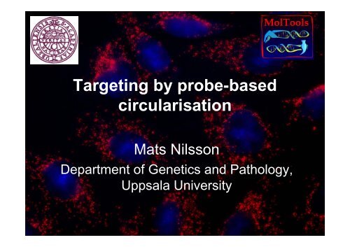
<strong>Targeting</strong> <strong>by</strong> <strong>probe</strong>-<strong>based</strong><br />
<strong>circularisation</strong><br />
Mats Nilsson<br />
Department of Genetics and Pathology,<br />
Uppsala University
Outline<br />
• Padlock <strong>probe</strong>s for multiplex l targeted t genotyping<br />
• Selector <strong>probe</strong>s for multiplex targeted genome<br />
analysis<br />
– sequencing (clinical sequencing)<br />
– CNV detection<br />
ti<br />
• Amplified single-molecule detection (digital RCA)<br />
• In situ genotyping
Padlock <strong>probe</strong>s<br />
• Daul-recognition provides specificity<br />
• Robust allele-distinction due to enzymatic ligation using standard<br />
reaction conditions<br />
•Probes become topologicaly linked<br />
•Unimolecular<br />
Nilsson, et al. (1994) Science, 265, 2085-2088.<br />
2088.<br />
Nilsson, et al. (1997) Nature Genetics, 16, 252-255.<br />
Nilsson, et al. (2000) Nature Biotechnol, 18, 791-793.
20,000-plex<br />
Genotyping<br />
Affymetrix targeted<br />
genotyping<br />
Hardenbol et al,<br />
Nature Biotechnol 2003;<br />
Hardenbol et al,<br />
Genome Res 2005<br />
ParAllele 20K GeneChip®
Targeted genome analysis<br />
using selector <strong>probe</strong>s<br />
Sequence-specific<br />
circularization<br />
Circle selection and amplification<br />
Universal sequence<br />
inserted
The Selection Process<br />
Direct ligation<br />
Structure<br />
specific cut
96-plex amplification of 96 selected<br />
genomic fragments<br />
• 89% efficiency<br />
(96%
Parallel sequencing of 177 exons in<br />
10 cancer genes<br />
454 sequencing<br />
59.000 reads per exp<br />
55%<br />
90% map to the selected<br />
sequence (49 kb) =><br />
Average enrichment:<br />
600.000<br />
10x<br />
508 <strong>probe</strong>s<br />
Dahl, et al. (2007) PNAS, 104, 9387-9392
Further development eop e<br />
• New <strong>probe</strong> design<br />
– reduced cost (4 EUR/<strong>probe</strong>)<br />
– More even representation ti (5-fold range)<br />
– Compatible with Solexa and SOLiD seq.<br />
• Double the size of selected fragments<br />
• Less input DNA (100 ng)<br />
• Applied to a cancer resequencing project<br />
with Tobias Sjöblom at UU.<br />
• Spun out a company:
Copy-number measurements<br />
Isaksson et al. Nucleic Acids Res., 35, e115 (2007)
Copy-number measurements<br />
Down Syndrome patient samples
Sizing a duplication in Rhodesian ridgeback<br />
dogs and mapping the break-point<br />
Breakpoint PCR<br />
Salmon-Hillbertz, et al. (2007)Nature Genet., 39, 1318
Signal amplification through<br />
rolling circle amplification (RCA)<br />
padlock <strong>probe</strong><br />
primer<br />
1500 nt/min<br />
t 1/2 11 hours<br />
Banér, et al. (1998) Nucleic Acids Res, 22, 5073-5078.
Homogenous detection of RCA products<br />
1000 repeats – 45 μm<br />
Random coiling<br />
[10 nM]<br />
1 μM]
Homogenous detection of RCA products<br />
Blab et al. (2004) Anal. Chem., 76, 495-8
Amplified single-molecule detection (digital RCA)<br />
Jarvius et al. (2006) Nature Methods, 3, 725-727727
Amplified single-molecule detection<br />
Advantages:<br />
• Ultimate detection sensitivity<br />
• Ultimate quantitative precision<br />
Spectral multiplexing
Characterization of ASMD key properties<br />
• Dynamic range 10 4<br />
– 30 s data acquisition<br />
• High quantitative precision<br />
– Typically 3 percent<br />
– Poisson limited (
In situ genotyping<br />
•Padlock probing<br />
Target strand<br />
•Rolling-circle amplification
Target-primed RCA
Detection of single nucleotide variation in the<br />
mitochondrial genome (A3243G)<br />
Larsson, C. et al. (2004) Nature Methods,1, 227-232.
Acknowledgements<br />
• Rudbeck lab, UU<br />
– Johan Banér<br />
– Mats Gullberg<br />
– Chatarina Larsson<br />
– Sara Henriksson<br />
– Jonas Jarvius<br />
– Jonas Melin<br />
– Henrik Johansson<br />
– Yuki Tanaka<br />
– Jenny Göransson<br />
– Fredrik Dahl<br />
– Magnus Isaksson<br />
– Johan Stenberg<br />
– Ulf Landegren<br />
Collaborators at Rudbeck<br />
– Ola Söderberg<br />
– Marie-Louise Bondeson<br />
– Lotta Thuresson<br />
– Niklas Dahl<br />
– Fredrik Öberg<br />
– Fredrik Pontén<br />
– Tobias Sjöblom<br />
– Marie Allen<br />
• SLU/UU/Broad Inst.<br />
Leif Andersson<br />
Göran Andersson<br />
Kerstin Lindblad-Toh<br />
• Ångström lab, UU<br />
Fredrik Nikolajeff<br />
Maria Strömme<br />
• Image Analysis, UU<br />
Carolina Wähl<strong>by</strong><br />
Ewert Bengtsson<br />
• Aarhus University<br />
Jörn Koch<br />
• Leiden University<br />
Ton Raap<br />
Thomas Schmidt<br />
• University of Tokyo<br />
Takehiko Kitamori<br />
• Nagoya University<br />
Yoshinobu Baba<br />
• Stanford/ParAllele/Affymetrix<br />
Paul Hardenbol et al.<br />
• Karolinska Institute<br />
Nils-Göran Larsson<br />
• EU FP-7 READNA<br />
• EU FP-6 ”COMICS”<br />
• Swedish Research<br />
Councils (M & NT)<br />
• Uppsala BioX<br />
• The Wallenberg<br />
Foundation<br />
• Swedish Defence<br />
Nanotech Program<br />
• VINNOVA/SSF/JST