CB August 06 - Hungarovet
CB August 06 - Hungarovet
CB August 06 - Hungarovet

You also want an ePaper? Increase the reach of your titles
YUMPU automatically turns print PDFs into web optimized ePapers that Google loves.
procedures pro<br />
DIAGNOSTICS<br />
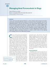
Abdominocentesis<br />
Daniel D. Smeak, DVM, Diplomate ACVS, Ohio State University<br />
PERCUTANEOUS SAMPLING OF PERITONEAL FLUID<br />
with a needle or small catheter, called abdominocentesis (sometimes<br />
also known as peritoneocentesis or abdominal paracentesis),<br />
is a simple, rapid, and safe diagnostic method. When physical<br />
examination or imaging indicates moderate to large amounts of peritoneal<br />
effusion in a patient, sampling the fluid for cytologic, microbiological,<br />
or biochemical analysis often helps the clinician to quickly<br />
make a diagnosis and then to initiate timely and appropriate therapy.<br />
Abdominocentesis is most often performed to determine whether a<br />
patient needs exploratory celiotomy, particularly for early diagnosis of<br />
peritonitis or serious injury.<br />
Before considering abdominocentesis, determine the relative value versus<br />
the risk of a blind puncture, particularly when a large vascular<br />
abdominal mass, an enlarged vascular organ, or a disorder affecting a<br />
distended hollow organ (such as a pyometra) is suspected, or in<br />
patients with severe coagulopathies. In these instances, consider ultrasound-guided<br />
sample collection if available. Abdominocentesis is best<br />
performed after plain radiography unless a rapid diagnosis needs to be<br />
made in a deteriorating patient. Free peritoneal air on radiographs may<br />
help the clinician identify hollow organ rupture or peritoneal perforation.<br />
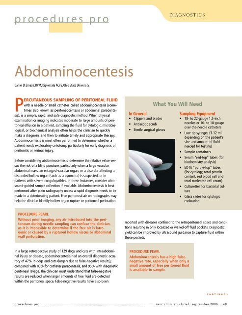
In General<br />
• Clippers and blades<br />
• Antiseptic scrub<br />
• Sterile surgical gloves<br />
What You Will Need<br />
Sampling Equipment<br />
• 18- to 22-gauge 1.5-inch<br />
needles or 16- to 18-gauge<br />
over-the-needle catheters<br />
• Luer tip syringes (3-12 ml<br />
depending on the patient’s<br />
size and amount of fluid<br />
needed for testing)<br />
• Sample containers<br />
• Serum “red-top” tubes (for<br />
biochemistry analysis)<br />
• EDTA “purple-top” tubes<br />
(for cytology, total protein<br />
content, red blood cell and<br />
total nucleated cell count)<br />
• Culturettes for bacterial culture<br />
• Glass slides for cytologic<br />
evaluation<br />
PROCEDURE PEARL<br />
Without prior imaging, any air introduced into the peritoneum<br />
during needle sampling can confuse the clinician,<br />
as it is impossible to determine if the free air is iatrogenic<br />
or caused by a ruptured hollow viscus or abdominal<br />
wall perforation.<br />
reported with diseases confined to the retroperitoneal space and conditions<br />
resulting in only localized or walled-off fluid pockets. Diagnostic<br />
yield can be improved by ultrasound guidance to capture fluid within<br />
these pockets.<br />
In a large retrospective study of 129 dogs and cats with intraabdominal<br />
injury or disease, abdominocentesis had an overall diagnostic accuracy<br />
of 47% in dogs and cats (largely due to false-negative results),<br />
compared with 83% for catheter paracentesis, and 95% with diagnostic<br />
peritoneal lavage. The clinician must understand that false-negative<br />
results are reduced when larger amounts of free fluid are detected<br />
within the peritoneal space. False-negative results have also been<br />
PROCEDURE PEARL<br />
Abdominocentesis has a high falsenegative<br />
rate, especially when only a<br />
small amount of free peritoneal fluid<br />
is available to sample.<br />
continues<br />
procedures pro ....................................................................................................................NAVC clinician’s brief...september.20<strong>06</strong>.....49
procedures pro C ONTINUED<br />
STEP BY STEP HOW TO PERFORM ABDOMINOCENTESIS<br />
Positioning & Skin<br />
Preparation<br />
Abdominocentesis is usually performed in conscious<br />
animals with physical restraint. If the<br />
patient is intractable, judicious use of chemical<br />
sedation should be considered.<br />
1. Clip and prepare the ventral abdominal<br />
area for aseptic fluid collection.<br />
2. Infiltrate the proposed sites with a local<br />
anesthetic agent if a larger over-the-needle<br />
catheter is chosen for sampling.<br />
3. For simple abdominocentesis, stand the<br />
patient or position it in sternal recumbency.<br />
Access the most dependent site on the<br />
abdomen. Alternatively, nonambulatory or<br />
unruly patients can be positioned in left lateral<br />
recumbency. First puncture the<br />
right side to help avoid hemorrhage or contamination<br />
of the sample from accidental<br />
aspiration of the spleen.<br />
Productive Puncture Sites<br />
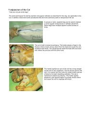
Simple Abdominocentesis<br />
With the prepared animal in left lateral<br />
recumbency, insert the needle or fenestrated<br />
over-the-needle catheter just caudal to the<br />
umbilicus at or within 1 to 2 cm right of midline.<br />
Direct the needle toward the dependent<br />
side, slightly caudal toward the pelvis. The<br />
abdominocentesis shown here is positive.<br />
Fluid is flowing from the fenestrated catheter<br />
hub into a blood collection tube (Inset). If this<br />
approach is unsuccessful, attempt a second<br />
stick 2 to 4 cm cranial to the umbilicus on or<br />
slightly right of midline, with the needle<br />
angled in a slight craniodependent direction.<br />
PROCEDURE PEARL<br />
I experience fewer<br />
“dry taps” when I use<br />
a 16-gauge over-theneedle<br />
catheter into<br />
which several staggered,<br />
side port holes<br />
have been added<br />
with a scalpel blade.<br />
Multiple side ports<br />
reduce obstruction<br />
from loose apposing<br />
tissue, particularly<br />
when small amounts<br />
of intraabdominal<br />
fluid are expected.<br />
Creating side ports in an over-the-needle catheter. Four or five staggered<br />
holes are made with a sharp no. 10 scalpel blade. Be sure the<br />
holes are no larger than one third of the catheter circumference because<br />
larger holes may weaken the shaft and accidental breakage may occur<br />
when removing the catheter from the abdomen.<br />
PROCEDURE PEARL<br />
If simple abdominocentesis is unsuccessful and immediate<br />
results are important, a four-quadrant tap may be<br />
productive. … Four sites for needle placement are used<br />
with the umbilicus as the center point.<br />
50...september.20<strong>06</strong>.....NAVC clinician’s brief .....................................................................................................................procedures pro
PROCEDURE PEARL<br />
Hemorrhagic effusion generally does<br />
not clot—blood from inadvertent<br />
splenic aspiration generally does.<br />
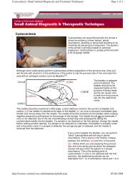
between the nipples (green dashed lines).<br />
These vessels should be avoided during<br />
puncture.<br />
Puncture Technique<br />
(Any Site)<br />
1. With gloved hands, gently insert the needle<br />
(without a syringe attached) through<br />
the abdominal wall, and observe for any<br />
fluid within the hub.<br />
2. Slowly rotating the needle, not “blind jabbing,”<br />
may help fluid to escape through<br />
the needle.<br />
3. When using a fenestrated over-the-needle<br />
catheter, observe for fluid escape once the<br />
stylet has been removed.<br />
4. Collect any fluid that flows from the needle<br />
in the appropriate container.<br />
5. If no fluid is obtained, attach an appropriately<br />
sized syringe and apply very gentle<br />
suction.<br />
Four-Quadrant Abdominocentesis<br />
If simple abdominocentesis is unsuccessful<br />
and immediate results are important, a fourquadrant<br />
tap may be productive. With the prepared<br />
animal in dorsal or left lateral<br />
recumbency, insert the needle or catheters<br />
described above. Four sites for needle placement<br />
are used with the umbilicus as the center<br />
point:<br />
• Right cranial quadrant<br />
• Left cranial quadrant<br />
• Right caudal quadrant<br />
• Left caudal quadrant<br />
Avoid the caudal superficial epigastric vessels<br />
during needle or catheter puncture. Stay away<br />
from an imaginary line drawn longitudinally<br />
along the nipples. Red Xs depict suggested<br />
quadrant tap sites. The caudal superficial and<br />
deep epigastric vessels lie paramedian in the<br />
neighborhood of a longitudinal line drawn<br />
PROCEDURE PEARL<br />
Applying undue suction with a<br />
syringe increases the incidence of<br />
obstruction of the needle with tissue<br />
and subsequent false-negative<br />
results.<br />
6. When one site is negative, repeat the needle<br />
puncture in another quadrant. A positive<br />
tap at any site completes the<br />
procedure.<br />
Do not automatically accept that a negative<br />
abdominocentesis means little to no free<br />
abdominal fluid. Remember, between 5 and 6<br />
ml/kg body weight of peritoneal fluid is<br />
continues<br />
procedures pro .....................................................................................................................NAVC clinician’s brief...september.20<strong>06</strong>.....51
procedures pro C ONTINUED<br />
COMING<br />
SOON<br />
to these pages…<br />
required to achieve positive results with<br />
abdominocentesis in experimental dogs. If<br />
there is sufficient suspicion of abdominal effusion,<br />
ultrasound-guided needle sampling or<br />
diagnostic peritoneal lavage may be the next<br />
step. If diagnostic peritoneal lavage is<br />
planned, fluid can be infused and removed<br />
through the fenestrated over-the-needle<br />
catheter.<br />
Complications<br />
Complications from abdominocentesis are limited<br />
and uncommon. Inadvertent puncture of<br />
the caudal superficial epigastric vessels may<br />
cause significant ventral abdominal bruising.<br />
Blind puncture can lead to hemorrhage or hollow<br />
organ laceration, but this complication is<br />
rare provided the patient is properly<br />
restrained. The most common complication is<br />
contamination of the sample with hemorrhage,<br />
or sampling of a hollow viscus, such as<br />
the bladder or intestine, and this may contribute<br />
to misdiagnosis. False-positive results<br />
can be expected in about 5% to 10% of<br />
attempts.<br />
Evaluation of Peritoneal<br />
Fluid<br />
A representative fluid sample should be thoroughly<br />
analyzed. The test results must be<br />
combined with the clinician’s assessment of<br />
the animal’s clinical condition and, in many<br />
cases, compared with peripheral blood values<br />
to obtain an accurate diagnosis of intraabdominal<br />
disease. The following is a list of tests<br />
that can be requested from the sample<br />
depending on the clinician’s index of suspicion<br />
for a particular disease process. Appropriate<br />
sample collection, handling, and preparation<br />
are essential to obtain an accurate diagnosis.<br />
Refer to Connally’s article (see Aids &<br />
Peritoneal Fluid Evaluation<br />
General Fluid Analysis<br />
• Total protein<br />
• Red and white cell count<br />
• Specific gravity<br />
• pH<br />
Cytologic Evaluation<br />
• White blood cell type and morphologic<br />
characteristics<br />
• Foreign matter<br />
• Intracellular organisms<br />
Biochemical Analysis<br />
• Glucose<br />
• Creatinine<br />
• Urea nitrogen<br />
• Potassium<br />
• Cholesterol<br />
• Triglycerides<br />
• Bilirubin<br />
• Albumin<br />
• Globulin<br />
• Amylase and lipase<br />
• Lactate<br />
• Potassium<br />
• PCO 2 , PO 2<br />
Culture & Susceptibility<br />
• Aerobic and anaerobic bacterial culture<br />
Resources, back page) for more information<br />
about sample preparation and which tests to<br />
consider for a variety of intraabdominal disease<br />
conditions. ■<br />
See Aids & Resources, back page, for<br />
references, contacts, and appendices.<br />
Complications: Fluid Therapy<br />
Patient Support:<br />
Open Wound Care<br />
Procedures Pro: Artificial<br />
Insemination<br />
Make Your Diagnosis: Sudden<br />
Collapse<br />
Ask the Expert: Practice<br />
Promotional Strategies<br />
What’s the Take-Home<br />
Hip Dysplasia<br />
Plus, of course, Capsules,<br />
Practice Hotline, Comparative<br />
Imagery, How to Refer, Applied<br />
Cytology, 1000 Words, and much<br />
more!<br />
52...september.20<strong>06</strong>.....NAVC clinician’s brief .....................................................................................................................procedures pro