Venipuncture of the Cat - Hungarovet
Venipuncture of the Cat - Hungarovet
Venipuncture of the Cat - Hungarovet

You also want an ePaper? Increase the reach of your titles
YUMPU automatically turns print PDFs into web optimized ePapers that Google loves.
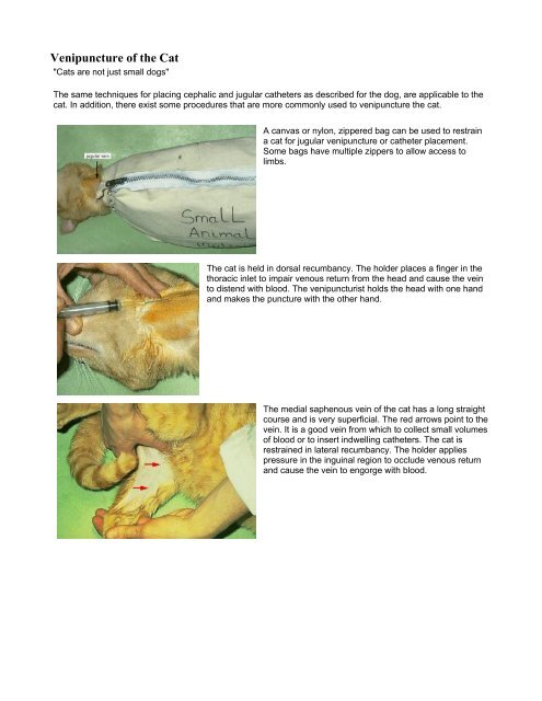
<strong>Venipuncture</strong> <strong>of</strong> <strong>the</strong> <strong>Cat</strong><br />
"<strong>Cat</strong>s are not just small dogs"<br />
The same techniques for placing cephalic and jugular ca<strong>the</strong>ters as described for <strong>the</strong> dog, are applicable to <strong>the</strong><br />
cat. In addition, <strong>the</strong>re exist some procedures that are more commonly used to venipuncture <strong>the</strong> cat.<br />
A canvas or nylon, zippered bag can be used to restrain<br />
a cat for jugular venipuncture or ca<strong>the</strong>ter placement.<br />
Some bags have multiple zippers to allow access to<br />
limbs.<br />
The cat is held in dorsal recumbancy. The holder places a finger in <strong>the</strong><br />
thoracic inlet to impair venous return from <strong>the</strong> head and cause <strong>the</strong> vein<br />
to distend with blood. The venipuncturist holds <strong>the</strong> head with one hand<br />
and makes <strong>the</strong> puncture with <strong>the</strong> o<strong>the</strong>r hand.<br />
The medial saphenous vein <strong>of</strong> <strong>the</strong> cat has a long straight<br />
course and is very superficial. The red arrows point to <strong>the</strong><br />
vein. It is a good vein from which to collect small volumes<br />
<strong>of</strong> blood or to insert indwelling ca<strong>the</strong>ters. The cat is<br />
restrained in lateral recumbancy. The holder applies<br />
pressure in <strong>the</strong> inguinal region to occlude venous return<br />
and cause <strong>the</strong> vein to engorge with blood.
cat's head to right<br />
Blood samples can be obtained from <strong>the</strong> medial saphenous vein.<br />
Because <strong>the</strong> vein has a small diameter, vigorous aspiration results in<br />
collapse <strong>of</strong> <strong>the</strong> vein. Therefore only slight suction can be applied to <strong>the</strong><br />
syringe when aspirating blood. The blood flows slowly so only a small<br />
volume (up to ~1ml) <strong>of</strong> blood can be obtained before <strong>the</strong> sample clots.<br />
At <strong>the</strong> completion <strong>of</strong> <strong>the</strong> venipuncture, as <strong>the</strong> needle is removed from<br />
<strong>the</strong> vein, <strong>the</strong> holder should release pressure from <strong>the</strong> inguinal region<br />
and place firm digital pressure at <strong>the</strong> puncture site for several minutes<br />
to prevent hematoma formation. Hematomas form readily and spread<br />
from <strong>the</strong> puncture site when venipuncture is unsuccessful. Therefore,<br />
start distal on <strong>the</strong> limb in case it is necessary to make more than one<br />
attempt at venipuncture.<br />
The medial saphenous vein can be ca<strong>the</strong>terized with short indwelling<br />
ca<strong>the</strong>ters or ca<strong>the</strong>ters marketed for jugular vein ca<strong>the</strong>terization. (See <strong>the</strong><br />
sections on jugular ca<strong>the</strong>ter placement and cephalic ca<strong>the</strong>ter placement<br />
for information on ca<strong>the</strong>ter types). A jugular ca<strong>the</strong>ter placed in <strong>the</strong> medial<br />
saphenous vein will have its tip in <strong>the</strong> posterior vena cava. Such<br />
ca<strong>the</strong>ters can be used for repetitive blood sampling (eg. serial blood<br />
glucose sampling used in diabetic regulation). Because <strong>the</strong> tip <strong>of</strong> <strong>the</strong><br />
ca<strong>the</strong>ter is in a large vein, hypertonic solutions such as hypertonic<br />
dextrose and total parenteral nutrition (TPN) solutions can be<br />
administered through ca<strong>the</strong>ters in this location. The picture demonstrates<br />
an Intrafuser jugular ca<strong>the</strong>ter placed in <strong>the</strong> medial saphenous vein. Note<br />
<strong>the</strong> bruising around <strong>the</strong> needle puncture site. This may occur when<br />
placing a through-<strong>the</strong>-needle ca<strong>the</strong>ter. The hole in <strong>the</strong> vein created by<br />
<strong>the</strong> needle is larger than <strong>the</strong> diameter <strong>of</strong> <strong>the</strong> ca<strong>the</strong>ter so when <strong>the</strong> needle<br />
is retracted from <strong>the</strong> vein, blood may leak from <strong>the</strong> puncture site into<br />
surrounding tissues. Correct bandaging <strong>of</strong> a medial saphenous ca<strong>the</strong>ter<br />
is crucial to its function. The procedure for bandaging <strong>the</strong> ca<strong>the</strong>ter in<br />
place is similar to <strong>the</strong> procedure for bandaging a jugular ca<strong>the</strong>ter in<br />
place, except that <strong>the</strong> bandage is applied in a direction parallel to <strong>the</strong><br />
needle guard, ra<strong>the</strong>r than perpendicular to <strong>the</strong> needle guard as with <strong>the</strong><br />
jugular ca<strong>the</strong>ter.<br />
A butterfly <strong>of</strong> one inch tape is applied to <strong>the</strong> needle guard. A piece <strong>of</strong> one inch tape is wrapped around <strong>the</strong> leg,<br />
catching both wings <strong>of</strong> <strong>the</strong> butterfly at <strong>the</strong> same time. A second piece <strong>of</strong> one inch tape is wrapped around <strong>the</strong><br />
limb at <strong>the</strong> level <strong>of</strong> <strong>the</strong> hub <strong>of</strong> <strong>the</strong> needle. Antiseptic or antibiotic ointment on a piece <strong>of</strong> gauze is applied to <strong>the</strong><br />
puncture site. Stretchy wrap such as cling is wrapped around <strong>the</strong> limb several times, around both pieces <strong>of</strong><br />
tape. This is followed by an outer wrap such as Vetwrap. The gauze pad used to elevate <strong>the</strong> needle guard from<br />
<strong>the</strong> neck when bandaging a jugular ca<strong>the</strong>ter is not used when taping in place a medial saphenous ca<strong>the</strong>ter. The<br />
use <strong>of</strong> an Intrafuser ca<strong>the</strong>ter with an extension set, allows you to place <strong>the</strong> extension set on <strong>the</strong> lateral side <strong>of</strong><br />
<strong>the</strong> limb so <strong>the</strong> ca<strong>the</strong>ter is easily accessible. It may be necessary to splint <strong>the</strong> leg to keep <strong>the</strong> ca<strong>the</strong>ter<br />
functioning.