Diabetic Ketoacidosis - Hungarovet
Diabetic Ketoacidosis - Hungarovet
Diabetic Ketoacidosis - Hungarovet

Create successful ePaper yourself
Turn your PDF publications into a flip-book with our unique Google optimized e-Paper software.
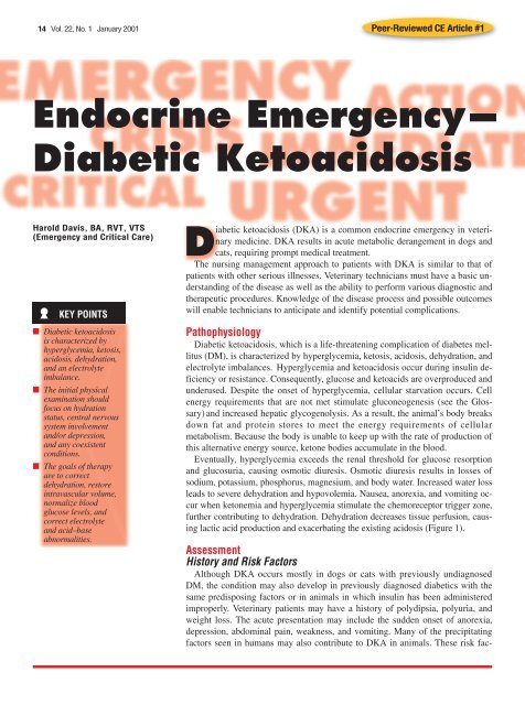
14 Vol. 22, No. 1 January 2001 Peer-Reviewed CE Article #1<br />
Endocrine Emergency—<br />
<strong>Diabetic</strong> <strong>Ketoacidosis</strong><br />
Harold Davis, BA, RVT, VTS<br />
(Emergency and Critical Care)<br />
KEY POINTS<br />
■ <strong>Diabetic</strong> ketoacidosis<br />
is characterized by<br />
hyperglycemia, ketosis,<br />
acidosis, dehydration,<br />
and an electrolyte<br />
imbalance.<br />
■ The initial physical<br />
examination should<br />
focus on hydration<br />
status, central nervous<br />
system involvement<br />
and/or depression,<br />
and any coexistent<br />
conditions.<br />
■ The goals of therapy<br />
are to correct<br />
dehydration, restore<br />
intravascular volume,<br />
normalize blood<br />
glucose levels, and<br />
correct electrolyte<br />
and acid–base<br />
abnormalities.<br />
<strong>Diabetic</strong> ketoacidosis (DKA) is a common endocrine emergency in veterinary<br />
medicine. DKA results in acute metabolic derangement in dogs and<br />
cats, requiring prompt medical treatment.<br />
The nursing management approach to patients with DKA is similar to that of<br />
patients with other serious illnesses. Veterinary technicians must have a basic understanding<br />
of the disease as well as the ability to perform various diagnostic and<br />
therapeutic procedures. Knowledge of the disease process and possible outcomes<br />
will enable technicians to anticipate and identify potential complications.<br />
Pathophysiology<br />
<strong>Diabetic</strong> ketoacidosis, which is a life-threatening complication of diabetes mellitus<br />
(DM), is characterized by hyperglycemia, ketosis, acidosis, dehydration, and<br />
electrolyte imbalances. Hyperglycemia and ketoacidosis occur during insulin deficiency<br />
or resistance. Consequently, glucose and ketoacids are overproduced and<br />
underused. Despite the onset of hyperglycemia, cellular starvation occurs. Cell<br />
energy requirements that are not met stimulate gluconeogenesis (see the Glossary)<br />
and increased hepatic glycogenolysis. As a result, the animal’s body breaks<br />
down fat and protein stores to meet the energy requirements of cellular<br />
metabolism. Because the body is unable to keep up with the rate of production of<br />
this alternative energy source, ketone bodies accumulate in the blood.<br />
Eventually, hyperglycemia exceeds the renal threshold for glucose resorption<br />
and glucosuria, causing osmotic diuresis. Osmotic diuresis results in losses of<br />
sodium, potassium, phosphorus, magnesium, and body water. Increased water loss<br />
leads to severe dehydration and hypovolemia. Nausea, anorexia, and vomiting occur<br />
when ketonemia and hyperglycemia stimulate the chemoreceptor trigger zone,<br />
further contributing to dehydration. Dehydration decreases tissue perfusion, causing<br />
lactic acid production and exacerbating the existing acidosis (Figure 1).<br />
Assessment<br />
History and Risk Factors<br />
Although DKA occurs mostly in dogs or cats with previously undiagnosed<br />
DM, the condition may also develop in previously diagnosed diabetics with the<br />
same predisposing factors or in animals in which insulin has been administered<br />
improperly. Veterinary patients may have a history of polydipsia, polyuria, and<br />
weight loss. The acute presentation may include the sudden onset of anorexia,<br />
depression, abdominal pain, weakness, and vomiting. Many of the precipitating<br />
factors seen in humans may also contribute to DKA in animals. These risk fac-
<strong>Diabetic</strong> <strong>Ketoacidosis</strong> Veterinary Technician January 2001 15<br />
tors may include too little insulin, infection, severe stress,<br />
hypokalemia, renal failure, inadequate fluid intake, and<br />
ingestion of drugs that decrease insulin secretion (e.g., β-<br />
blockers, thiazides) or cause insulin resistance (e.g., glucocorticoids,<br />
progestational agents). 1 Many disorders<br />
have been reported to coexist with DKA, 1,2 including<br />
Cushing’s syndrome, inflammatory bowel disease, pancreatitis,<br />
pneumonia, pyoderma, pyometra, renal insufficiency,<br />
and urinary tract infection.<br />
Physical Examination<br />
The initial physical examination should focus on hydration<br />
status, central nervous system involvement and/or depression,<br />
and any coexistent conditions. Physical findings<br />
may include dehydration, depression, weakness, tachycardia,<br />
and hypotension. Affected animals may be tachypneic<br />
or may experience slow, deep breathing patterns (i.e.,<br />
Kussmaul respiration). Patients may also have an acetone<br />
odor on the breath.<br />
Laboratory Diagnostics<br />
Laboratory diagnostics (e.g., blood and urine glucose<br />
Glossary<br />
levels) will be needed to confirm or support a tentative diagnosis<br />
of DKA as well as to monitor therapy. Blood glucose<br />
(BG) values in dogs and cats have ranged from 200<br />
to over 1000 mg/dl (mean, 500 mg/dl). 3 Ketonuria or ketonemia<br />
should be documented to differentiate DKA from<br />
uncomplicated DM. Electrolyte (e.g., sodium, potassium,<br />
chloride, magnesium) and acid–base (e.g., blood gases,<br />
total CO 2 ) disorders, which are major components of the<br />
disease process, should also be monitored. A complete<br />
blood count, serum chemistry profile, urinalysis, and<br />
urine culture should be conducted to determine any coexistent<br />
problems.<br />
Nursing Management<br />
The goals of therapy in DKA are to correct dehydration,<br />
restore intravascular volume, normalize BG levels,<br />
and correct electrolyte and acid–base abnormalities.<br />
Restoring Fluid Volume Deficit<br />
The fluid volume deficit results from decreased circulating<br />
volume secondary to hyperglycemia and its induced<br />
Insulin<br />
deficiency or resistance<br />
Chemoreceptor trigger zone—Area of the brain that results in<br />
vomiting when activated; potential stimulants of this zone<br />
include drugs (e.g., apomorphine, cardiac glycosides),<br />
toxins, uremia, metabolic derangements, and infections<br />
Gluconeogenesis—Formation of glucose from noncarbohydrate<br />
sources such as amino acids<br />
Glycogenolysis—Breakdown of glycogen to glucose<br />
Glycosuria—Presence of glucose in the urine<br />
Hepatomegaly—Enlargement of the liver<br />
Hyponatremia—Deficiency of sodium in the blood<br />
Ins and outs—The comparison of fluid intake (i.e., water,<br />
intravenous fluids) versus output (i.e., urine, diarrhea,<br />
vomiting, abdominal or chest fluid)<br />
Ketonemia—Abnormal increase of ketone bodies in the<br />
circulating blood<br />
Ketonuria—Presence of excess ketone bodies in the urine<br />
Lipolysis—Release of free fatty acids from adipose tissue<br />
Polydipsia—Excessive or abnormal thirst<br />
Polyuria—Excessive urination<br />
Proteolysis—Splitting of proteins by hydrolysis of the peptide<br />
bonds, with formation of smaller polypeptides<br />
Somogyi effect—Overtreatment with insulin in diabetes<br />
mellitus induces hypoglycemia, which in turn results in<br />
rebound hyperglycemia and ketosis<br />
Tachypneic—Experiencing quick, shallow breathing<br />
Physical and<br />
mental stress<br />
(infection/inflammation)<br />
Increased glucagon/cortisol/<br />
epinephrine/growth<br />
hormone<br />
Gluconeogenesis<br />
Lipolysis<br />
Increased ketogenesis<br />
Ketonemia<br />
Ketosis and<br />
metabolic acidosis<br />
Proteolysis<br />
Glycosuria<br />
and<br />
osmotic<br />
diuresis<br />
Decreased cellular<br />
use of glucose<br />
Hyperglycemia<br />
Vomiting/<br />
diarrhea;<br />
lack of H 2 O<br />
Electrolyte imbalance<br />
(sodium, potassium,<br />
phosphate, magnesium);<br />
dehydration/hypovolemia<br />
Decreased perfusion<br />
Lactic acid<br />
production<br />
Figure 1—Pathophysiology of diabetic ketoacidosis.
16 Veterinary Technician January 2001 <strong>Diabetic</strong> <strong>Ketoacidosis</strong><br />
osmotic diuresis. Fluid loss may also be caused by vomiting<br />
and lack of fluid intake. Within 20 to 24 hours after<br />
initiating fluid therapy, the goal is to return the patient to<br />
a normovolemic state evidenced by normal blood pressure;<br />
normal heart rate; normal central venous pressure;<br />
balanced ins and outs; sufficient urine production; normal<br />
skin turgor; and pink, moist mucous membranes.<br />
A central venous catheter should be placed so that periodic<br />
blood samples can be easily obtained and central venous<br />
pressure measurements can be taken to help guide<br />
fluid therapy. Initially, the fluid type to be administered<br />
will be dictated by electrolyte status. Because<br />
many DKA patients also have hyponatremia,<br />
0.9% saline is often given.<br />
DKA patients typically experience potassium<br />
depletion; therefore, fluids that are administered<br />
should be potassium enriched.<br />
Fluid rates and volumes will depend on the<br />
severity of dehydration, the animal’s maintenance<br />
needs, and abnormal ongoing losses<br />
(i.e., vomiting, diarrhea, diuresis). Caution<br />
should be exercised when giving<br />
hypotonic sodium solutions because these<br />
fluids have an increased amount of free<br />
water relative to isotonic solutions. Too<br />
much free water will put the patient at risk<br />
for developing cerebral edema. When the<br />
patient’s glucose declines to 250 mg/dl or<br />
less, 50% dextrose should be added to the<br />
fluid therapy to make a final dextrose concentration<br />
of 2.5% to 5%.<br />
Normalizing Blood Glucose Levels<br />
Regular crystalline insulin is recommended<br />
for treating DKA. Insulin therapy<br />
will decrease BG levels by driving glucose<br />
into the cells, thereby providing them with<br />
an alternative energy source other than<br />
ketone-producing fatty acids (see Effects of<br />
Insulin). Insulin therapy also drives potassium<br />
into the cells and will result in<br />
decreased serum potassium levels, thus<br />
unmasking a total body potassium deficit.<br />
Insulin protocols include<br />
intermittent intramuscular<br />
(IM) and<br />
continuous low-dose<br />
intravenous (IV) infusion<br />
techniques.<br />
When using the IM<br />
technique, insulin should<br />
Insulin Overdose and Clinical Signs of Hypoglycemia<br />
■ Overt hypoglycemia<br />
Lethargy, weakness,<br />
head tilting, ataxia, seizures<br />
Effects of Insulin<br />
■ Promotes glucose uptake<br />
by target cells<br />
■ Provides for glucose<br />
storage<br />
■ Prevents fat and glycogen<br />
breakdown<br />
■ Inhibits gluconeogenesis<br />
■ Increases protein synthesis<br />
Figure 2—An example of piggybacking<br />
fluids. In the inset, a secondary<br />
(piggybacked) line is attached to the<br />
primary infusion line. (Copyright ©<br />
Harold Davis, BA, RVT, VTS [ECC];<br />
Davis, CA)<br />
■ Insulin resistance<br />
■ Insulin-induced hyperglycemia<br />
(Somogyi effect)<br />
be administered hourly; the dose should be adjusted based<br />
on the rate of declining glucose levels. Once the animal’s<br />
BG level approaches 250 mg/dl, the hourly IM insulin dose<br />
should be administered subcutaneously every 4 to 6 hours.<br />
When the patient begins eating and drinking and is no<br />
longer ketotic or vomiting, long-acting insulin may be<br />
used.<br />
In the continuous low-dose IV infusion technique, the<br />
regular insulin dose should be diluted in 250 ml of 0.9%<br />
saline solution. The insulin infusion should be piggybacked<br />
(Figure 2) onto the primary fluid line and administered<br />
with a fluid infusion pump. Because<br />
insulin binds to glass and plastic, the<br />
first 50 ml of the infusion should be discarded.<br />
Regardless of the insulin protocol used,<br />
serum glucose levels should not be allowed<br />
to drop too fast; otherwise, the patient<br />
is at risk for developing cerebral edema.<br />
Cerebral edema occurs when an<br />
osmotic gradient develops between the<br />
brain and the extravascular fluid space.<br />
In addition to insulin, fluid therapy can<br />
be used to reduce serum glucose concentration.<br />
Fluids enhance glucose excretion by<br />
increasing glomerular filtration and urine<br />
flow. Fluids also decrease the secretion of<br />
diabetogenic hormones (i.e., epinephrine,<br />
norepinephrine, cortisol, glucagon). Diabetogenic<br />
hormones stimulate hyperglycemia.<br />
3<br />
Initially, BG concentration should be<br />
monitored every 1 to 2 hours. BG concentration<br />
should decline by 50 to 100 mg/<br />
dl/hour. 3 Patients should be monitored for<br />
hypoglycemia, which can be characterized<br />
by lethargy, depression, ataxia, weakness,<br />
seizures, or coma (see Insulin Overdose<br />
and Clinical Signs of Hypoglycemia).<br />
Therapy should include IV bolus administration<br />
of 0.25 g/kg (0.5 ml/kg) of 50%<br />
dextrose followed by a continuous infusion<br />
of 2.5% to 5% dextrose.<br />
Correcting<br />
Electrolyte and<br />
Acid–Base<br />
Abnormalities<br />
As mentioned, sodium<br />
loss through diuresis<br />
can be correct-
<strong>Diabetic</strong> <strong>Ketoacidosis</strong> Veterinary Technician January 2001 17<br />
ed with fluid therapy. Although the animal’s serum potassium<br />
concentration may initially be decreased, normal, or<br />
elevated, the patient’s total body stores of potassium may<br />
actually be depleted. Insulin therapy will drive serum<br />
potassium into the cells, causing a lowering of serum<br />
potassium concentration and possibly the development of<br />
hypokalemia. Correction of dehydration and metabolic<br />
acidosis can also cause hypokalemia by dilutional effects<br />
and redistribution, respectively. Potassium supplementation<br />
will be needed; therefore, potassium chloride should<br />
be added to the fluids when serum potassium levels are<br />
known to be low (Table I).<br />
If potassium levels are unknown, hypokalemia may be<br />
expected when the fluid deficit is caused by gastrointestinal<br />
loss, diuresis, and anorexia. Based on the magnitude<br />
of these losses, potassium depletion<br />
can be classified as<br />
mild, moderate, or severe;<br />
fluids should, therefore, be<br />
supplemented with 20, 30, or<br />
40 mEq/L of potassium, respectively.<br />
If the patient is hypophosphatemic,<br />
deficit requirements<br />
of potassium and<br />
phosphorus may be replaced<br />
by fluid supplements consisting<br />
of divided doses of potassium<br />
phosphate and potassium<br />
chloride. Care should be<br />
exercised when administering<br />
potassium supplements. The<br />
maximum potassium administration<br />
rate is 0.5 mEq/kg/<br />
hour. Initially, electrolytes<br />
should be monitored every 2<br />
to 4 hours. Ultimately, the patient’s<br />
condition will dictate<br />
the frequency of monitoring.<br />
In the absence of electrolyte<br />
measurements, clinical<br />
signs and electrocardiographic<br />
monitoring may be<br />
used. Clinical signs<br />
of hypokalemia include<br />
severe muscle<br />
weakness, cervical<br />
ventroflexion, and<br />
arrhythmias. Clinical<br />
signs of hyperkalemia<br />
include bradycardia,<br />
weakness, and<br />
TABLE I<br />
Supplementation Guidelines<br />
Potassium (mEq/L)<br />
Measured<br />
Suggested<br />
Concentration<br />
Supplementation<br />
>5.5 0<br />
3.5–5.5 5<br />
3.0–3.4 20<br />
2.5–2.9 30<br />
2.0–2.4 40<br />
18 Veterinary Technician January 2001 <strong>Diabetic</strong> <strong>Ketoacidosis</strong><br />
hypoglycemia (i.e., lethargy, depression, ataxia, weakness,<br />
seizures, coma) recur. Owners should be instructed to<br />
monitor water intake, urine output, body weight, and appetite.<br />
When these factors are normal, diabetic patients are<br />
usually adequately controlled. 5<br />
Initial Plan<br />
Based on history and physical<br />
examination findings, the<br />
veterinarian ordered a complete<br />
blood count to rule out<br />
inflammation and dehydration.<br />
A serum chemistry<br />
panel (including electrolytes)<br />
was conducted to assess metabolic<br />
and electrolyte derangements<br />
(Table II). Urinalysis and culture samples<br />
were taken to rule out glycosuria, ketonuria, and infection.<br />
An ultrasonography was obtained to assess abdominal<br />
pain. A multilumen jugular catheter was placed and a<br />
fluid therapy plan initiated.<br />
Case Report<br />
Lulu, a 6-kg, 8-year-old spayed miniature poodle, presented<br />
with a 2-week history of vomiting that occurred<br />
two to three times per day for<br />
the first week. Although the<br />
poodle did not appear to be lethargic,<br />
it seemed to be getting<br />
progressively worse. The dog<br />
began losing its appetite 1<br />
week earlier and became<br />
anorectic 4 days before admission.<br />
There was a history<br />
of polyuria and polydipsia before<br />
the onset of vomiting.<br />
Lulu was not on any medications,<br />
and the animal’s vaccinations<br />
were current.<br />
On physical examination,<br />
the poodle was depressed but<br />
responsive. Temperature was<br />
103.5°F, with a respiratory<br />
rate of 60 breaths/minute, a<br />
body condition score of 6 out<br />
of 9, decreased skin elasticity<br />
(about 8% dehydrated), and<br />
increased breath sounds in all<br />
lung fields. The midabdomen<br />
was painful; large, firm intestines<br />
were palpated with<br />
hepatomegaly present. The<br />
right prescapular lymph<br />
nodes were enlarged at 1 to<br />
1.5 cm. The dog’s breath had<br />
an acetone smell.<br />
TABLE II<br />
Lulu’s Abnormal Laboratory Values<br />
and Reference Ranges<br />
Reference<br />
Parameters Lulu’s Values Range<br />
Metamyelocytes 750/µl 0<br />
Bands 9750/µl 0–300/µl<br />
Leukocytes 37,500/µl 6000–17,000/µl<br />
Hematocrit 57% 37%–55%<br />
Total protein 9.6 g/dl 6.0–8.0 g/dl<br />
Serum glucose 741 mg/dl 70–120 mg/dl<br />
Potassium 2.1 mmol/L 4.1–5.3 mmol/L<br />
Phosphorus 2.9 mg/dl 3.0–6.2 mg/dl<br />
Total carbon 11 mmol/L 16–26 mmol/L<br />
dioxide<br />
Nursing Actions/Considerations<br />
■ Place a jugular catheter.<br />
■ Initiate a fluid therapy plan.<br />
■ Monitor resolution of dehydration and restoration of<br />
intravascular volume.<br />
■ Monitor resolution of hyperglycemia and ketonuria.<br />
■ Monitor and document fluid “ins and outs.”<br />
■ Consider actions to be taken if pain, fluid overload,<br />
hypoglycemia, evidence of infection, hypo- or<br />
hyperkalemia, or catheter-related problems occur.<br />
Calculation of Lulu’s Fluid Therapy<br />
Replacement volume (6 kg x 0.08) = 0.48 L<br />
Maintenance volume (6 kg x 75 ml/kg/day) = 0.45 L<br />
Total volume<br />
= 0.93 L or 930 ml<br />
930 ml ÷ 24 hours = 39 ml/hour<br />
Note: Abnormal losses (i.e., vomiting, diarrhea, polyuria)<br />
should be made up by adding the previous hour’s abnormal<br />
losses to the next hour’s fluid input.<br />
Nursing Plan/Goals<br />
When placing the jugular catheter, aseptic technique<br />
was used. Steps must be taken to minimize the risk of<br />
bacterial entry when placing<br />
or caring for indwelling<br />
catheters. Likewise, laboratory<br />
samples should be obtained<br />
with minimal stress to<br />
the animal. The goal of the<br />
nursing team in this case was<br />
to develop a fluid therapy<br />
plan using lactated Ringer’s<br />
solution assuming 8% dehydration<br />
(see Calculation of<br />
Lulu’s Fluid Therapy), correct<br />
the dehydration over 24<br />
hours, and replace any abnormal<br />
fluid losses over the next<br />
few hours (see Nursing Actions/Considerations).<br />
Based on laboratory and<br />
imaging results, the poodle’s<br />
problem list included DKA<br />
and pancreatitis. Serum glucose<br />
was 741 mg/dl, and glycosuria<br />
and ketonuria were<br />
present. In addition, Lulu had<br />
hypokalemia, hypophosphatemia,<br />
and metabolic acidosis.<br />
The complete blood count<br />
revealed hemoconcentration<br />
and leukocytosis with a left<br />
shift and marked toxicity. Ultrasonography<br />
of the abdomen<br />
was compatible with<br />
pancreatitis.<br />
Treatment and<br />
Response<br />
Initially, IM regular insulin<br />
therapy was initiated at 1<br />
U/hour and then decreased to<br />
0.5 U/hour. BG was measured<br />
hourly; fluids were supplemented<br />
with dextrose to
20 Veterinary Technician January 2001 <strong>Diabetic</strong> <strong>Ketoacidosis</strong><br />
2.5%. When the BG level approached 250 mg/dl, the veterinarian<br />
was consulted (see Potential Adverse Conditions<br />
in <strong>Diabetic</strong> <strong>Ketoacidosis</strong>). The lactated Ringer’s solution<br />
was supplemented with 40 mEq/L potassium (split half<br />
with potassium chloride and half with potassium phosphate).<br />
IV piperacillin/tazobactam sodium (168 mg) was<br />
administered every 6 hours. IV famotidine (3 mg) was given<br />
every 24 hours. Serum electrolytes and venous blood<br />
gases were monitored every 4 hours. The poodle was given<br />
nothing by mouth. Temperature, pulse, and respiration<br />
were checked every 4 hours (for the first 24 hours) and<br />
then every 6 hours. IM oxymorphone (0.05 mg/kg) was<br />
administered every 4 hours as needed for pain.<br />
Lulu’s glucose reached 220 mg/dl within 12 hours, and<br />
the ketonuria was resolved. With regard to the fluid therapy,<br />
50% dextrose was added to make a 2.5% solution.<br />
The patient was started on NPH insulin. At the time of<br />
discharge (the sixth hospital day), Lulu was regulated on<br />
NPH insulin twice daily.<br />
Summary<br />
<strong>Diabetic</strong> ketoacidosis is a complex endocrine/metabolic<br />
disorder. After animals are diagnosed with DKA, a therapeutic<br />
plan should be implemented and the patient’s response<br />
to therapy monitored.<br />
An IV catheter should be placed and fluids should be<br />
administered as directed by the veterinarian. Technicians<br />
must be capable of monitoring vital signs as well as the<br />
resolution of hypovolemia and dehydration and must also<br />
Potential Adverse Conditions<br />
in <strong>Diabetic</strong> <strong>Ketoacidosis</strong><br />
A veterinarian should be consulted if the patient experiences:<br />
■ A heart rate 160 beats/min<br />
■ Systolic blood pressure
Veterinary Technician January 2001 21<br />
2. _____________ occurs when hyperglycemia exceeds the<br />
renal threshold.<br />
a. Potassium resorption<br />
b. Sodium resorption<br />
c. Osmotic diuresis<br />
d. Water retention<br />
3. Which of the following is a potential risk factor for DKA<br />
a. too little insulin<br />
b. increased insulin secretion<br />
c. increased ketone metabolism<br />
d. a high-fiber diet<br />
4. ____________ has been reported to coexist with DKA.<br />
a. Respiratory distress syndrome<br />
b. A portosystemic shunt<br />
c. Glaucoma<br />
d. Cushing’s syndrome<br />
5. Which of the following is not a goal of DKA therapy<br />
a. reduction of total CO 2<br />
b. correction of dehydration<br />
c. normalization of BG level<br />
d. correction of serum electrolyte abnormalities<br />
6. Which type of fluid places diabetic patients at risk for<br />
cerebral edema<br />
a. plasma c. hypotonic<br />
b. isotonic d. whole blood<br />
7. Insulin administration<br />
a. increases serum glucose.<br />
b. drives potassium into the cells.<br />
c. drives potassium out of the cells.<br />
d. increases ketone production.<br />
8. Glucose reduction should not exceed ________ mg/dl/<br />
hour.<br />
a. 25<br />
b. 50<br />
c. 75<br />
d. 100<br />
9. What is the likely cause if the following electrocardiographic<br />
changes occur: bradycardia, decreased P-wave<br />
amplitude, prolonged P-R interval and QRS complex,<br />
and spiked T wave<br />
a. hypokalemia<br />
b. hyperkalemia<br />
c. hypercalcemia<br />
d. hypocalcemia<br />
10. Which acid–base disturbance does NaHCO 3 correct<br />
a. respiratory alkalosis<br />
b. respiratory acidosis<br />
c. metabolic acidosis<br />
d. metabolic alkalosis