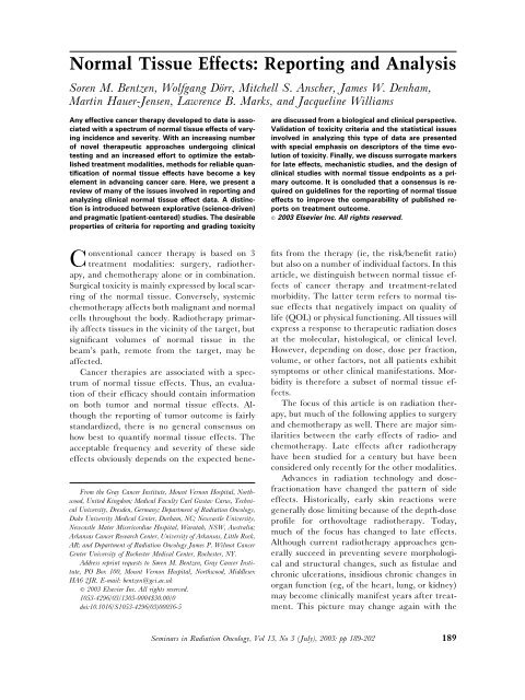
Normal Tissue Effects: Reporting and Analysis
Normal Tissue Effects: Reporting and Analysis
Normal Tissue Effects: Reporting and Analysis

You also want an ePaper? Increase the reach of your titles
YUMPU automatically turns print PDFs into web optimized ePapers that Google loves.
<strong>Normal</strong> <strong>Tissue</strong> <strong>Effects</strong>: <strong>Reporting</strong> <strong>and</strong> <strong>Analysis</strong><br />
Soren M. Bentzen, Wolfgang Dörr, Mitchell S. Anscher, James W. Denham,<br />
Martin Hauer-Jensen, Lawrence B. Marks, <strong>and</strong> Jacqueline Williams<br />
Any effective cancer therapy developed to date is associated<br />
with a spectrum of normal tissue effects of varying<br />
incidence <strong>and</strong> severity. With an increasing number<br />
of novel therapeutic approaches undergoing clinical<br />
testing <strong>and</strong> an increased effort to optimize the established<br />
treatment modalities, methods for reliable quantification<br />
of normal tissue effects have become a key<br />
element in advancing cancer care. Here, we present a<br />
review of many of the issues involved in reporting <strong>and</strong><br />
analyzing clinical normal tissue effect data. A distinction<br />
is introduced between explorative (science-driven)<br />
<strong>and</strong> pragmatic (patient-centered) studies. The desirable<br />
properties of criteria for reporting <strong>and</strong> grading toxicity<br />
From the Gray Cancer Institute, Mount Vernon Hospital, Northwood,<br />
United Kingdom; Medical Faculty Carl Gustav Carus, Technical<br />
University, Dresden, Germany; Department of Radiation Oncology,<br />
Duke University Medical Center, Durham, NC; Newcastle University,<br />
Newcastle Mater Misericordiae Hospital, Waratah, NSW, Australia;<br />
Arkansas Cancer Research Center, University of Arkansas, Little Rock,<br />
AR; <strong>and</strong> Department of Radiation Oncology James P. Wilmot Cancer<br />
Center University of Rochester Medical Center, Rochester, NY.<br />
Address reprint requests to Søren M. Bentzen, Gray Cancer Institute,<br />
PO Box 100, Mount Vernon Hospital, Northwood, Middlesex<br />
HA6 2JR. E-mail: bentzen@gci.ac.uk<br />
© 2003 Elsevier Inc. All rights reserved.<br />
1053-4296/03/1303-0004$30.00/0<br />
doi:10.1016/S1053-4296(03)00036-5<br />
are discussed from a biological <strong>and</strong> clinical perspective.<br />
Validation of toxicity criteria <strong>and</strong> the statistical issues<br />
involved in analyzing this type of data are presented<br />
with special emphasis on descriptors of the time evolution<br />
of toxicity. Finally, we discuss surrogate markers<br />
for late effects, mechanistic studies, <strong>and</strong> the design of<br />
clinical studies with normal tissue endpoints as a primary<br />
outcome. It is concluded that a consensus is required<br />
on guidelines for the reporting of normal tissue<br />
effects to improve the comparability of published reports<br />
on treatment outcome.<br />
© 2003 Elsevier Inc. All rights reserved.<br />
Conventional cancer therapy is based on 3<br />
treatment modalities: surgery, radiotherapy,<br />
<strong>and</strong> chemotherapy alone or in combination.<br />
Surgical toxicity is mainly expressed by local scarring<br />
of the normal tissue. Conversely, systemic<br />
chemotherapy affects both malignant <strong>and</strong> normal<br />
cells throughout the body. Radiotherapy primarily<br />
affects tissues in the vicinity of the target, but<br />
significant volumes of normal tissue in the<br />
beam’s path, remote from the target, may be<br />
affected.<br />
Cancer therapies are associated with a spectrum<br />
of normal tissue effects. Thus, an evaluation<br />
of their efficacy should contain information<br />
on both tumor <strong>and</strong> normal tissue effects. Although<br />
the reporting of tumor outcome is fairly<br />
st<strong>and</strong>ardized, there is no general consensus on<br />
how best to quantify normal tissue effects. The<br />
acceptable frequency <strong>and</strong> severity of these side<br />
effects obviously depends on the expected benefits<br />
from the therapy (ie, the risk/benefit ratio)<br />
but also on a number of individual factors. In this<br />
article, we distinguish between normal tissue effects<br />
of cancer therapy <strong>and</strong> treatment-related<br />
morbidity. The latter term refers to normal tissue<br />
effects that negatively impact on quality of<br />
life (QOL) or physical functioning. All tissues will<br />
express a response to therapeutic radiation doses<br />
at the molecular, histological, or clinical level.<br />
However, depending on dose, dose per fraction,<br />
volume, or other factors, not all patients exhibit<br />
symptoms or other clinical manifestations. Morbidity<br />
is therefore a subset of normal tissue effects.<br />
The focus of this article is on radiation therapy,<br />
but much of the following applies to surgery<br />
<strong>and</strong> chemotherapy as well. There are major similarities<br />
between the early effects of radio- <strong>and</strong><br />
chemotherapy. Late effects after radiotherapy<br />
have been studied for a century but have been<br />
considered only recently for the other modalities.<br />
Advances in radiation technology <strong>and</strong> dosefractionation<br />
have changed the pattern of side<br />
effects. Historically, early skin reactions were<br />
generally dose limiting because of the depth-dose<br />
profile for orthovoltage radiotherapy. Today,<br />
much of the focus has changed to late effects.<br />
Although current radiotherapy approaches generally<br />
succeed in preventing severe morphological<br />
<strong>and</strong> structural changes, such as fistulae <strong>and</strong><br />
chronic ulcerations, insidious chronic changes in<br />
organ function (eg, of the heart, lung, or kidney)<br />
may become clinically manifest years after treatment.<br />
This picture may change again with the<br />
Seminars in Radiation Oncology, Vol 13, No 3 (July), 2003: pp 189-202 189
190 Bentzen et al<br />
Table 1. Typical Characteristics of Pragmatic <strong>and</strong> Explorative Scoring Systems<br />
Routine Studies<br />
Multiple endpoints often merged into a single grade<br />
Easy to assess clinically<br />
Complete: any clinical occurrence of a late effect<br />
should be gradable <strong>and</strong> recordable using the<br />
dictionary<br />
Symptoms <strong>and</strong> QoL: these endpoints are most<br />
important because of their relevance to the<br />
patient.<br />
The focus is often on severely debilitating toxicity<br />
Statistical methods: Kaplan-Meier, Prevalence,<br />
Cumulative incidence<br />
Follow patients for their lifetime<br />
Endpoints selected based on clinical relevance<br />
Focus on the patient<br />
Toxicity-Specific Studies<br />
The emphasis is on a specific endpoint, varying grade<br />
May require specific diagnostic procedures<br />
Selective: the focus is on a specific biological effect of<br />
therapy that may be quantified <strong>and</strong> subjected to<br />
mechanistic studies<br />
Analytic <strong>and</strong> objective signs are the preferred<br />
endpoints as these can be validated across centres/<br />
observers<br />
Less severe toxicity is of interest as the increased<br />
incidence improves the statistical power of a study<br />
Statistical method: Kaplan-Meier<br />
Follow patients long enough to ensure statistical<br />
power<br />
Endpoints are selected to provide high sensitivity <strong>and</strong><br />
high specificity<br />
Focus on biology<br />
recent <strong>and</strong> expected advances in radiation <strong>and</strong><br />
cancer biology, functional imaging, <strong>and</strong> treatment<br />
planning <strong>and</strong> delivery that may facilitate<br />
high local doses.<br />
Routine Versus Toxicity-Specific<br />
Investigations<br />
The study of normal tissue side effects has 3 main<br />
aims: (1) to serve as an integral part of quality<br />
assurance in routine practice, including ongoing<br />
management of morbidity; (2) to establish the<br />
type, incidence <strong>and</strong> severity of effects for specific<br />
therapies to inform the decision making by patients,<br />
physicians, <strong>and</strong> health care managers; <strong>and</strong><br />
(3) to investigate the pathobiology underlying<br />
these effects, <strong>and</strong> thereby develop strategies for<br />
their prevention or amelioration. These are toxicity-specific<br />
studies with an in-depth assessment<br />
of one or more toxicity items as primary study<br />
endpoints. This may improve the QOL of cancer<br />
survivors <strong>and</strong>/or allow an intensification of therapy<br />
to improve cure rates.<br />
Corresponding to these aims, there are 2<br />
trends in normal tissue effects research, one being<br />
pragmatic <strong>and</strong> patient centered <strong>and</strong> the other<br />
being mechanistic <strong>and</strong> biology centered. Table 1<br />
summarizes a number of methodological aspects<br />
that tend to differ or at least are emphasized to a<br />
varying extent with the 2 types of studies.<br />
Clearly, careful clinical observation <strong>and</strong> documentation<br />
of side effects, early as well as late, is<br />
required in any case. The data obtained should<br />
ideally be reported in a format that facilitates<br />
comparison between studies <strong>and</strong> allow analysis by<br />
other researchers. This requires comprehensive,<br />
validated, <strong>and</strong> widely accepted scoring systems.<br />
Arguably, the failure to devise such a system is an<br />
impediment to normal tissue effects research. On<br />
the other h<strong>and</strong>, it is not obvious that a single<br />
system could be devised that would be equally<br />
useful for the various research aims.<br />
Also, establishing a set of toxicity criteria will<br />
not suffice. There are a number of outst<strong>and</strong>ing<br />
issues in the design <strong>and</strong> analysis of normal tissue<br />
studies that need clarification to improve their<br />
scientific value, <strong>and</strong> these will be discussed further.<br />
Distinguishing Early Versus Late <strong>Effects</strong><br />
From a biological <strong>and</strong> clinical point of view, it is<br />
useful to have a nominal division of normal tissue<br />
effects into early <strong>and</strong> late effects. Early effects<br />
are expressed during or immediately after the<br />
end of therapy, whereas late effects may become<br />
manifest after latent periods of months to years.<br />
Early reactions are usually reversible <strong>and</strong> therefore<br />
often considered less relevant for limiting<br />
treatment intensity. However, a recent review<br />
found that there may be an association between<br />
the occurrence of early reactions <strong>and</strong> the risk of
<strong>Normal</strong> <strong>Tissue</strong> <strong>Effects</strong>: <strong>Reporting</strong> <strong>and</strong> <strong>Analysis</strong> 191<br />
subsequent late effects in a number of organs. 1 In<br />
contrast, late effects are generally considered irreversible<br />
<strong>and</strong> progressive. Denham <strong>and</strong> colleagues<br />
2 recently proposed to replace the simple<br />
early/late classification of normal tissue effects<br />
with a more mechanistically based classification.<br />
Although this change of paradigm seems highly<br />
desirable in terms of advancing the development<br />
of strategies for prevention <strong>and</strong> treatment of normal<br />
tissue effects, it may be premature to discard<br />
the distinction between early <strong>and</strong> late effects<br />
when reporting toxicity.<br />
There is no consensus in the literature concerning<br />
the exact definition of early <strong>and</strong> late<br />
effects. Often an operational definition is used,<br />
simply classifying effects based on an arbitrary<br />
cutoff for their latent period. One cutoff that has<br />
been used is 90 days after the onset of the treatment.<br />
It has, however, been proposed to define<br />
late effects as those that occur or have not healed<br />
by 90 days after the end of therapy. 3 This latter<br />
definition seems more appropriate with many<br />
combined modality therapies extending the period<br />
of active therapy over several months. Unfortunately,<br />
there is no general biological justification<br />
for any of these conventions. However, a<br />
classification of early <strong>and</strong> late effects is clinically<br />
relevant, <strong>and</strong> therefore it is essential for comparability<br />
between studies that a common definition<br />
is agreed on. Recently, new guidelines have been<br />
proposed for clarifying early versus late effects<br />
based on aggregate data review. The application<br />
<strong>and</strong> precise methods for this have yet to be fully<br />
described. Ideally, it would be desirable if this<br />
definition could be endpoint specific based on a<br />
biological consideration of the actual time course<br />
of effects for a given normal tissue effect.<br />
Recording of Early <strong>Effects</strong><br />
As noted, early effects of cancer therapy usually<br />
occur during or shortly after the treatment (ie, at<br />
a time when the patient is still under close observation<br />
by the treating physician). Moreover,<br />
early effects (eg, in skin) have been used as a<br />
biological dosimeter in the early decades of radiotherapy;<br />
just like hematological toxicity has<br />
been dose limiting for many cytotoxic drugs.<br />
Thus, there is a fairly comprehensive literature<br />
on early morbidity after both radiotherapy <strong>and</strong><br />
chemotherapy. This has resulted in the development<br />
<strong>and</strong> implementation of scoring systems designed<br />
for radiotherapy, like the Radiation Therapy<br />
Oncology Group/European Organisation for<br />
Research <strong>and</strong> Treatment of Cancer system<br />
(RTOG/EORTC), <strong>and</strong> chemotherapy, like the<br />
Common Toxicity Criteria or the World Health<br />
Organization system. Recently, these have been<br />
amalgamated in the Common Toxicity Criteria<br />
version 2.0, 4 which is aimed at creating a common<br />
system applicable for each of the 2 modalities<br />
alone or in combination. Although developed<br />
specifically for reporting early effects in clinical<br />
trials, they have also been widely applied in routine<br />
practice.<br />
More elaborate scoring systems have been<br />
used in clinical studies focussing on a specific<br />
early side effect. Some of these have been extensions<br />
or modifications of the RTOG/EORTC system,<br />
like for example the inclusion of the affected<br />
area in the evaluation of oral mucositis 5 ;<br />
others have increased the level of detail on the<br />
more functional, subjective consequences of the<br />
reaction for the patient, like the morbidity scoring<br />
system by Dische 6 (which is available as a<br />
draft but unfortunately has never been published<br />
in its entirety). Clearly, the use of more in-depth<br />
toxicity scoring instruments will typically increase<br />
the time spent on each follow-up of a<br />
patient, but this may be justified by the additional<br />
information obtained.<br />
Recording of Late <strong>Effects</strong><br />
Toxicity criteria for late effects are much less<br />
established than those for early effects. The most<br />
ambitious attempt so far to develop a comprehensive<br />
system for the grading <strong>and</strong> recording of<br />
late radiation effects was the Subjective, Objective,<br />
Management, Analytic/Late <strong>Effects</strong> <strong>Normal</strong><br />
<strong>Tissue</strong> (SOMA/LENT) system, first published in<br />
1995. 7,8 We will herein consider the features of<br />
this system in some detail as an example of some<br />
of the general issues involved in late morbidity<br />
recording. The SOMA system was devised in the<br />
early 1990s, when working parties formed by the<br />
RTOG in the United States <strong>and</strong> the EORTC<br />
joined forces in an ambitious attempt to devise a<br />
rational, comprehensive scoring system for use in<br />
radiation oncology As suggested by its acronym,<br />
the characteristic feature of the SOMA/LENT<br />
system is that each toxicity item is classified by
192 Bentzen et al<br />
Table 2. Example of LENT/SOMA scale: Toxicity Items for the Lung<br />
Grade 1 Grade 2 Grade 3 Grade 4<br />
Subjective<br />
Cough Occasional Intermittent Persistent Refractory<br />
Dyspnea Breathless on<br />
intense<br />
exertion<br />
Breathless on<br />
mild<br />
exertion<br />
Breathless at<br />
rest, limits<br />
all activities<br />
Prevents any<br />
physical<br />
activity<br />
Chest pain/<br />
discomfort<br />
Objective<br />
Pulmonary<br />
fibrosis<br />
Lung<br />
function<br />
Management<br />
Pain<br />
Occasional &<br />
minimal<br />
Radiological<br />
abnormality<br />
10%-25%<br />
reduction of<br />
respiration<br />
volume <strong>and</strong>/<br />
or diffusion<br />
capacity<br />
Intermittent &<br />
tolerable<br />
Patchy dense<br />
abnormalities<br />
on<br />
radiograph<br />
25%-50%<br />
reduction of<br />
respiration<br />
volume <strong>and</strong>/<br />
or diffusion<br />
capacity<br />
Persistent &<br />
intense<br />
Dense<br />
confluent<br />
radiographic<br />
changes<br />
limited to<br />
radiation<br />
field<br />
50%-75%<br />
reduction of<br />
respiration<br />
volume <strong>and</strong>/<br />
or diffusion<br />
capacity<br />
Occasional<br />
non-narcotic<br />
Regular nonnarcotic<br />
Regular<br />
narcotic<br />
Cough Non-narcotic Narcotic,<br />
Intermittent<br />
corticosteroids<br />
Dyspnea Occasional O 2 Continuous O 2<br />
Analytic<br />
PFT<br />
DLCO<br />
%O 2 /CO 2<br />
saturation<br />
CT/MRI<br />
Perfusion<br />
scan<br />
Lung lavage<br />
Refractory &<br />
excruciating<br />
Dense fibrosis,<br />
severe<br />
scarring &<br />
major<br />
retraction of<br />
normal lung<br />
75% redu<br />
ction of<br />
respiration<br />
volume <strong>and</strong>/<br />
or diffusion<br />
capacity<br />
Surgical<br />
intervention<br />
Respirator,<br />
continuous<br />
corticosteroids<br />
Decrease to<br />
75%-90%<br />
of preTx<br />
value<br />
Decrease to<br />
50%-75%<br />
of preTx<br />
value<br />
Decrease to<br />
25%-50%<br />
of preTx<br />
value<br />
Decrease to<br />
25% of<br />
preTx value<br />
Decrease to<br />
Decrease to<br />
Decrease to Decrease to<br />
75%-90%<br />
50%-75%<br />
25%-50% 25% of<br />
of preTx<br />
of preTx<br />
of preTx preTx value<br />
value<br />
value<br />
value<br />
70% O 2 ,<br />
60% O 2 ,<br />
50% O 2 , 50% O 2 ,<br />
50% CO 2 60% CO 2 70% CO 2 70% CO 2<br />
Assessment of lung volume <strong>and</strong> zones of fibrosis<br />
Assessment of pulmonary blood flow <strong>and</strong> alveolar filling<br />
Assessment of cells <strong>and</strong> cytokines<br />
its subjective symptoms, objective signs, <strong>and</strong><br />
management-related or analytical measures.<br />
Table 2 shows the toxicity items for lung in the<br />
SOMA/LENT system. Subjective symptoms for<br />
this organ are cough, dyspnea, <strong>and</strong> chest pain/<br />
discomfort. A patient presenting with occasional<br />
cough <strong>and</strong> who is breathless on mild exertion<br />
would thus have a grade 1 cough <strong>and</strong> a grade 2<br />
dyspnea. Objective signs would be radiologic<br />
changes on a chest radiograph or reduced lung<br />
function. The Management would depend on the<br />
symptom but includes prescription of narcotic or<br />
nonnarcotic drugs, supplemental oxygen, corticosteroids,<br />
respirator, or surgical interventions. In<br />
addition to the S, O, M components, a number of<br />
specific analytic tests are suggested. In case of<br />
lung function, some test outcomes are graded<br />
(eg, pulmonary functional tests), whereas others<br />
are not (eg, pulmonary lavage <strong>and</strong> computed tomography/magnetic<br />
resonance imaging scans).
<strong>Normal</strong> <strong>Tissue</strong> <strong>Effects</strong>: <strong>Reporting</strong> <strong>and</strong> <strong>Analysis</strong> 193<br />
Figure 1 Schematic representation of the trade-off<br />
between specificity <strong>and</strong> patient relevance of various<br />
dimensions of normal tissue effects.<br />
Figure 1 shows in schematic form the tradeoff<br />
between the specificity of toxicity items <strong>and</strong> the<br />
relevance for the patient. At one end of the spectrum,<br />
QOL is the patient’s systematic self-assessment<br />
of various aspects of physical <strong>and</strong> social<br />
function. Specific instruments have been designed<br />
for the assessment of QOL, such as the<br />
EORTC QLQ-C30 questionnaire. Quality of life<br />
is affected by a large number of factors; some are<br />
health related <strong>and</strong> others that are not. Although<br />
it is of major interest to study the impact of<br />
treatment morbidity on QOL, this is not a specific<br />
indication of the biological effect of cancer<br />
therapy per se. At the other extreme, a change in<br />
the magnetic resonance imaging T1 relaxation<br />
time may have biological significance but may not<br />
cause any symptoms for the patient. A final issue<br />
in this context is the importance of subclinical<br />
toxicities. To use the gut as an example, vitamin<br />
B12 malabsorption after irradiation has been<br />
shown in a number of clinical studies. 9 This is<br />
likely to be important for the patient, even<br />
though it may not cause identifiable subjective<br />
symptoms. Attempts to combine these varied dimensions<br />
into aggregate or summary scores have<br />
met with little success because of fundamental<br />
qualitative differences (discussed later).<br />
Ranking <strong>and</strong> Classifying the Severity of<br />
Adverse <strong>Effects</strong><br />
Most clinical manifestations of normal tissue effects<br />
of radiotherapy may be ranked according to<br />
their severity, <strong>and</strong> this grading (also called scoring)<br />
should ideally be the subject of validation<br />
studies. One example is late bladder toxicity assessed<br />
by urination frequency 10 in which grade 1<br />
to 4 are defined as 3- to 4-hour intervals, 2- to<br />
3-hour intervals, 1- to 2-hour intervals, <strong>and</strong><br />
hourly, respectively. Increasing grades are defined<br />
so that they correspond to increasing levels<br />
of biological effect <strong>and</strong>/or increasing detriment to<br />
the QOL or physical functioning. Statistically, a<br />
graded-scale endpoint is also referred to as an<br />
ordinal variable. No assumptions are required in<br />
regards to a numerical relationship between the<br />
different grades (eg, a grade 4 reaction is not in<br />
any sense twice as bad as a grade 2); the only<br />
assumption is that an ordering of the categories<br />
is possible <strong>and</strong> meaningful.<br />
Several reported studies dichotomize graded<br />
endpoints into, say, none-mild <strong>and</strong> moderate or<br />
severe reactions. This procedure is evidently associated<br />
with a loss of information <strong>and</strong> should<br />
generally be avoided unless there are clinical or<br />
biological arguments for such dichotomization.<br />
Simulation studies have shown that the number<br />
of patients required to achieve a certain statistical<br />
precision of an estimate of a radiobiological<br />
parameter in a clinical study may be up to 3 times<br />
higher using a dichotomous endpoint rather than<br />
a graded scale. 11 Graded response data for late<br />
effects may be analyzed using a generalization of<br />
the Cox model or the mixture model. 12 For<br />
graded endpoints without censoring, ordinal logistic<br />
regression may be used. 13<br />
Similarly, a cystometric assessment of bladder<br />
volume 14 will effectively produce a continuum of<br />
reduced bladder capacity after radiotherapy.<br />
Again, a continuous scale endpoint may be reduced—at<br />
the price of a loss of information—to<br />
an ordinal endpoint by defining a limited number<br />
of grades corresponding to varying ranges of<br />
bladder volume.<br />
Although the reduction of continuous scale to<br />
ordinal <strong>and</strong> again to dichotomous endpoints is<br />
associated with a loss of information, this may<br />
convey some advantages in terms of computation<br />
or in terms of the interpretation of the findings of<br />
a study. Most importantly, binary data form the<br />
basis for the analysis of radiation dose-response<br />
relationships. The typical sigmoid dose-response<br />
curves are, more precisely, dose-incidence curves<br />
representing the proportion of patients who have<br />
reached a certain threshold response as a func-
194 Bentzen et al<br />
tion of dose. 15 From the dose-response curve, the<br />
effective dose (ED) after which the endpoint is<br />
reached in, say, 50%, 5%, or 3% of the patients<br />
(ie, the ED 50 , ED 5 , or ED 3 ) can be estimated<br />
together with an estimate of the statistical uncertainty<br />
in these quantities. The steepness of<br />
the dose-response curve is most often quantified<br />
by the value (ie, the normalized dose-response<br />
gradient at the steepest part of the curve). For a<br />
st<strong>and</strong>ard logistic dose-response curve, this is at<br />
the 50% response level, <strong>and</strong> therefore the steepness<br />
of the curve is specified as 50 .<br />
Summary <strong>Reporting</strong> of Late <strong>Effects</strong><br />
From a pragmatic point of view, there have been<br />
attempts to combine the information about all<br />
the many manifestations of treatment effect into<br />
a single figure, an overall toxicity grade. Proponents<br />
of this idea made the analogy with the<br />
grouping of T, N, <strong>and</strong> M categories for human<br />
tumors into clinical stages. There is, however, an<br />
important difference between these 2 situations.<br />
Clinical tumor stage groupings are defined based<br />
on similarities in cause-specific survival. Using<br />
the terminology of numerical taxonomy, causespecific<br />
survival is the “operational taxonomic<br />
unit” (OTU) that will allow deciding if, say, a<br />
T 3 N 0 <strong>and</strong>aT 2 N 1 tumor are similar (ie, whether<br />
they should be grouped in the same clinical<br />
stage). In the case of toxicity grading, there is no<br />
immediately obvious OTU. One possible OTU<br />
would appear to be the patient’s QOL as assessed<br />
by a validated instrument. In other words, specific<br />
signs <strong>and</strong> symptoms would be grouped together<br />
if they had a similar effect on quality of<br />
life. However, such a system would mix the actual<br />
manifestations of normal tissue damage with how<br />
well the patient copes with these.<br />
In the original publication of the LENT/<br />
SOMA system, it was suggested to add the individual<br />
item scores for an organ <strong>and</strong> then calculate<br />
the average score. This is clearly not a very<br />
good idea. In the case of lung reactions (Table 2),<br />
it was proposed to add the 8 items in the SOM<br />
part of the scale <strong>and</strong> divide by 8. Thus, a patient<br />
dying (grade 5 per definition) from restricted<br />
pulmonary function without having recorded any<br />
of the other 7 signs or symptoms or management<br />
interventions for pulmonary injury would get a<br />
score of 5/80.625 (ie, less than a patient who<br />
had grade 1 signs <strong>and</strong> symptoms for all the<br />
items). This flaw of the original suggestion for<br />
using the SOMA/LENT system was almost immediately<br />
recognized, <strong>and</strong> the suggestion was<br />
withdrawn in a remarkable double publication of<br />
letters to the editors of the International Journal of<br />
Radiation Oncology Biology Physics <strong>and</strong> Radiotherapy<br />
<strong>and</strong> Oncology. 16,17 Interestingly, the original suggestion<br />
has never been replaced by a more definitive<br />
suggestion from the groups formulating the<br />
SOMA system. One obvious idea is to record the<br />
maximum grade of any toxicity item for a specific<br />
organ/tissue <strong>and</strong> use this as the grade of toxicity.<br />
18 Thus, a patient experiencing, say, a grade 3<br />
loss of sphincter control after radiotherapy will<br />
be recorded as having experienced a grade 3<br />
rectal complication. Although this suggestion appears<br />
to show more common sense than the original<br />
SOMA/LENT proposal, it still involves a<br />
number of assumptions regarding the comparability<br />
of grades across toxicity items. From a biological<br />
perspective, it seems illogical even to<br />
attempt to pool various components of morbidity<br />
<strong>and</strong> arrive at a single number quantifying late<br />
effects.<br />
Validation of Toxicity Criteria<br />
Ideally, a proposed set of toxicity criteria should<br />
undergo a systematic <strong>and</strong> scientific study of their<br />
feasibility, reliability, validity, responsiveness <strong>and</strong><br />
specificity for the treatment before it is applied in<br />
clinical practice or in clinical research. This, however,<br />
is a major undertaking, <strong>and</strong> it has been<br />
largely neglected for the systems developed so<br />
far. Each aspect of validity has several subcomponents<br />
that should be addressed, <strong>and</strong> some of<br />
these are listed in Table 3. Some elements can<br />
only be tested in a well-designed, prospective<br />
study. Others are soft qualities that may be difficult<br />
to quantify rigorously but nevertheless are<br />
important for widespread acceptance of a scoring<br />
system. Many of the concepts involved have been<br />
developed in social sciences <strong>and</strong> psychology, <strong>and</strong><br />
there is rich literature on these topics. In cancer<br />
outcomes research, most of the literature in this<br />
area is concerned with the validation of quality of<br />
life instruments. There is some variability in the<br />
terminology used, but we have tried to focus on a
<strong>Normal</strong> <strong>Tissue</strong> <strong>Effects</strong>: <strong>Reporting</strong> <strong>and</strong> <strong>Analysis</strong> 195<br />
Table 3. Elements of Assessing the Performance of a Toxicity Scale<br />
Element Subelement Comments, Typical Metrics<br />
Feasibility Compliance The proportion of toxicity items that are assessed (<strong>and</strong> judged to<br />
have a valid score) at a single follow-up or at a number of<br />
consecutive follow-ups<br />
Cost<br />
The cost in monetary <strong>and</strong>/or time units of completing a toxicity<br />
assessment for a given patient<br />
Convenience<br />
The ease of using a toxicity scale <strong>and</strong> the convenience to the<br />
patient/rater. This includes consideration of invasiveness, any<br />
health risks associated with the use of diagnostic ionising<br />
radiation, pain <strong>and</strong> psychological distress<br />
Reliability<br />
Intra-rater<br />
reproducibility<br />
Inter-rater<br />
concordance<br />
Test-retest reliability<br />
Temporal stability<br />
Internal consistency<br />
The ability of a single observer to reproduce the toxicity items <strong>and</strong><br />
grades when presented with the same clinical or analytical<br />
information, e.g. a laboratory test or a photograph of breast<br />
appearance<br />
The concordance between the toxicity grades evaluated by<br />
multiple raters when presented with the same information - this<br />
should be tested within departments, between departments <strong>and</strong><br />
between countries<br />
The reliability of toxicity grades when re-testing the same patient<br />
with a minimal time interval between tests. Again, intra- <strong>and</strong><br />
inter-rater reliability is of interest.<br />
The reliability of toxicity grades when re-testing the same patient<br />
with a specified time interval between tests. Some physiological<br />
toxicity assays may vary with time of the day or from day to day<br />
Items relating to the same aspect of toxicity should produce<br />
consistent results. Internal consistency may be checked by<br />
deliberate including repeat or slightly modified items on the<br />
scale<br />
Validity Face validity Clinical relevance, common sense, biological <strong>and</strong><br />
pathophysiological relevance<br />
Content validity The ability of the scale to cover the range of morbidities seen in a<br />
population of patients after a specific therapy<br />
Criterion validity The correlation between the toxicity item <strong>and</strong> some criterion<br />
variable of interest. An example could be the correlation<br />
between a dysphagia grade <strong>and</strong> weight loss or between a lung<br />
toxicity measure <strong>and</strong> the persons actual physical activity<br />
Responsiveness<br />
Convergent validity<br />
Sensitivity to<br />
treatment<br />
intensity<br />
modification<br />
Threshold sensitivity<br />
The correlation between a toxicity item <strong>and</strong> other assessments of<br />
the same aspect of toxicity using previously validated scales<br />
The ability to detect a dose-response relationship or changes in<br />
irradiated volume, or the addition of chemotherapy or surgery.<br />
Sensitivity to interventions for morbidity.<br />
The minimal change in the patient’s health status that can be<br />
detected on the scale.<br />
number of elements that would be of particular<br />
importance in evaluating toxicity criteria.<br />
Feasibility refers to the practicality <strong>and</strong> cost of<br />
using the scale as well as its acceptability to<br />
patients <strong>and</strong> physicians. There are several examples<br />
of scoring systems that never came into<br />
widespread use because they, rightly or wrongly,<br />
were thought to be impractical, complicated to<br />
use, or excessively time consuming.<br />
Reliability is the ability to reproduce the same<br />
toxicity scores within or between subjects or observers<br />
(Table 3). There are 3 types of reliability<br />
used in validation of grading instruments. Testretest<br />
designs are used to determine if responses<br />
are consistent within the same observer or subject.<br />
Interobserver reliability is a critical feature<br />
of measuring consistency in grading between 2<br />
observers. Obviously, blinding is essential in re-
196 Bentzen et al<br />
liability studies. Lastly, internal consistency measures<br />
various dimensions relating to the same<br />
aspect of toxicity. Reliability is often quantified<br />
by a statistical measure of concordance such as<br />
Chronbach’s alpha.<br />
Validity refers to whether the scale measures<br />
what it is supposed to measure. Face <strong>and</strong> content<br />
validity (see Table 3) may be difficult to quantify<br />
but will typically be tested through some kind<br />
of consensus development process. Widespread<br />
practical use of a system will indirectly depend<br />
on, <strong>and</strong> help in establishing, its face <strong>and</strong> content<br />
validity. Criterion validity is the correlation between<br />
a toxicity item <strong>and</strong> some criterion variable<br />
of interest. For example, the peak grade of mucositis<br />
after head <strong>and</strong> neck radiotherapy has been<br />
shown to correlate with functional dysphagia <strong>and</strong><br />
with the requirement for prescribed analgesics. 13<br />
Somewhat related to this is convergent validity,<br />
which is correlation between a particular toxicity<br />
item <strong>and</strong> the same aspect of toxicity assessed on<br />
a previously validated scale. Both criterion <strong>and</strong><br />
convergent validity should be tested in well-designed<br />
prospective studies with an estimation of<br />
the sample size required to test a specific hypothesis<br />
concerning the strength of the association<br />
between 2 scales or between a toxicity item <strong>and</strong> a<br />
criterion variable.<br />
Responsiveness measures sensitivity for detecting<br />
true clinical change over time <strong>and</strong> is an<br />
important aspect of any toxicity criteria. In radiation<br />
oncology, this may be interpreted as the<br />
ability of a given toxicity item to detect a radiation<br />
dose-response relationship, but it might also<br />
be established as the ability to reflect any other<br />
treatment intensification or even an intervention<br />
to relieve morbidity. Again, for a toxicity item to<br />
be of practical value, this item should exhibit<br />
responsiveness over the clinically relevant range<br />
of biologically effective doses.<br />
Another important consideration for a given<br />
endpoint is the specificity for treatment. For example,<br />
many functional tests may be affected by<br />
the patient’s age or by intercurrent disease. This<br />
does not in itself make these endpoints invalid or<br />
even less interesting. Often, specificity may be<br />
enhanced by analyzing the change relative to a<br />
pretreatment baseline rather than the absolute<br />
measure. There are, however, statistical difficulties<br />
in quantifying the impact of toxicities having<br />
high baseline prevalence. For example, even if<br />
they are important clinically, it is very difficult to<br />
estimate the impact of accelerated coronary atherosclerosis<br />
after irradiation to the heart or of<br />
accelerated, malabsorption-related osteoporosis<br />
after radiotherapy of the abdomen or pelvis.<br />
Very few prospective, rationally designed validation<br />
studies have been conducted on normal<br />
tissue effects scales. Most studies, allegedly addressing<br />
validity, have in reality considered feasibility<br />
or, in a few cases, convergent validity. 18,19<br />
Reliability is rarely systematically tested. Responsiveness<br />
has in some cases been established<br />
as a byproduct of studies designed with a therapeutic<br />
aim rather than in a prospective validation<br />
study. If radioresponsiveness is established from<br />
a pooling of patients with different histology or<br />
different stage of disease, special care should be<br />
taken to consider whether interpatient differences<br />
might confound the analysis.<br />
Analyzing <strong>and</strong> <strong>Reporting</strong> the Time<br />
Evolution of <strong>Normal</strong> <strong>Tissue</strong> <strong>Effects</strong><br />
Early <strong>Effects</strong><br />
Most early effects of cancer therapy show a dynamic<br />
pattern of onset <strong>and</strong> resolution over time.<br />
After large single doses, the time course of the<br />
expression of injury is dominated by the tissue<br />
biology, <strong>and</strong> treatment-related parameters are<br />
almost irrelevant. For fractionated therapy, however,<br />
the time of occurrence of the maximum<br />
grade of reaction <strong>and</strong> the time for this to resolve<br />
will depend on the intensity of the treatment<br />
schedule as well. 13 This raises the question of the<br />
optimal frequency of follow-ups required to get<br />
the information necessary for a sensible analysis.<br />
As an example, for specific studies of oral mucositis<br />
Maciejewski (oral communication, September<br />
2000) has suggested that daily observations are<br />
desirable. However, once or twice weekly scoring<br />
is generally considered sufficient for this as well<br />
as for the majority of early effects. There are<br />
pragmatic issues here too. In a busy clinic, more<br />
frequent observations are not practical.<br />
Another problem relates to the question of<br />
what is the optimal analysis <strong>and</strong> reporting of<br />
early effects data. Several descriptors have been<br />
used in the literature, <strong>and</strong> again the lack of a<br />
consensus hampers the direct comparison between<br />
studies. Among the suggestions to overcome<br />
this problem are using the average score
<strong>Normal</strong> <strong>Tissue</strong> <strong>Effects</strong>: <strong>Reporting</strong> <strong>and</strong> <strong>Analysis</strong> 197<br />
over a given time period or even the entire area<br />
under the response-time curve. From both an<br />
interpretational <strong>and</strong> a statistical point of view,<br />
there is a difficulty with calculating averages of<br />
ordinal parameters. The problem is that the average<br />
is typically not defined on the scale <strong>and</strong><br />
that taking the average involves the implicit assumption<br />
that 2 patients with a grade 2 reaction<br />
equal 1 patient with a grade 4 reaction. (The<br />
analogy would be to characterize a patient population<br />
with the average tumor stage). Admittedly,<br />
there are situations in which the area under the<br />
response-time curve may serve as a useful summary<br />
of morbidity burden. However, the usefulness<br />
of such a description—<strong>and</strong> the potential for<br />
misleading conclusions—should be carefully considered<br />
in each case.<br />
For routine reporting, the prevalence of a specific<br />
grade of morbidity as a function of time is<br />
easier to interpret. The following terminology<br />
has been proposed. 13 The peak prevalence is simply<br />
the maximum proportion of patients presenting<br />
with that grade of reaction at any specific<br />
follow-up. The incidence of the reaction, in contrast,<br />
is the proportion of patients who develop<br />
this grade of reaction at any point in time. As a<br />
supplement to these quantities, the time over<br />
which a defined score is experienced may be a<br />
useful descriptor. Note, that for this parameter<br />
taking averages or differences between treatment<br />
groups is immediately meaningful.<br />
Late <strong>Effects</strong><br />
Longitudinal studies of the temporal evolution of<br />
late effects of cancer therapy have shown a wide<br />
range of latent times between irradiation <strong>and</strong> the<br />
occurrence of a specific endpoint. Telangiectasia<br />
of the skin is a classical example in which both<br />
the cumulative proportion of patients who have<br />
experienced a specific grade of reaction is increasing<br />
with time 20 <strong>and</strong> also the severity or<br />
grade of reaction will progress over time in many<br />
individuals. 21<br />
With the more widespread use of survival (or<br />
actuarial) statistics in cancer outcome studies, it<br />
was realized that quantification of the occurrence<br />
of any endpoint requiring prolonged observation<br />
of the patient requires some adjustment for the<br />
actual number of patients at risk (ie, still under<br />
observation) as a function of time after therapy.<br />
In the case of late effects after radiotherapy, the<br />
use of actuarial statistics was advocated by<br />
Hatlevoll et al 22 <strong>and</strong> Abratt 23 in the early 1980s.<br />
Cumulative incidence estimates have been<br />
proposed as an alternative to the Kaplan-Meier<br />
method for quantifying morbidity as a function of<br />
time. 24 The problem is, however, that these estimates<br />
are not specific to morbidity. 25 This means<br />
that the cumulative incidence of morbidity after<br />
strictly identical treatments will be higher in a<br />
group of patients with a good prognosis than in<br />
an otherwise comparable group with a poor prognosis.<br />
Prevalence estimates as a function of time<br />
have also been shown to provide useful information<br />
on the burden of late morbidity after radiotherapy.<br />
26 These estimates represent the proportion<br />
of patients having a specific type <strong>and</strong> grade<br />
of morbidity among all patients who are still alive<br />
<strong>and</strong> under observation at that point in time.<br />
The relative merits of these estimates as a<br />
means of quantifying morbidity have been the<br />
topic of some discussion. In reality, these methods<br />
are not alternative but rather complementary<br />
descriptors of the time evolution of morbidity. To<br />
improve the comparability of reports, actuarial<br />
estimates should be calculated <strong>and</strong> reported as a<br />
minimum requirement. 25 As a supplement, especially<br />
in pragmatic studies (Table 1), it may be<br />
useful to estimate cumulative incidence or prevalence<br />
of morbidity. Clearly, prevalence is an<br />
important measure of the burden of chronic toxicity<br />
on patients, their families, the health care<br />
system, <strong>and</strong> the society. Much more research is<br />
needed on the long-term evolution of morbidity<br />
in cancer survivors.<br />
Several recent studies have been concerned<br />
with the incidence of late effects after 10 to 20<br />
years of follow-up. 27-29 In a recent paper, Jung<br />
<strong>and</strong> colleagues 30 presented the results of a<br />
quantitative analysis of published data on the<br />
time-incidence relationship for a number of late<br />
endpoints. Jung et al 30 proposed a simple classification<br />
of these relationships into 4 categories<br />
<strong>and</strong> observed that the observations from many<br />
published studies seemed to be well fitted by an<br />
exponential latent-time distribution. This would<br />
imply that expression of injury is a r<strong>and</strong>om process<br />
with a constant probability per unit time of<br />
expressing the injury among patients who have<br />
not already done so by that time (ie, a constant<br />
hazard rate). At first glance, this might seem to
198 Bentzen et al<br />
indicate that very long follow-up times would be<br />
required to obtain a precise estimate of the toxicity<br />
of a given therapy. From a statistical point of<br />
view, this is actually not the case, the reason<br />
being that if the latent-time distribution is exponential,<br />
most of the events (carrying most of the<br />
information) will have occurred within the first<br />
few half times of expression. 31 There are also<br />
good data sets from experimental animals suggesting<br />
that the hazard rate is not generally constant<br />
for a variety of endpoints. Ironically, this<br />
may actually lead to a need for very long observation<br />
times anyway. This would depend on the<br />
actual shape <strong>and</strong> position of the latent-time distribution<br />
for the endpoint of interest. Some endpoints,<br />
most notably treatment-induced cancer,<br />
require long follow-up to be reliably quantified.<br />
Adverse <strong>Effects</strong> <strong>Analysis</strong> in Oncology<br />
Clinical Trials<br />
Traditionally, clinical trials of cytotoxic drugs are<br />
classified as belonging to 1 of 3 phases. A phase I<br />
trial is the first testing in humans of a new drug<br />
aimed to establish the maximum acceptable dose<br />
of the drug to be taken into phase II trials. Phase<br />
II trials screen new compounds or combinations<br />
of compounds for biological effect, most often<br />
using tumour volume shrinkage (ie, clinical response)<br />
as the endpoint. Such trials are typically<br />
designed to allow early trial termination if it<br />
becomes apparent that a clinically relevant target<br />
efficacy is unlikely to be reached. Drugs that<br />
show promising response rates in phase II trials<br />
are then considered for testing in r<strong>and</strong>omized<br />
controlled phase III trials aimed to test therapeutic<br />
combinations including the new compound<br />
against current best st<strong>and</strong>ard therapy.<br />
This classification scheme <strong>and</strong> the prototypical<br />
study design associated with the 3 phases of trials<br />
do not work well for radiation therapy trials.<br />
Phase I studies of cytostatic drugs typically involve<br />
relatively rapid escalation of drug doses,<br />
treating for example 3 patients at each dose level<br />
before deciding whether to escalate dose. Late<br />
chemoinduced effects are not considered. In radiotherapy<br />
trials, this may not be feasible, simply<br />
because late morbidity is generally regarded as<br />
intensity limiting <strong>and</strong> this is expressed after a<br />
long latent period. Even if the study is focussing<br />
on early morbidity, there is generally not a specific<br />
dose-limiting toxicity after radiotherapy. Instead,<br />
to judge whether a modified radiotherapy<br />
schedule is clinically acceptable requires a precise<br />
estimate of incidence <strong>and</strong> severity of early<br />
effects that again means that the sample size will<br />
need to be large. Phase II trials of cytotoxic drugs<br />
use tumor volume regression as the primary endpoint.<br />
With radiotherapy, most tumors show partial<br />
or complete regression <strong>and</strong> it is therefore<br />
ultimate local tumour control that is the most<br />
important endpoint for tumour effect. Rather<br />
than sequential phase I <strong>and</strong> II studies, most modified<br />
radiotherapy schedules are piloted in feasibility<br />
studies, often comprising as many as between<br />
50 <strong>and</strong> 100 patients when the primary<br />
endpoints are early <strong>and</strong> late morbidity <strong>and</strong> locoregional<br />
tumor control.<br />
The design of r<strong>and</strong>omized controlled phase III<br />
trials is more uniform across treatment modalities.<br />
Local tumor control <strong>and</strong> overall survival are<br />
the primary endpoints of treatment efficacy <strong>and</strong><br />
the target sample size for such trials will be<br />
estimated so that the trial has sufficient statistical<br />
power to resolve what is judged to be a clinically<br />
relevant improvement in these parameters.<br />
From a normal tissue effects perspective, there<br />
are 2 major concerns. First, morbidity, especially<br />
late, is frequently inadequately recorded <strong>and</strong> reported.<br />
The lack of st<strong>and</strong>ardized scoring systems<br />
<strong>and</strong> the often poor quality control of morbidity<br />
scores limits the value of much of the published<br />
literature. Second, even when morbidity is reported,<br />
the statistical power of the trial is not<br />
sufficient to resolve a clinically important change<br />
in this endpoint. Thus, in most trials, the possible<br />
change in therapeutic gain cannot be reliably<br />
judged. This is clearly a field in which more research<br />
is urgently needed.<br />
Surrogate Markers <strong>and</strong> Endpoints for<br />
Late <strong>Effects</strong><br />
The long latent period <strong>and</strong> the problems with low<br />
statistical power in many trials with late effects<br />
as a primary endpoint have stimulated interest in<br />
surrogate markers for late effects. It is useful to<br />
distinguish between surrogate markers <strong>and</strong> surrogate<br />
endpoints for late effects. A surrogate<br />
marker is a biological effect of treatment that,<br />
when it occurs in an individual patient, changes<br />
the probability that this patient develops a sub-
<strong>Normal</strong> <strong>Tissue</strong> <strong>Effects</strong>: <strong>Reporting</strong> <strong>and</strong> <strong>Analysis</strong> 199<br />
sequent clinically relevant late effect. The ideal<br />
surrogate marker would have both a high positive<br />
predictive value <strong>and</strong> a high negative predictive<br />
value for the clinical endpoint of interest. Also,<br />
detection of the surrogate marker should offer a<br />
clinically relevant lead time allowing a rational<br />
selection of cases for modification of ongoing primary<br />
therapy or for early interventions to relieve<br />
or prevent subsequent late morbidity.<br />
One example of a surrogate marker is plasma<br />
transforming growth factor- (TGF-) as a<br />
marker for pulmonary injury after radiotherapy.<br />
32 A normalization of the plasma level of this<br />
marker toward the end of a course of radiotherapy<br />
was shown to yield a positive predictive value<br />
of 90% for identifying patients who did not develop<br />
radiation pneumonitis. In a series of 38<br />
patients with inoperable non–small-cell lung cancer,<br />
Anscher et al 33 used changes in TGF- level<br />
as a criterion for escalating radiation dose in the<br />
individual cases. Twenty-four patients had persistently<br />
abnormal TGF- levels <strong>and</strong> received a total<br />
dose of 73.6 Gy. Among 14 patients whose<br />
TGF- levels were normal after 73.6 Gy, 8 were<br />
escalated to 80 Gy <strong>and</strong> 6 were escalated to 86.4<br />
Gy. In the 86.4-Gy group, dose-limiting toxicity<br />
was reached because there were 2 (33%) grade 3<br />
late toxicities. The authors concluded, from this<br />
uncontrolled study, that it may be feasible to use<br />
plasma TGF- levels to select patients for radiation<br />
dose escalation.<br />
Another example is from a prospective longitudinal<br />
study of clinical, endoscopic, <strong>and</strong> histopathologic<br />
rectal toxicity in 33 patients during<br />
ongoing pelvic radiation therapy. 34 Although clinical<br />
symptoms of early rectal morbidity progressed<br />
toward the end of the 6-week treatment<br />
course, endoscopic pathology was maximal at 2<br />
weeks <strong>and</strong> stabilized thereafter. Also, histologic<br />
changes were consistently more pronounced at 2<br />
weeks than at 6 weeks. These observations could<br />
point to possible surrogate markers of late effects<br />
<strong>and</strong> may have implications for the design <strong>and</strong><br />
timing of prophylactic <strong>and</strong> therapeutic interventions<br />
to reduce radiation proctitis.<br />
The long latent period of late effects must also<br />
be taken into consideration when estimating the<br />
positive <strong>and</strong> negative predictive value of a surrogate<br />
marker. A patient expressing the marker<br />
who dies before reaching the clinical endpoint is<br />
not necessarily a false-positive; again, it is necessary<br />
to use actuarial methods to correct for censoring.<br />
A surrogate endpoint, on the other h<strong>and</strong>, does<br />
not necessarily affect the probability of an individual<br />
expressing the clinical endpoint of concern<br />
but is indicative of treatment toxicity in a population<br />
of patients. For such an endpoint to be<br />
useful, it should provide a considerable lead time,<br />
should offer advantages in terms of reliable quantification<br />
<strong>and</strong>/or improved statistical power, <strong>and</strong><br />
should be responsive to modifications of therapy.<br />
As an example, the incidence of confluent mucositis<br />
after radiation therapy in the head <strong>and</strong><br />
neck region has been found to be a good indication<br />
of the overall biological intensity with respect<br />
to early morbidity. It is not a good surrogate<br />
endpoint for late effects as illustrated for example<br />
by the significant increase in the prevalence<br />
of confluent mucositis, but significant decrease of<br />
the incidence of late effects, after continuous<br />
hyperfractionated accelerated radiotherapy as<br />
compared with conventional radiotherapy for<br />
squamous cell carcinomas of the head <strong>and</strong> neck<br />
region.<br />
Mechanistic Studies<br />
Biological research on normal tissue effects ultimately<br />
aims to prevent or ameliorate the side<br />
effects of cancer therapy. Over the past decade, it<br />
has become clear that both early <strong>and</strong> late effects<br />
in normal tissues are a result of a complicated<br />
network of signaling molecules (cytokines) <strong>and</strong><br />
receptors initiated at the time of radiation injury<br />
<strong>and</strong> progressing until the clinical endpoints are<br />
manifest. 35,36 This recognition led to a rapid increase<br />
in biological studies attempting to identify<br />
the primary effectors in late-effect induction<br />
pathways that may then serve as targets for ameliorative<br />
treatments. However, the simple overexpression<br />
(or activation) of a moiety is not necessarily<br />
indicative of a causal relationship or even<br />
its role in the pathogenesis of a late effect. Indeed,<br />
the multifunctional nature of many cytokines,<br />
chemokines, <strong>and</strong> so on means that some<br />
may serve as both good guys <strong>and</strong> bad guys, being<br />
involved in both the induction of late effects, but<br />
also serving as necessary components of healing<br />
<strong>and</strong> repair in the normal tissue. A primary example<br />
of such a molecule is the previously mentioned<br />
TGF-. 37-38 In addition, many cytokines
200 Bentzen et al<br />
exist in a state of homeostasis, with the overexpression<br />
of one being balanced by the expression<br />
of another; such compensation mechanisms have<br />
been frequently observed <strong>and</strong> discussed among<br />
members of the interleukin family. 39 Finally, consideration<br />
must be given to the observation that<br />
no 2 tissues have identical expression of late<br />
effects. This differentiation probably occurs as a<br />
result of variations in cell populations in each<br />
tissue, leading to different target cells. Thus, no<br />
assessment of the potential usefulness of a signaling<br />
factor can be made without some recognition<br />
of the spatial component (ie, the cells <strong>and</strong><br />
tissue environment that are involved).<br />
All of these considerations pose a great challenge<br />
to preclinical research in model systems.<br />
Obviously, results from in vitro studies in permanent<br />
cell lines must be transferred to primary cell<br />
lines or tissue cultures. In any case, conclusions<br />
from these studies have to be validated in vivo in<br />
experimental animals, using relevant endpoints<br />
<strong>and</strong> well-designed experimental protocols. The<br />
same applies to results from molecular biology<br />
studies. In many cases, in vivo experiments reveal<br />
that intervention in 1 pathogenic pathway, which<br />
has been shown to modulate the cellular response<br />
in vitro, is ineffective in vivo because of either<br />
alternative pathways or because of interactions<br />
between different cell populations present in a<br />
complete tissue.<br />
Predictive Assays<br />
The large patient-to-patient variability in the response<br />
to a course of radiotherapy has stimulated<br />
interest in predictive assays of both tumour <strong>and</strong><br />
normal tissue radioresponsiveness. Again, it is<br />
useful to distinguish between surrogate markers<br />
for effect <strong>and</strong> predictive assays. Surrogate markers<br />
in vivo could in principle form the basis for a<br />
predictive assay. Such an approach would require<br />
that at least part of the therapy be delivered to<br />
the patient, which may limit its value in clinical<br />
decision making. Likewise, although the outcome<br />
of a predictive assay would affect our knowledge<br />
of the probability of a patient reaching a specific<br />
endpoint, it is not a surrogate marker of biological<br />
effect as discussed earlier.<br />
Although several assays have been evaluated<br />
<strong>and</strong> at least some of these have shown promise,<br />
there is still no predictive assay that is anywhere<br />
near implementation in the clinical routine. In<br />
the absence of direct evidence for the utility of<br />
normal tissue predictive assays, it is of interest to<br />
estimate the relative importance of deterministic<br />
versus stochastic effects in explaining patient-topatient<br />
variability in normal tissue responsiveness.<br />
A recent article 40 analyzed the occurrence<br />
of telangiectasia of the skin in patients treated<br />
with bilateral internal mammary fields <strong>and</strong> found<br />
that as much as 90% (with 95% confidence limits<br />
65% <strong>and</strong> 100%) of the variability in the radioresponsiveness<br />
in the right-sided field was explained<br />
by the radioresponsiveness in the leftsided<br />
field. This suggests that deterministic<br />
factors may dominate <strong>and</strong> stimulates further research<br />
to define these determinants.<br />
Combined Modality or Intensified<br />
Radiotherapy<br />
Novel combinations of therapies <strong>and</strong> new dosevolume<br />
combinations, radically different from<br />
those giving rise to most of our historical normal<br />
tissue complication data, may considerably<br />
change the incidence <strong>and</strong> type of morbidities<br />
seen. Interestingly, in the present context, this<br />
may lead to a need for changing the toxicity<br />
criteria themselves.<br />
A concrete example is the Fox-Chase modification<br />
of the SOMA/LENT scale for rectal toxicity.<br />
41 This modification was suggested based on<br />
long-term follow-up of a group of patients with<br />
prostate cancer included in a nonr<strong>and</strong>omized<br />
dose-escalation study. The investigators observed<br />
a number of cases of chronic rectal bleeding requiring<br />
at least 1 blood transfusion <strong>and</strong>/or more<br />
than 2 coagulations after high-dose conformal<br />
radiotherapy for prostate cancer <strong>and</strong> proposed to<br />
classify this as grade 3 rectal toxicity. This manifestation<br />
of rectal damage was not included in<br />
the original SOMA/LENT system, simply because<br />
it is very rarely seen with conventional dose levels.<br />
Similarly, when 3-dimensional conformal<br />
radiotherapy is used to escalate the dose to smallvolume<br />
lung cancers, it is likely that the doselimiting<br />
toxicity will be structural damage to the<br />
lung <strong>and</strong> major vessels, rather than the restricted<br />
lung-function that limits the dose of conventional<br />
radiotherapy.<br />
More recently, multimodality therapies with<br />
novel agents, including biologically targeted ther-
<strong>Normal</strong> <strong>Tissue</strong> <strong>Effects</strong>: <strong>Reporting</strong> <strong>and</strong> <strong>Analysis</strong> 201<br />
apies, are currently undergoing early clinical<br />
testing. There is little doubt that these strategies<br />
will improve tumor control <strong>and</strong> consequently also<br />
improve survival rates. However, this may come<br />
at a price: many of these new treatment regimens<br />
do involve an implicit or explicit acceptance of an<br />
increased incidence of treatment-related morbidity.<br />
Also, it can be expected that the morbidity<br />
profile may change as dose distributions are radically<br />
changed or drugs with new mechanisms of<br />
action are introduced.<br />
Conclusion<br />
An increasing number of new therapies <strong>and</strong> modifications<br />
of established therapies originate from<br />
advances in basic <strong>and</strong> clinical cancer research.<br />
The translation of these into clinical practice is a<br />
huge challenge also to the systematic scientific<br />
study of normal tissue effects. A therapeutic gain<br />
cannot be achieved without carefully balancing<br />
tumour cure <strong>and</strong> survival rates against morbidity<br />
<strong>and</strong> quality of life. Development of st<strong>and</strong>ardized<br />
common toxicity criteria <strong>and</strong> a widespread adoption<br />
of these in clinical trials would be a major<br />
step forward for clinical cancer research. For late<br />
effects, we suggest that the actuarial incidence is<br />
reported as a minimum <strong>and</strong> that this is supplemented<br />
by prevalence or cumulative incidence<br />
estimates if this is regarded relevant. Subjective<br />
symptoms, objective signs, management of toxicity,<br />
<strong>and</strong> analytical measures are complementary<br />
dimensions of toxicity, <strong>and</strong> we propose that these<br />
are reported <strong>and</strong> analyzed separately. In mechanistic<br />
studies or studies testing interventions to<br />
prevent or ameliorate morbidity specific biological<br />
endpoints are preferable. An involvement of<br />
normal tissue radiation biologists should be<br />
sought to optimize the design of clinical instruments<br />
for treatment effect assessment. A consensus<br />
is needed for the development of guidelines<br />
for the analysis <strong>and</strong> reporting of normal tissue<br />
effects.<br />
References<br />
1. Dorr W, Hendry JH: Consequential late effects in normal<br />
tissues. Radiother Oncol 61:223-231, 2001<br />
2. Denham JW, Hauer-Jensen M, Peters LJ: Is it time for a<br />
new formalism to categorize normal tissue radiation injury<br />
Int J Radiat Oncol Biol Phys 50:1105-1106, 2001<br />
3. Bentzen SM, Overgaard J: Clinical manifestations of normal-tissue<br />
damage, in Steel GG (ed): Basic Clinical Radiobiology.<br />
London, Arnold, 1997, pp 87-97<br />
4. Trotti A, Byhardt R, Stetz J: Common toxicity criteria:<br />
version 2.0: An improved reference for grading the acute<br />
effects of cancer treatment: Impact on radiotherapy. Int J<br />
Radiat Oncol Biol Phys 47:13-47, 2000<br />
5. Maciejewski B, Zajusz A, Pilecki B: Acute mucositis in the<br />
stimulated oral mucosa of patients during radiotherapy<br />
for head <strong>and</strong> neck cancer. Radiother Oncol 22:7-11, 1991<br />
6. Dische S, Warburton MF, Jones D: The recording of<br />
morbidity related to radiotherapy. Radiother Oncol 16:<br />
103-108, 1989<br />
7. Pavy J-J, Denekamp J, Letschert J: Late effects damage<br />
scoring: The SOMA scale. Radiother Oncol 35:11-60,<br />
1995<br />
8. Rubin P, Constine LS, Fajardo LF: Overview: Late effects<br />
of normal tissues (LENT) scoring system. Int J Radiat<br />
Oncol Biol Phys 31:1041-1042, 1995<br />
9. Yeoh E, Horowitz M, Russo A: Effect of pelvic irradiation<br />
on gastrointestinal function: A prospective longitudinal<br />
study. Am J Med 95:397-406, 1993<br />
10. Marks LB, Carroll PR, Dugan TC: The response of the<br />
urinary bladder, urethra, <strong>and</strong> ureter to radiation <strong>and</strong><br />
chemotherapy. Int J Radiat Oncol Biol Phys 31:1257-<br />
1280, 1995<br />
11. Taylor JMG: Properties of maximum likelihood estimates<br />
of the ratio of parameters in ordinal response regression<br />
models. Commun Statist-Simula 19:469-480, 1990<br />
12. Bentzen SM, Thames HD, Travis EL: Direct estimation<br />
of latent time for radiation injury in late-responding normal<br />
tissues: Gut, lung <strong>and</strong> spinal cord. Int J Radiat Biol<br />
55:27-43, 1989<br />
13. Bentzen SM, Saunders MI, Dische S: Radiotherapy-related<br />
early morbidity in head <strong>and</strong> neck cancer: Quantitative<br />
clinical radiobiology as deduced from the CHART<br />
trial. Radiother Oncol 60:123-135, 2001<br />
14. Lajer H, Thranov IR, Bagi P: Evaluation of urologic morbidity<br />
after radiotherapy for cervical carcinoma by urodynamic<br />
examinations <strong>and</strong> patient voiding schemes: A prospective<br />
study. Int J Radiat Oncol Biol Phys 54:1362-1368,<br />
2002<br />
15. Bentzen SM: Dose-response relationships in radiotherapy,<br />
in Steel GG (ed): Basic Clinical Radiobiology. London,<br />
Arnold, 2002, pp 94-104<br />
16. Denekamp J, Bartelink H, Rubin P: Correction for the use<br />
of the SOMA LENT tables. Radiother Oncol 39:191-191,<br />
1996<br />
17. Denekamp J, Bartelink H, Rubin P: Correction for the use<br />
of the SOMA LENT tables. Int J Radiat Oncol Biol Phys<br />
35:417, 1996<br />
18. Anacak Y, Yalman D, Ozsaran Z: Late radiation effects to<br />
the rectum <strong>and</strong> bladder in gynecologic cancer patients:<br />
The comparison of LENT/SOMA <strong>and</strong> RTOG/EORTC<br />
late-effects scoring systems. Int J Radiat Oncol Biol Phys<br />
50:1107-1112, 2001<br />
19. Livsey JE, Routledge J, Burns M: Scoring of treatmentrelated<br />
late effects in prostate cancer. Radiother Oncol<br />
65:109-121, 2002<br />
20. Bentzen SM, Turesson I, Thames HD: Fractionation sensitivity<br />
<strong>and</strong> latency of telangiectasia after postmastec-
202 Bentzen et al<br />
tomy radiotherapy: A graded response analysis. Radiother<br />
Oncol 18:95-106, 1990<br />
21. Turesson I: Individual variation <strong>and</strong> dose dependency in<br />
the progression rate of skin telangiectasia. Int J Radiat<br />
Oncol Biol Phys 19:1569-1574, 1990<br />
22. Hatlevoll R, Host H, Kaalhus O: Myelopathy following<br />
radiotherapy of bronchial carcinoma with large single<br />
fractions: A retrospective study. Int J Radiat Oncol Biol<br />
Phys 9:41-44, 1983<br />
23. Abratt RP: <strong>Reporting</strong> the late complication rates of cancer<br />
therapy. Br J Radiol 56:348-350, 1983<br />
24. Caplan RJ, Pajak TF, Cox JD: <strong>Analysis</strong> of the probability<br />
<strong>and</strong> risk of cause-specific failure. Int J Radiat Oncol Biol<br />
Phys 29:1183-1186, 1994<br />
25. Bentzen SM, Vaeth M, Pedersen D: Why actuarial estimates<br />
should be used in reporting late normal-tissue<br />
effects of cancer treatment. . .NOW. Int J Radiat Oncol<br />
Biol Phys 32:1531-1534, 1995<br />
26. Haie-Meder C, Kramar A, Lambin P: <strong>Analysis</strong> of complications<br />
in a prospective r<strong>and</strong>omized trial comparing two<br />
brachytherapy low dose rates in cervical carcinoma. Int J<br />
Radiat Oncol Biol Phys 29:953-960, 1994<br />
27. Johansson S, Svensson H, Denekamp J: Timescale of evolution<br />
of late radiation injury after postoperative radiotherapy<br />
of breast cancer patients. Int J Radiat Oncol Biol<br />
Phys 48:745-750, 2000<br />
28. Lundby L, Jensen VJ, Overgaard J: Long-term colorectal<br />
function after postoperative radiotherapy for colorectal<br />
cancer. Lancet 350:564, 1997<br />
29. Eifel PJ, Levenback C, Wharton JT: Time course <strong>and</strong><br />
incidence of late complications in patients treated with<br />
radiation therapy for FIGO stage IB carcinoma of the<br />
uterine cervix. Int J Radiat Oncol Biol Phys 32:1289-1300,<br />
1995<br />
30. Jung H, Beck-Bornholdt HP, Svoboda V: Quantification of<br />
late complications after radiation therapy. Radiother Oncol<br />
61:233-246, 2001<br />
31. Bentzen SM, Dische S: Late morbidity: The Damocles<br />
sword of radiotherapy Radiother Oncol 61:219-221, 2001<br />
32. Anscher MS, Kong FM, Andrews K: Plasma transforming<br />
growth factor beta1 as a predictor of radiation pneumonitis.<br />
Int J Radiat Oncol Biol Phys 41:1029-1035, 1998<br />
33. Anscher MS, Marks LB, Shafman TD: Using plasma<br />
transforming growth factor beta-1 during radiotherapy to<br />
select patients for dose escalation. J Clin Oncol 19:3758-<br />
3765, 2001<br />
34. Hovdenak N, Fajardo LF, Hauer-Jensen M: Acute radiation<br />
proctitis: A sequential clinicopathologic study during<br />
pelvic radiotherapy. Int J Radiat Oncol Biol Phys 48:1111-<br />
1117, 2000<br />
35. Rubin P, Johnston CJ, Williams JP: A perpetual cascade of<br />
cytokines postirradiation leads to pulmonary fibrosis. Int<br />
J Radiat Oncol Biol Phys 33:99-109, 1995<br />
36. Denham JW, Hauer-Jensen M: The radiotherapeutic injury—A<br />
complex ’wound’. Radiother Oncol 63:129-145,<br />
2002<br />
37. Barcellos-Hoff MH: How do tissues respond to damage at<br />
the cellular level The role of cytokines in irradiated<br />
tissues. Radiat Res 150:S109-S120, 1998<br />
38. Barcellos-Hoff MH: Radiation-induced transforming<br />
growth factor <strong>and</strong> subsequent extracellular matrix reorganization<br />
in murine mammary gl<strong>and</strong>. Cancer Res 53:<br />
3880-3886, 1993<br />
39. Fantuzzi G: Lessons from interleukin-deficient mice: The<br />
interleukin-1 system. Acta Physiol Sc<strong>and</strong> 173:5-9, 2001<br />
40. Safwat A, Bentzen SM, Turesson I: Deterministic rather<br />
than stochastic factors explain most of the variation in<br />
the expression of skin telangiectasia after radiotherapy.<br />
Int J Radiat Oncol Biol Phys 52:198-204, 2002<br />
41. Hanlon AL, Schultheiss TE, Hunt MA: Chronic rectal<br />
bleeding after high-dose conformal treatment of prostate<br />
cancer warrants modification of existing morbidity scales.<br />
Int J Radiat Oncol Biol Phys 38:59-63, 1997