
Create successful ePaper yourself
Turn your PDF publications into a flip-book with our unique Google optimized e-Paper software.
The drug discovery process not only develops new drugs that cure,<br />
control, or prevent disease . This process must also include rigorous<br />
testing for severe side effects . This is particularly true for drugs that<br />
are intended for long-term or even lifetime use, such as anti-rejection<br />
drugs for transplant patients .<br />
Long before human trials can proceed, drugs must be tested for their<br />
efficacy and safety . The Preclinical Safety unit at Novartis (Basel,<br />
Switzerland) has the responsibility of determining adverse side effects<br />
of all new molecular entities (drugs) in vitro (i .e ., in cultured cells)<br />
and in vivo (animal testing) . Besides other toxicological studies, the<br />
researchers perform genetic toxicology assays (tests) to assess the<br />
genotoxic potential of new drug candidates . Drug effects on mammalian<br />
cells are studied, using chromosomal abnormalities (besides other<br />
cellular and nuclear endpoints) to assess the compound’s potential<br />
hazards . When such abnormalities occur, it means that the drug in<br />
question has interacted with the cell’s genome . Dr . Wilfried Frieauff,<br />
Laboratory Manager of the Preclinical Safety unit says that when a<br />
statistically and biologically significant increase in these events are<br />
observed, there’s a potential for the drug candidate to induce cancer .<br />
Frieauff manages the labs, and he is responsible for the image analysis<br />
platform – its development, system maintenance, and support . The<br />
Preclinical Safety unit runs hundreds of tests annually on literally<br />
millions of cells . Given the numbers, it’s no surprise that the analysis<br />
process is one that benefits from automation .<br />
At the end of the 1980s, Frieauff’s lab began to use Leitz MIAS image<br />
analyzers for that purpose . But by the late 1990s, Dr . Frieauff realized<br />
that the systems were seriously outdated . “It was a stand-alone system<br />
that could not be part of a network . Replacement parts were no longer<br />
available,” he explains . These early image analysis systems had a<br />
maximum capacity of processing only 16 slides in a row . “Sometimes we<br />
would load the slides on Friday, and the system would finish and sit idling<br />
until Monday morning .” More importantly, Frieauff expected the future<br />
would bring more image analysis development projects and he “realized<br />
that it was time to totally re-engineer and modernize our image analysis .”<br />
Case Study: Novartis<br />
After extensive research of the market, Frieauff selected several<br />
components that would comprise the new ROBIAS, the Robotic Image<br />
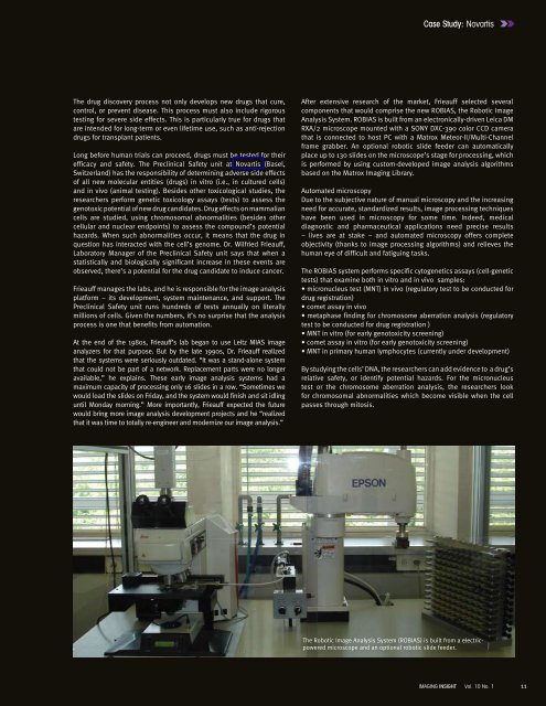
Analysis System . ROBIAS is built from an electronically-driven Leica DM<br />
RXA/2 microscope mounted with a SONY DXC-390 color CCD camera<br />
that is connected to host PC with a <strong>Matrox</strong> Meteor-II/Multi-Channel<br />
frame grabber . An optional robotic slide feeder can automatically<br />
place up to 130 slides on the microscope’s stage for processing, which<br />
is performed by using custom-developed image analysis algorithms<br />
based on the <strong>Matrox</strong> Imaging Library .<br />
Automated microscopy<br />
Due to the subjective nature of manual microscopy and the increasing<br />
need for accurate, standardized results, image processing techniques<br />
have been used in microscopy for some time . Indeed, medical<br />
diagnostic and pharmaceutical applications need precise results<br />
– lives are at stake – and automated microscopy offers complete<br />
objectivity (thanks to image processing algorithms) and relieves the<br />
human eye of difficult and fatiguing tasks .<br />
The ROBIAS system performs specific cytogenetics assays (cell-genetic<br />
tests) that examine both in vitro and in vivo samples:<br />
• micronucleus test (MNT) in vivo (regulatory test to be conducted for<br />
drug registration)<br />
• comet assay in vivo<br />
• metaphase finding for chromosome aberration analysis (regulatory<br />
test to be conducted for drug registration )<br />
• MNT in vitro (for early genotoxicity screening)<br />
• comet assay in vitro (for early genotoxicity screening)<br />
• MNT in primary human lymphocytes (currently under development)<br />
By studying the cells’ DNA, the researchers can add evidence to a drug’s<br />
relative safety, or identify potential hazards . For the micronucleus<br />
test or the chromosome aberration analysis, the researchers look<br />
for chromosomal abnormalities which become visible when the cell<br />
passes through mitosis .<br />
The Robotic Image Analysis System (ROBIAS) is built from a electricpowered<br />
microscope and an optional robotic slide feeder .<br />
IMAGING INSIGHT Vol. 10 No. 1<br />
11