Research Day - UBC Dentistry - University of British Columbia
Research Day - UBC Dentistry - University of British Columbia
Research Day - UBC Dentistry - University of British Columbia

Create successful ePaper yourself
Turn your PDF publications into a flip-book with our unique Google optimized e-Paper software.
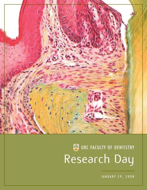
<strong>UBC</strong> FACULTY OF DENTISTRY<br />
<strong>Research</strong> <strong>Day</strong><br />
J A N U A R Y 2 9 , 2 0 0 8
“<br />
To advance oral health through<br />
outstanding education, research,<br />
and community service.<br />
”<br />
<strong>UBC</strong> FACULTY OF DENTISTRY MISSION STATEMENT<br />
Table <strong>of</strong> Contents<br />
Message from the Dean 1<br />
Message from the Associate Dean <strong>of</strong> <strong>Research</strong> 2<br />
<strong>Research</strong> <strong>Day</strong> Schedule 3<br />
Presenters 4<br />
Poster Abstracts 13<br />
<strong>Research</strong> Clusters 35<br />
Summer Student Employment Opportunities 38<br />
<strong>Research</strong> Laboratories 41<br />
Notes 47
<strong>UBC</strong> FACULTY OF DENTISTRY RESEARCH DAY | 1<br />
MESSAGE FROM THE DEAN > Welcome to the first <strong>Research</strong> <strong>Day</strong> for the<br />
<strong>University</strong> <strong>of</strong> <strong>British</strong> <strong>Columbia</strong> Faculty <strong>of</strong> <strong>Dentistry</strong>. A yearly event to highlight<br />
the research accomplishments <strong>of</strong> the faculty has been one <strong>of</strong> our goals for several<br />
years. We are pleased that in 2008 we have achieved this objective and<br />
organized our first annual research event.<br />
<strong>Research</strong> discovery is one <strong>of</strong> the five pillars <strong>of</strong> <strong>UBC</strong>’s TREK 2010 vision and <strong>UBC</strong> is<br />
recognized internationally as an outstanding research-intensive university. The<br />
Faculty <strong>of</strong> <strong>Dentistry</strong> has an exceptional history <strong>of</strong> research achievement directly<br />
addressing the TREK 2010 objectives. One <strong>of</strong> the strategic objectives essential<br />
to our planning for the next five years is to amplify our research productivity.<br />
<strong>Research</strong> <strong>Day</strong> provides a forum to highlight our current research accomplishments<br />
and begin to define the future opportunities for new investigation.<br />
<strong>Research</strong> in the health sciences is frequently initiated by a question derived from some aspect related to a patient<br />
presentation. Our curriculum is organized in a way to facilitate asking or eliciting these types <strong>of</strong> questions about<br />
patient presentations and empowering students to find the information necessary to make an evidence-based<br />
decision. The <strong>Research</strong> <strong>Day</strong> program is organized to use these types <strong>of</strong> patient-based questions to demonstrate the<br />
origin <strong>of</strong> basic biomedical research projects and the application <strong>of</strong> their findings through translation to new clinical<br />
therapies. Clinical problems or questions related to loss <strong>of</strong> teeth, loss <strong>of</strong> bone, and replacement <strong>of</strong> the dentition occupy<br />
a majority <strong>of</strong> the practice experience for dentists. These situations can present significant challenges for both the patient<br />
and the dentist. Considerable progress has been made in the past few years to develop new clinical approaches based<br />
on scientific discovery to manage these clinical problems in the best possible way. Thus, clinical questions arising<br />
from the loss <strong>of</strong> teeth, bone and their replacement have been selected as the focus for the presentations on this<br />
first <strong>Research</strong> <strong>Day</strong>.<br />
The term “Bench to Bedside” has <strong>of</strong>ten been used to describe the necessary translation <strong>of</strong> basic research findings to<br />
patient applications. Examples <strong>of</strong> that process, however, have not <strong>of</strong>ten been presented in a comprehensive fashion.<br />
The goal <strong>of</strong> today’s program is to demonstrate how the Faculty <strong>of</strong> <strong>Dentistry</strong> is making contributions in basic biomedical<br />
research that are leading to improved treatment options for patients.<br />
The Faculty <strong>of</strong> <strong>Dentistry</strong> has established several basic biomedical research themes that have achieved international<br />
recognition. It is significant that the Faculty <strong>of</strong> <strong>Dentistry</strong> has two Tier I Canada <strong>Research</strong> Chairs, Pr<strong>of</strong>essors Dieter<br />
Brömme and Christopher Overall, and their research achievements in basic processes <strong>of</strong> tissue degradation and bone<br />
resorption will open the day’s program and highlight significant problems in oral health care. It is also significant that<br />
one <strong>of</strong> our alumni, Dr. Sonia Leziy, is an internationally-recognized expert in the clinical application <strong>of</strong> implants and<br />
she will close the program with the ultimate application <strong>of</strong> basic research discovery—new and improved approaches to<br />
patient care. Through this <strong>Research</strong> <strong>Day</strong> program, it should become apparent that basic research is a critical element for<br />
the improvement <strong>of</strong> patient care and that the <strong>UBC</strong> Faculty <strong>of</strong> <strong>Dentistry</strong> plays a leading role in these advances.<br />
I would like to thank the outstanding set <strong>of</strong> speakers who have agreed to participate in this inaugural <strong>Research</strong> <strong>Day</strong><br />
and to thank the members <strong>of</strong> the <strong>Research</strong> <strong>Day</strong> organizing committee, Ingrid Ellis, Viki Koulouris, Hannu Larjava, Ed<br />
Putnins, Laura Rosenthal, and Andrea Wink, who contributed so much to make this event a success.<br />
I hope you enjoy the program. I know that I am looking forward to an exciting day <strong>of</strong> scientific advances leading to<br />
new and improved patient care applications.<br />
Thank you for your participation.<br />
Charles Shuler, D.M.D., Ph.D.<br />
Pr<strong>of</strong>essor and Dean, <strong>UBC</strong> Faculty <strong>of</strong> <strong>Dentistry</strong>
2 | <strong>UBC</strong> FACULTY OF DENTISTRY RESEARCH DAY<br />
MESSAGE FROM THE ASSOCIATE DEAN OF RESEARCH > I would also<br />
like to welcome you all to the first <strong>Research</strong> <strong>Day</strong> <strong>of</strong> the Faculty <strong>of</strong> <strong>Dentistry</strong><br />
at the <strong>University</strong> <strong>of</strong> <strong>British</strong> <strong>Columbia</strong>. I am confident that this new tradition<br />
will continue in the foreseeable future, exposing our students, staff members,<br />
part-time instructors and eventually the public to the variety <strong>of</strong> research<br />
topics addressed in our faculty.<br />
Today, it is no longer acceptable to contain research and the researchers to silos<br />
with no information leaking to the outside world. This has become a mandatory<br />
process as our granting agencies require researchers to publicize their<br />
research findings. In addition, there is a wide lag between basic science findings<br />
and their applications to clinical practice.<br />
We are making a conscientious effort to get the research that is performed in<br />
our faculty to the public domain. <strong>Research</strong> <strong>Day</strong> is one <strong>of</strong> the many ways we are<br />
planning to tackle this task.<br />
The <strong>UBC</strong> Faculty <strong>of</strong> <strong>Dentistry</strong> <strong>Research</strong> <strong>Day</strong> is theme-based, focusing on one theme at a time and the theme will<br />
change every year, exposing the audience to new faces and new research and clinical problems that our researchers<br />
are trying to address. This year’s theme focuses on implant therapy at a very broad level and combines information<br />
from basic science <strong>of</strong> tissue degradation and cell interactions with implants to clinical applications.<br />
During the break in the middle <strong>of</strong> the day, you are welcome to visit the poster session where our undergraduate and<br />
graduate students, post-doctoral fellows and faculty members will present their research projects. During the poster<br />
session, you will be exposed to a larger variety <strong>of</strong> research topics investigated in our Faculty.<br />
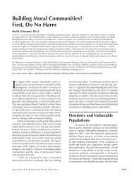
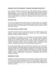
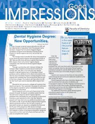
The 2008 <strong>Research</strong> <strong>Day</strong> theme revolves around clinical problems that Mrs. Istu Te is experiencing. She is attending a<br />
dental <strong>of</strong>fice and tries to ask many questions to make an informed decision as to how to proceed with her treatment<br />
and then improve her quality <strong>of</strong> life. These questions are highlighted at the beginning <strong>of</strong> each presentation. Her chief<br />
complaint is that she had an old problem with one <strong>of</strong> her front teeth for many years that recently became acute and<br />
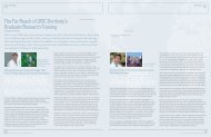
caused her to see a dentist; she presented with an abscess. The tooth was pulled and during the tissue healing, the gums<br />
(gingiva) collapsed and now she presents with a large gap between her front teeth that needs to be restored.<br />
THE CASE OF MRS. ISTU TE<br />
Case Learning Objectives:<br />
At the end <strong>of</strong> <strong>Research</strong> <strong>Day</strong>, participants are expected to be able to:<br />
• describe the pathophysiology <strong>of</strong> s<strong>of</strong>t and hard tissue degradation around natural teeth;<br />
• understand the functions <strong>of</strong> metalloproteinases and cathepsins in the above processes;<br />
• describe the clinical applications <strong>of</strong> bone augmentation for restoration <strong>of</strong> the alveolar<br />
ridge in implant placement;<br />
• describe how the cells interact with various implant materials and surface topographies;<br />
• explain the indications and limitations <strong>of</strong> immediate implant placement;<br />
• understand the management <strong>of</strong> edentulous patients with or without implants;<br />
• recognize the importance <strong>of</strong> proper management <strong>of</strong> anterior implant esthetics; and<br />
• understand the principles <strong>of</strong> successful implant therapy at the anterior esthetic zone.<br />
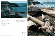
A. Endodontic problem<br />
with tooth #21 that lead<br />
to vertical fracture and<br />
hopeless prognosis<br />
I hope you enjoy this <strong>Research</strong> <strong>Day</strong> and the Case <strong>of</strong> Mrs. Istu Te.<br />
Hannu Larjava, D.D.S., Ph.D., Dip. Perio.<br />
Associate Dean <strong>of</strong> <strong>Research</strong> and Graduate Studies<br />
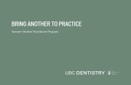
B.-C. Edentulous area after<br />
extraction <strong>of</strong> #21
<strong>UBC</strong> FACULTY OF DENTISTRY RESEARCH DAY | 3<br />
<strong>Research</strong> <strong>Day</strong> Schedule<br />
Tuesday, January 29, 2008<br />
at the <strong>UBC</strong> Student Union Building Ballroom<br />
IMPLANTS: From basic science <strong>of</strong> tissue destruction<br />
to predictable esthetic implant placement<br />
7:30 – 8:00 Registration & Continental Breakfast<br />
8:00 – 8:15 Welcome<br />
Dr. Charles Shuler, Dean<br />
8:15 – 8:30 Case <strong>of</strong> ‘Istu Te’: “I have a big hole in my gums after the tooth was pulled and<br />
cannot smile; why, and what can be done”<br />
Introduction <strong>of</strong> the Clinical Case<br />
Dr. Hannu Larjava, Associate Dean <strong>of</strong> <strong>Research</strong> and Graduate Studies<br />
8:30 – 9:00 “I was told that I lost my gum because <strong>of</strong> an infected tooth.”<br />
New roles for matrix metalloproteinases in controlling inflammation<br />
Dr. Christopher Overall, Canada <strong>Research</strong> Chair in Metalloproteinase Proteomics & Systems Biology<br />
9:00 – 9:30 “I was told that my bone shrunk away after the tooth was pulled.”<br />
Basic mechanisms <strong>of</strong> bone resorption<br />
Dr. Dieter Brömme, Canada <strong>Research</strong> Chair in Proteases & Diseases<br />
9:30 – 10:00 “Can I have implants How do they interact in my jaw bone”<br />
Improving the implant surface by controlling cell behaviour using surface topography<br />
Dr. Donald Brunette, Pr<strong>of</strong>essor <strong>of</strong> Oral Biology<br />
10:00 – 10:15 C<strong>of</strong>fee Break<br />
10:15 – 10:45 “My upper tooth has to be pulled. Can I have an implant placed right away”<br />
Immediate implant placement in fresh extraction sockets in the esthetic zone<br />
Dr. Tassos Irinakis, Graduate Periodontics Program Director<br />
10:45 – 11:15 “What about the big hole How can a metal root go in and look nice”<br />
Ridge augmentation<br />
Dr. Ian Matthew, Oral & Maxill<strong>of</strong>acial Surgery Chair<br />
11:15 – 12:15 “Some <strong>of</strong> my lower teeth are also bad. If I loose them all, can I have implants there as well”<br />
Managing edentulism with or without implant prostheses<br />
Dr. Joanne Walton, Pr<strong>of</strong>essor <strong>of</strong> Prosthodontics<br />
Dr. Ross Bryant, Assistant Pr<strong>of</strong>essor <strong>of</strong> Prosthodontics<br />
12:15 – 1:45 <strong>Research</strong> poster presentations<br />
by undergraduate students, graduate students, post-doctoral fellows,<br />
research associates and faculty members<br />
[box lunch provided]<br />
1:45 – 4:30 “How will these new implants look”<br />
The science and art <strong>of</strong> aesthetic implant treatment: A blueprint for success<br />
Dr. Sonia Leziy, Certified Specialist in Periodontics<br />
[3:15 – 3:30 Break]<br />
4:30 – 5:00 Discussion; case wrap-up; poster prize winners<br />
Dr. Hannu Larjava, Associate Dean <strong>of</strong> <strong>Research</strong> and Graduate Studies
4 | <strong>UBC</strong> FACULTY OF DENTISTRY RESEARCH DAY<br />
PRESENTERS<br />
Bios and Synopses<br />
• Christopher M. Overall, B.D.S., B.Sc. (Dent.)(Hons.), M.D.S. Ph.D.<br />
• Dieter Brömme, Ph.D.<br />
• Donald M. Brunette, Ph.D.<br />
• Tassos Irinakis, D.D.S., M.Sc., Dip.Perio, F.R.C.D.(C.)<br />
• Ian Matthew, Ph.D., M.Dent.Sc., B.D.S., F.D.S.R.C.S.(Eng.), F.D.S.R.C.S.(Ed.)<br />
• Joanne N. Walton, D.D.S., Cert. Pros., F.R.C.D.(C.)<br />
• Ross Bryant, D.D.S., Ph.D., F.R.C.D.(C.)<br />
• Sonia S. Leziy, D.D.S., Dip.Perio
<strong>UBC</strong> FACULTY OF DENTISTRY RESEARCH DAY | 5<br />
Christopher M. Overall, B.D.S., B.Sc. (Dent.)(Hons.), M.D.S., Ph.D.<br />
Dr. Overall is a Pr<strong>of</strong>essor and Tier 1 Canada <strong>Research</strong> Chair in<br />
Metalloproteinase Proteomics and Systems Biology at the <strong>University</strong><br />
<strong>of</strong> <strong>British</strong> <strong>Columbia</strong>. He completed his B.D.S., B.Sc. (Dent)(Honours)<br />
and Masters degrees at the <strong>University</strong> <strong>of</strong> Adelaide, South<br />
Australia; his Ph.D. in Biochemistry at the <strong>University</strong> <strong>of</strong> Toronto;<br />
and was a MRC Centennial Fellow in his post-doctoral work with<br />
Dr. Michael Smith Nobel Laureate, Biotechnology Laboratory.<br />
He won the CIHR Award for <strong>Research</strong> Excellence in Oral Health,<br />
Institute <strong>of</strong> Musculoskeletal Health & Arthritis in 2002 and the<br />
2002 CIHR Scientist <strong>of</strong> the Year, the <strong>University</strong> <strong>of</strong> <strong>British</strong> <strong>Columbia</strong><br />
Killam Senior <strong>Research</strong>er Award (Science) 2005, and was the Chair<br />
<strong>of</strong> the 2003 MMP and the 2010 Protease Gordon <strong>Research</strong> Conferences. On Sabbatical in 1997-<br />
1998 he was a Visiting Scientist at <strong>British</strong> Biotech Pharmaceuticals, Oxford, UK and in 2004-2005<br />
he was a Visiting Scientist at the Expert Protease Platform, Novartis Pharma, Basel, Switzerland. He is the<br />
pioneer <strong>of</strong> Degradomics, with five Nature Review Papers on this, protease genomics, drug target<br />
validation, MMP therapeutics, and substrate discovery and has developed many new quantitative<br />
approaches to elucidate the protease and substrate degradomes.<br />
New roles for matrix metalloproteinases<br />
in controlling inflammation<br />
We discovered that chemokines are a new class <strong>of</strong> substrate for matrix metalloproteinases (MMPs) (McQuibban<br />
et al 2000, Science 289,1202-1206). Monocyte chemotactic protein-3 (MCP-3) was cleaved by MMP-2 to generate a<br />
potent receptor antagonist to block cell responses to a variety <strong>of</strong> chemokines in vitro. In vivo, this was manifest as<br />
a pr<strong>of</strong>ound reduction in monocyte accumulation and by a 40% reduction in experimental peritonitis. Hence, MMPs<br />
also exhibit pr<strong>of</strong>ound anti-inflammatory properties. We also identified MMP-8, the polymorphonuclear (PMN)<br />
leukocyte collagenase, as a critical mediator initiating lipopolysaccharide (LPS)-responsiveness in vivo. PMN infiltration<br />
towards LPS is abrogated in MMP8-null mice. MMP-8 cleaves LPS-induced CXC chemokine (LIX) at Ser4~Val5 and<br />
Lys79~Arg80 to increase bioactivity. However, with biochemical redundancy between MMPs 1, 2, 9, and 13, which<br />
also cleave LIX at position 4~5, it was surprising to observe such a markedly reduced PMN infiltration towards<br />
LPS and LIX in MMP8 -/- mice. Hence, rather than collagen, these PMN chemoattractants are important MMP-8<br />
substrates in vivo; PMN-derived MMP-8 cleaves and activates LIX to execute an in cis PMN-controlled feed-forward<br />
mechanism to orchestrate the initial inflammatory response and promote LPS responsiveness in tissue.<br />
Through the activity <strong>of</strong> macrophage-specific MMP-12 we found that macrophages then dampen the LPS-induced<br />
influx <strong>of</strong> PMNs—so providing a new mechanism for the termination <strong>of</strong> PMN recruitment in acute inflammation.<br />
MMP-12 specifically cleaves and inactivates human ELR+ CXC chemokines (CXCL1, 2, 3, 5, and 8) at E-LR, the critical<br />
receptor-binding motif, or for CXCL6, carboxy-terminal to it. Furthermore, MMP-12 processed and inactivated<br />
monocyte chemotactic proteins CCL2, 7, 8, and 13 at position 4-5 generating CCR antagonists. We propose the<br />
macrophage, specifically through MMP-12, assists in orchestrating the regulation <strong>of</strong> acute inflammatory<br />
responses by precise proteolysis <strong>of</strong> ELR+ CXC and CC chemokines. So rather than extracellular matrix<br />
proteins as substrates, MMPs are critical regulators <strong>of</strong> innate immunity through processing <strong>of</strong> chemokines.
6 | <strong>UBC</strong> FACULTY OF DENTISTRY RESEARCH DAY<br />
Dieter Brömme, Ph.D.<br />
Ph.D. in Biochemistry from the Martin-Luther Universität Halle-<br />
Wittenberg/GDR in 1983 • <strong>Research</strong> Associate at the Institute <strong>of</strong><br />
Biochemistry in Halle/GDR from 1983-1990 • <strong>Research</strong> Officer at the<br />
Biotechnology <strong>Research</strong> Institute in Montreal/QC from 1991-1993 •<br />
Senior Scientist with Khepri Pharmaceuticals, Inc. (later acquired<br />
by Arris/Axys Pharmaceuticals) in South San Francisco/CA from<br />
1993-1997 • Doctor <strong>of</strong> Science from the Friedrich Schiller Universität<br />
Jena/Germany in 1997 • Associate/Full Pr<strong>of</strong>essor at the Mount Sinai<br />
School <strong>of</strong> Medicine in New York from 1997-2004 • Full Pr<strong>of</strong>essor and<br />
Canada <strong>Research</strong> Chair at the Faculty <strong>of</strong> <strong>Dentistry</strong> <strong>of</strong> the <strong>University</strong><br />
<strong>of</strong> <strong>British</strong> <strong>Columbia</strong> in Vancouver, B.C. since 2004.<br />
Basic mechanisms <strong>of</strong> bone resorption<br />
Our skeleton is replaced every 7-10 years by a process called bone remodeling. Bone remodeling is a fine balance<br />
between bone resorption and bone formation. It happens on a major scale after bone fractures, tooth extraction,<br />
and other traumatic events but on a smaller scale throughout life to grow bone and to repair micr<strong>of</strong>ractures<br />
caused by everyday physical stress on the skeleton. Bone is primarily formed by osteoblasts and degraded by<br />
multinucleated osteoclasts. Osteoclast-mediated bone resorption can be subdivided into two processes: (1)<br />
demineralization and (2) resorption <strong>of</strong> the organic bone matrix, which consists <strong>of</strong> 90% type I collagen. During the<br />
demineralization step, osteoclasts acidify the underlaying bone, which leads to the dissolution <strong>of</strong> the inorganic,<br />
mostly phosphoapatite content <strong>of</strong> bone. The bone salts are responsible for the strength and hardness <strong>of</strong> the<br />
bone, whereas the collageneous matrix forms the scaffold <strong>of</strong> the bone and is responsible for its overall stability.<br />
The organic matrix is subsequently degraded by proteases secreted by the osteoclast. The result <strong>of</strong> this proteolytic<br />
action <strong>of</strong> the osteoclast is the formation <strong>of</strong> a resorption pit. The major proteolytic activity <strong>of</strong> osteoclasts is the<br />
cysteine protease, cathepsin K. Cathepsin K is the only known mammalian collagenase capable <strong>of</strong> cleaving inside<br />
the triple helical collages at multiple sites and to completely break down the collagen fibrils into small soluble<br />
pieces. To be active as a collagenase, cathepsin K forms specific complexes with bone-resident glycosaminoglycans.<br />
Other collagenases <strong>of</strong> the matrix metalloproteinase family appear to help in the “cleaning” <strong>of</strong> remaining collagen<br />
fibril remnants in the resorption pits prior to the invasion <strong>of</strong> osteoblasts, which will refill the pit with new bone.<br />
Excessive bone resorption is a major problem <strong>of</strong> aging and is mostly manifested in a disease called osteoporosis.<br />
Recent therapeutic efforts to slow down bone resorption are focused on the development <strong>of</strong> highly potent<br />
cathepsin K inhibitors which are presently in clinical phase II trials.
<strong>UBC</strong> FACULTY OF DENTISTRY RESEARCH DAY | 7<br />
Donald M. Brunette, Ph.D.<br />
Donald Maxwell Brunette, Ph.D. (Medical Biophysics) is a Pr<strong>of</strong>essor<br />
in the Department <strong>of</strong> Oral Biological and Medical Sciences, and a<br />
former Head <strong>of</strong> the Departments <strong>of</strong> Oral Biology and Oral Biological<br />
& Medical Sciences, as well as a former Associate Dean <strong>of</strong> <strong>Research</strong><br />
and Graduate Studies in the Faculty <strong>of</strong> <strong>Dentistry</strong> at the <strong>University</strong><br />
<strong>of</strong> <strong>British</strong> <strong>Columbia</strong>. A founding member <strong>of</strong> the Medical <strong>Research</strong><br />
Council (MRC Canada) Group in Periodontal Physiology directed<br />
by Dr. A.H. Melcher at the <strong>University</strong> <strong>of</strong> Toronto, Dr. Brunette has<br />
published over 90 articles in refereed journals and has won awards<br />
from the International Association <strong>of</strong> Dental <strong>Research</strong>, Association<br />
<strong>of</strong> Canadian Faculties <strong>of</strong> <strong>Dentistry</strong> (W.W. Wood Teaching Award)<br />
and the American Medical Writers Association (for the first edition <strong>of</strong> Critical Thinking). His research<br />
has been continuously supported by MRC/CIHR for over 30 years and has encompassed several<br />
fields, including citation analysis and clinical studies <strong>of</strong> breath odour, but is mainly concerned with<br />
cell-biomaterial interactions. He serves as a consultant to three biomaterial-related journals as well<br />
as to the American Dental Association’s Council on Scientific Affairs and has been a member, chair or<br />
scientific <strong>of</strong>ficer <strong>of</strong> grant evaluation committees <strong>of</strong> the Canadian Institutes <strong>of</strong> Health <strong>Research</strong> as well<br />
as the National Science Foundation (USA).<br />
Improving the implant surface by controlling<br />
cell behaviour using surface topography<br />
Commercially-available dental implants vary widely in their surface properties, with each manufacturer touting<br />
the advantages <strong>of</strong> their systems. This diversity <strong>of</strong> available choices indicates that an optimum surface has yet to<br />
be developed, and improved surfaces will be required as dental implants are placed in more and more demanding<br />
situations. It is clear that surface properties, including topography and chemistry, are <strong>of</strong> prime importance in establishing<br />
the response <strong>of</strong> tissues to biomaterials. Our objective is to develop principles <strong>of</strong> cell response to topography<br />
that could be used to design surfaces for tissue interaction on implanted devices. We have studied the effects <strong>of</strong><br />
topography on cell behaviour using micr<strong>of</strong>abricated surfaces produced by techniques used in the microelctronics<br />
industry as well as those produced by industrial processes in implant fabrication such as grit blasting or acid etching.<br />
Micr<strong>of</strong>abrication techniques have enabled the production <strong>of</strong> precisely-controlled surfaces in silicon (Si) with<br />
features designed to affect cell behaviour. The surfaces originally produced in Si or titanium (Ti) can be replicated in<br />
epoxy, and coated with Ti and used as substrata for cell culture or implanted in vivo. Processes relevant to implant<br />
performance that have been found to be affected by surface topography include epithelial and fibroblast locomotion<br />
and protease production, fibroblast gene activity, connective tissue attachment, cell signaling, osteoblast differentiation,<br />
and cytokine secretion by macrophages. There is a good correlation between the effects <strong>of</strong> the surfaces<br />
in vivo and in vitro for some processes such as direction <strong>of</strong> cell migration and bone formation. Surface topographic<br />
features can act synergistically to achieve their effects. Only a few <strong>of</strong> the many possible topographies that can be<br />
produced by micr<strong>of</strong>abrication techniques have been investigated, and there is considerable potential for optimizing<br />
cell responses to implanted devices by topographic control <strong>of</strong> cell behaviour.
8 | <strong>UBC</strong> FACULTY OF DENTISTRY RESEARCH DAY<br />
Tassos Irinakis, D.D.S., M.Sc., Dip.Perio., F.R.C.D.(C.)<br />
Dr. Anastasios (Tassos) Irinakis completed his dental degree in Athens,<br />
Greece. After working in private practice he continued his education<br />
at the <strong>University</strong> <strong>of</strong> <strong>British</strong> <strong>Columbia</strong> where he received his specialty<br />
training in Periodontics and Implant Surgery. He was awarded the<br />
“McAnulty Memorial Prize” for Excellence in Periodontics in 2001. His<br />
postgraduate education led to a Diploma in Periodontics and a Masters<br />
degree in Oral Biology. He continued to obtain his Fellowship with<br />
the Royal College <strong>of</strong> Dentists <strong>of</strong> Canada.<br />
Dr. Irinakis currently works at <strong>UBC</strong> as a Clinical Associate Pr<strong>of</strong>essor in<br />
the Division <strong>of</strong> Periodontics & Dental Hygiene. He is the Director <strong>of</strong><br />
Graduate Periodontics & Implant Surgery. He has established a Bone<br />
Grafting & Implant Clinic at <strong>UBC</strong> where he <strong>of</strong>fers his services weekly. He is a Periodontal Consultant<br />
for Vancouver General Hospital and for the College <strong>of</strong> Dental Surgeons <strong>of</strong> <strong>British</strong> <strong>Columbia</strong>. He also<br />
maintains part-time private practice, performing a variety <strong>of</strong> periodontal and implant surgeries in<br />
Vancouver and Coquitlam, B.C. and Calgary, Alberta, Canada.<br />
Dr. Irinakis was recently awarded the “2005 Educator Award” from the American Academy <strong>of</strong><br />
Periodontology. He serves the Academy as a member <strong>of</strong> the Review Panel for the Journal <strong>of</strong> Periodontology.<br />
He has also founded an Institute for Continuing Dental Education, i.e. I.D.E.A.S.; Institute for Dental<br />
Education & Advanced Surgeries (www.dentalideas.ca). Dr. Irinakis lectures actively in study clubs,<br />
conferences and hands-on courses for dental pr<strong>of</strong>essionals nationally and internationally.<br />
Immediate implant placement in fresh extraction<br />
sockets in the esthetic zone<br />
We are commonly faced with the dilemma between a conservative approach and an assertive response when<br />
evaluating a compromised or failing tooth. Should we proceed with complicated endodontic therapy or should<br />
we consider extraction and restorative rehabilitation with and implant This presentation will focus on the second<br />
approach and provide insight in order for the clinician to respond appropriately to such cases with a successful<br />
diagnosis and treatment plan. Key factors will be briefly mentioned on when to proceed with socket preservation<br />
(and bone grafting and delayed implant placement) and when to proceed with immediate implant placement in<br />
fresh extraction sites. On one hand, the practitioner can extract the tooth and fill the socket with grafting material<br />
to preserve the architecture <strong>of</strong> the socket and proceed with delayed implant placement at a later time. On the other<br />
hand, it may be prudent and desirable to immediately place an implant into a fresh extraction socket at the time<br />
<strong>of</strong> tooth removal, thus saving the patient money and time. Advantages and challenges <strong>of</strong> each procedure will be<br />
mentioned, along with tips to allow easier decision making. Esthetic considerations are always a crucial determining<br />
factor and helpful guidelines will be <strong>of</strong>fered throughout this presentation.
<strong>UBC</strong> FACULTY OF DENTISTRY RESEARCH DAY | 9<br />
Ian Matthew, Ph.D., M.Dent.Sc., B.D.S., F.D.S.R.C.S.(Eng.), F.D.S.R.C.S.(Ed.)<br />
Dr. Ian Matthew is a certified specialist in oral surgery within the<br />
European Union, and is currently Chair, Division <strong>of</strong> Oral Surgery at<br />
the <strong>UBC</strong> Faculty <strong>of</strong> <strong>Dentistry</strong>. His principal interest in the field <strong>of</strong><br />
implantology is in the provision <strong>of</strong> a “hands-on” educational<br />
forum for undergraduates, general practice residents, graduate<br />
periodontics residents, and graduate dentists. Dr. Matthew has over<br />
20 years experience in implantology using Straumann ITI implants,<br />
and more recently, the Nobel Biocare implant system.<br />
Ridge augmentation<br />
Dental implants require adequate bone substrate to achieve osseointegration. Bone loss occurs naturally following<br />
tooth extraction, oral cancer, or facial trauma. Bone grafting can augment bone at implant sites with inadequate<br />
bone structure due to previous extractions, gum disease or injuries. <strong>Research</strong> into osteoporosis has provided a<br />
greater insight into a condition which is prevalent among the population and can also impact on implant placement.<br />
Distraction osteogenesis, an alternative to ridge augmentation with grafted bone, is based on the pioneering<br />
work <strong>of</strong> Ilizarov, and is <strong>of</strong> particular value in restoring large jaw defects.<br />
Graft selection is based on the clinical findings and other factors such as clinician training, graft availability, cost,<br />
personal preference, and patient choice. It is possible to augment a small defect using synthetic graft materials<br />
based on hydroxyapatite, the basic inorganic substrate <strong>of</strong> bone. Cadaveric bone graft material from a tissue bank is<br />
also available. However, the gold standard is the patient’s own bone harvested from a local or distant site. Maxillary<br />
sinus grafts are also performed as an alternative to ridge augmentation, to replace bone in the posterior upper<br />
jaw. A resorbable membrane may be indicated to protect the bone graft and encourage bone regeneration (guided<br />
tissue regeneration).<br />
Major bone grafts are typically performed to repair more extensive jaw defects. This bone is harvested from various<br />
sites depending on the size <strong>of</strong> the defect: the hip (iliac crest), lateral knee (tibia), and skull (cranium). These procedures<br />
are routinely performed in an operating room and may require a hospital stay. Techniques in pain control now permit<br />
the patient to have the surgery as an out-patient and return home the same day.<br />
The pursuit <strong>of</strong> technical excellence in bone grafting relies heavily on meticulous pre-operative planning, aided by<br />
advanced radiology techniques and s<strong>of</strong>tware, for example Nobel Biocare’s NobelGuide. The aim <strong>of</strong> this presentation<br />
is to illustrate the key points involved in treatment planning for bone grafting procedures. The objectives are to<br />
describe indications for bone grafting, choices and selection <strong>of</strong> graft material, clinical and radiological requirements<br />
for treatment planning, and grafting techniques for ridge augmentation. <strong>Research</strong> into grafting techniques will<br />
also be covered.
10 | <strong>UBC</strong> FACULTY OF DENTISTRY RESEARCH DAY<br />
Joanne N. WaltoN, D.D.S., Cert. Pros., F.R.C.D.(C.)<br />
Dr. Joanne Walton completed her dental education at the <strong>University</strong><br />
<strong>of</strong> Alberta and practiced general dentistry for four years before<br />
specializing in prosthodontics at Walter Reed Medical Center in<br />
Washington, D.C. After six years in full-time prosthodontic practice,<br />
including two years as a part-time clinical instructor at the <strong>University</strong><br />
<strong>of</strong> <strong>British</strong> <strong>Columbia</strong> Faculty <strong>of</strong> <strong>Dentistry</strong>, she became a full-time<br />
faculty member at <strong>UBC</strong>, maintaining a part-time prosthodontic<br />
practice. Since joining <strong>UBC</strong>, Dr. Walton has conducted research<br />
related to both implant prosthodontics and dental education. She<br />
has collaborated extensively with Dr. Michael MacEntee to examine<br />
prosthodontic outcomes with implant-retained overdentures,<br />
currently completing the first clinical trial to compare patient satisfaction, treatment costs and<br />
prosthodontic maintenance with dentures retained by a single implant to those retained by the<br />
standard two implants.<br />
Managing edentulism with or without<br />
implant prostheses – Part I<br />
Today’s presentation focuses on results <strong>of</strong> the clinical trials conducted by Drs. Walton and MacEntee investigating<br />
patient satisfaction, prosthodontic maintenance, altered sensation, implant angulation, and implant treatment<br />
choices, all related to implant overdentures (IOD) in an older population. Recognizing that many edentulous<br />
patients are satisfied with conventional dentures, the overall goal <strong>of</strong> this research has been to provide sound<br />
evidence for clinicians seeking the least expensive, least invasive treatment alternatives for those patients who<br />
may be struggling with conventional complete dentures.
<strong>UBC</strong> FACULTY OF DENTISTRY RESEARCH DAY | 11<br />
Ross Bryant, D.D.S., Ph.D., F.R.C.D.(C.)<br />
Dr. Bryant is a clinician, researcher and educator in the ELDERS<br />
(Elders’ Link with Dental Education, <strong>Research</strong> and Service) Group<br />
at the <strong>University</strong> <strong>of</strong> <strong>British</strong> <strong>Columbia</strong>, with teaching responsibilities<br />
in implant dentistry and fixed and removable prosthodontics. His<br />
research interests, and presentations and publications, focus on quality<br />
<strong>of</strong> life assessment and management <strong>of</strong> the biological and psychosocial<br />
impact <strong>of</strong> tooth loss, oral prostheses and dental implants. He treats<br />
patients in the Oral Implant Clinic at the <strong>UBC</strong> Specialty Clinic, and<br />
he has for several years served on the executive <strong>of</strong> the B.C. Society <strong>of</strong><br />
Prosthodontists and the Dental Specialist Society <strong>of</strong> B.C.<br />
Managing edentulism with or without<br />
implant prostheses - Part II<br />
Being unable preserve the remaining lower dentition begs an open discussion <strong>of</strong> treatment options with the<br />
patient, including a conventional denture, and either a fixed or removable implant prosthesis. However, the problem<br />
remains that it is not clear what the optimal strategy is for management <strong>of</strong> edentulism – with or without<br />
implants. This presentation reviews evidence <strong>of</strong> biological and psychosocial outcomes for managing complete<br />
tooth loss in adults. The biological outcome <strong>of</strong> implants depends on selection <strong>of</strong> patients with stable general and<br />
oral health and adequate bone status. Unfavorable jawbone condition can impair implant success, particularly in<br />
the atrophic edentulous maxilla. However, aging itself does not seem to detract from oral implant success, despite<br />
age-related systemic illness including osteoporosis. Generally, both implant prostheses and complete dentures are<br />
associated with favorable physiological and psychosocial outcomes. Interestingly, edentate older adults may prefer<br />
the ease <strong>of</strong> cleaning associated with removable compared to fixed prostheses, whereas this may not be the case for<br />
younger adults. Substantial differences also exist in the initial fiscal costs <strong>of</strong> the various options, and some efforts<br />
are being made to reduce costs to improve accessibility. However, there is no consensus on how or even whether<br />
to assess the long-term cost-benefit comparing the various options, particularly when considering the impact on<br />
quality <strong>of</strong> life <strong>of</strong> patients. It is generally accepted that tooth loss diminishes quality <strong>of</strong> life through impaired oral<br />
function and appearance and the resulting impact on psychological and social experiences. It is also increasingly<br />
clear that conventional dentures can be acceptable substitutes for many individuals with missing teeth. Indeed,<br />
contrary to current opinions, elders with or without implants can use very positive language to describe their quality<br />
<strong>of</strong> life outcomes, suggesting factors other than the physical impairment can be influencing how individuals adapt<br />
positively to tooth loss. For some, oral implants can <strong>of</strong>fer the hope <strong>of</strong> stable artificial teeth with improved satisfaction.<br />
The scientific challenge in managing adults with terminal dentitions or edentulism in at least one jaw remains<br />
the need to distinguish the influence <strong>of</strong> patient-mediated expectations on the physiological and psychosocial<br />
outcome <strong>of</strong> treatment.
12 | <strong>UBC</strong> FACULTY OF DENTISTRY RESEARCH DAY<br />
Sonia S. Leziy, D.D.S., Dip.Perio.<br />
Sonia S. Leziy received her dental degree from McGill <strong>University</strong>. Her<br />
post-graduate degree in periodontics was completed at the <strong>University</strong><br />
<strong>of</strong> <strong>British</strong> <strong>Columbia</strong> in 1993. Dr. Leziy is a Fellow <strong>of</strong> the Royal College<br />
<strong>of</strong> Dentists <strong>of</strong> Canada, a Fellow <strong>of</strong> the ICOI, and a member <strong>of</strong> the<br />
<strong>British</strong> <strong>Columbia</strong> Society <strong>of</strong> Periodontists, the Canadian Academy<br />
<strong>of</strong> Periodontists, the American Academy <strong>of</strong> Periodontists and is past<br />
president <strong>of</strong> the <strong>British</strong> <strong>Columbia</strong> Society <strong>of</strong> Periodontists. Dr. Leziy is<br />
a member <strong>of</strong> the advisory board <strong>of</strong> the Journal Spectrum Dialogue,<br />
a member <strong>of</strong> the Nobel Biocare Global Scientific Committee and a<br />
member <strong>of</strong> the Nobel Biocare NobelKnowledge web expert panel.<br />
She is an assistant clinical pr<strong>of</strong>essor and sessional lecturer at the<br />
<strong>University</strong> <strong>of</strong> <strong>British</strong> <strong>Columbia</strong> and co-mentors the VIP study club, a Vancouver chapter <strong>of</strong> the Seattle<br />
Study Club. She has published and lectures internationally on implants and aesthetics/periodontal<br />
plastic surgery. Dr. Leziy maintains a full-time private practice with Dr. Brahm Miller (prosthodontist)<br />
and fellow periodontists Dr. Priscilla Walsh and Dr. Andrea Csiszar, specializing in periodontics<br />
and implant surgery.<br />
The science and art <strong>of</strong> aesthetic implant treatment:<br />
a blueprint for success<br />
Conventional concepts in aesthetic restorative treatment generally focus on modifying tooth form and colour.<br />
Today, it is unquestionably recognized that the interplay with the surrounding gingival framework plays an equally<br />
important role in the aesthetic outcome <strong>of</strong> treatment. An understanding <strong>of</strong> how to control the tissue architecture<br />
around implant restorations is a key element to a successful aesthetic outcome.<br />
This presentation will focus on the following key subjects:<br />
• Treatment planning considerations in the esthetic zone: from extraction through restoration.<br />
• Engineering hard and s<strong>of</strong>t tissues.<br />
• 3D implant positioning and design-influence: impact on tissue architecture.<br />
• Provisionalization as the final ‘surgical’ step.<br />
• Inter-implant papilla: surgical considerations for enhanced esthetics.
<strong>UBC</strong> FACULTY OF DENTISTRY RESEARCH DAY | 13<br />
POSTER ABSTRACTS<br />
Poster Competition Judges<br />
• Dr. Dieter Brömme<br />
• Dr. Ross Bryant<br />
• Dr. Virginia Diewert<br />
• Dr. Leeni Koivisto<br />
• Dr. Anthony McCullagh<br />
• Dr. Gethin Owen<br />
• Dr. Steve Pelech, Faculty <strong>of</strong> Medicine<br />
• Dr. Clive Roberts<br />
• Dr. Ravi Shah, Chair <strong>of</strong> Poster Competition<br />
• Dr. David Sweet<br />
• Dr. Doug Waterfield<br />
• Dr. Chris Wyatt
14 | <strong>UBC</strong> FACULTY OF DENTISTRY RESEARCH DAY<br />
1. Effect Of Implant Surface Roughness On The NFkB Signaling<br />
Pathway In Macrophages<br />
Ali TA*, Brunette DM, Waterfield JD<br />
Department <strong>of</strong> Oral Biological & Medical Sciences, The <strong>University</strong> <strong>of</strong> <strong>British</strong> <strong>Columbia</strong>, Vancouver, Canada<br />
Objectives: The aim <strong>of</strong> this study is to examine the effect <strong>of</strong> implant surface topography on activating the NFkB signalling pathway<br />
in Macrophages. The further understanding <strong>of</strong> the signalling pathways will lay a solid foundation for the development <strong>of</strong><br />
methods for controlling inflammation in the form <strong>of</strong> down regulating the inflammatory response and up regulating the healing<br />
and in turn Osseointegration.<br />
Methods: Murine macrophage-like cells (RAW 264.7 cell line) were used in this study. They were cultured on epoxy replicas <strong>of</strong><br />
Smooth, Acid Etched and SLA surfaces. The macrophages were analysed for their ability to both adhere and translocate NFkB to the<br />
nucleus under a number <strong>of</strong> experimental conditions [with and without serum, LPS and membrane cholesterol].<br />
Results: A. Macrophage Activation: 1. Different surface topography causes activation <strong>of</strong> the NFkB pathway differently. The Smooth<br />
surface showed the highest level <strong>of</strong> activation. 2. Addition <strong>of</strong> the suboptimal concentrations <strong>of</strong> LPS mildly enhanced the response by<br />
signalling through the Toll receptor. 3. Triggering <strong>of</strong> the macrophages occurred in the absence <strong>of</strong> fetal calf sera although to a lesser<br />
extent. This indicates that components <strong>of</strong> sera were partially responsible for activation <strong>of</strong> the NFkB pathway. 4. Disruption <strong>of</strong> the<br />
lipid rafts in the membrane through removal <strong>of</strong> the cholesterol content affected the triggering and signalling <strong>of</strong> the NFkB pathway.<br />
It appears that it may be concentration dependent. B. Adherence: 1. Smooth surfaces bound more macrophages in the 30 minute<br />
assay. 2. Serum components were also found to increase binding <strong>of</strong> macrophages to the various test surfaces. 3. Removal <strong>of</strong><br />
cholesterol did not affect the ability <strong>of</strong> macrophage to bind their respective surfaces.<br />
Conclusions: This in vitro study has demonstrated that surface topography modulated activation <strong>of</strong> the NFkB signalling pathway<br />
in a time-dependent manner. Further research will be required to determine mechanisms regulating specificity and selectivity <strong>of</strong><br />
NFkB function, as well as its role in different cell types.<br />
2. Influence In The Quality Of Life And Efficacy Of Support Therapy<br />
For TMD As An Adjunctive Therapy For Oral Appliance Treatment<br />
Cunali PA 1 , Almeida FR 2 *, Garbuio S 1 , Santos CD 1 , Nascimento LS 1 , Valdrighi NY 1 , Tufik S 1 , Bittencourt LRA 1<br />
1<br />
Federal <strong>University</strong> <strong>of</strong> São Paulo – UNIFESP, Brazil; 2 Faculty <strong>of</strong> <strong>Dentistry</strong>, The <strong>University</strong> <strong>of</strong> <strong>British</strong> <strong>Columbia</strong>, Vancouver, Canada<br />
Objectives: Pain in the mastication muscles and/or in the temporomandibular joints (Temporomandibular Dysfunction-TMD)<br />
can be a contra-indication <strong>of</strong> Oral Appliance (OA) therapy or can lead to non-compliance <strong>of</strong> OA as a treatment for Obstructive Sleep<br />
Apnea Syndrome (OSAS). The quality <strong>of</strong> life is reduced in OSAS patients and can be worsened if the patient happens to also suffer<br />
from Temporomandibular Dysfunction (TMD). Support therapy (ST), in the means <strong>of</strong> jaw exercises, is the first choice <strong>of</strong> treatment<br />
for TMD. The aim <strong>of</strong> the study is to follow a population with pre-existing TMD symptoms and evaluate the influence <strong>of</strong> ST on the<br />
patient’s quality <strong>of</strong> life and to assess the efficacy <strong>of</strong> ST in reducing pain symptoms related to TMD during OA therapy.<br />
Methods: Patients with mild to moderate OSAS and TMD symptoms who were referred to OA therapy have been included in the<br />
study. The patients were divided in two groups: support therapy (ST) and placebo therapy (PT). The study was double blinded and<br />
patients were randomized prior to performing ST or PT. Quality <strong>of</strong> life (SF 36) and the Fletcher & Luckett sleep questionnaire were<br />
used prior and after 120 days <strong>of</strong> therapy for all patients. TMD symptoms were scored following the <strong>Research</strong> Diagnostic Criteria for<br />
Temporomandibular Disorders – RDC. The RDC was score as presence or absence <strong>of</strong> pain related to TMD prior treatment and after<br />
OA titration. OA compliance was assessed one week after insertion and after titration.<br />
Results: Fifteen patients received ST and 14 patients received PT. Both groups showed significant reduction in the AHI. After titration,<br />
SF 36 showed a significant improvement in 5 <strong>of</strong> its 8 domains in the ST group (pain, limitation <strong>of</strong> emotional aspects, mental sanity<br />
p
<strong>UBC</strong> FACULTY OF DENTISTRY RESEARCH DAY | 15<br />
3. Differential Response Of Fibroblasts And Osteoblasts<br />
To A Roughness-Gradient<br />
Baharloo RB 1 *, Zink C 2 , Yokoyama T 1 , Spencer ND 2 , Brunette DM 1<br />
1<br />
Faculty <strong>of</strong> <strong>Dentistry</strong>, The <strong>University</strong> <strong>of</strong> <strong>British</strong> <strong>Columbia</strong>, Vancouver, Canada; 2 Laboratory for Surface Science and Technology, Department<br />
<strong>of</strong> Materials, ETH, Zürich, Switzerland<br />
Objectives: To examine the effect <strong>of</strong> titanium surfaces with roughness gradient on the behaviour <strong>of</strong> different cell types.<br />
Methods: Multi microcolonies <strong>of</strong> green rat dermal fibroblast cells (GDF), rat calvarial osteoblast cells (RCO), and pig periodontal<br />
ligament epithelial cells (PLE) were stamped on the surfaces with a wide range <strong>of</strong> roughness values (Ra=~1µm-6µm). The responses<br />
<strong>of</strong> the microcolonies <strong>of</strong> cells were studied using epifluorescent light microscopy, confocal laser scanning microscopy, scanning<br />
electron microscopy, and live cell imaging up to day-7 post culture.<br />
Results: RCO in contrast to GDF and PLE microcolonies exhibited a preference to move towards increasing roughness. Parameters<br />
such as microcolonies’ projected area, microcolonies’ direction <strong>of</strong> migration along gradient, and cell polarity in different regions <strong>of</strong><br />
the gradient will be reported.<br />
Conclusions: This study shows that a roughness gradient has the potential to differentially affect different cell types leading to<br />
the possibility <strong>of</strong> designing implant surfaces <strong>of</strong> varying roughness that are specialized for specific cell types.<br />
Acknowledgements: We would like to thank the Canadian Institutes <strong>of</strong> Health <strong>Research</strong> for funding and Mr. Leon Cheng for<br />
technical assistance.<br />
4. Lessons Learned From Oral Cancer Patients –<br />
Experiences From Detection To Diagnosis<br />
Biggar H 1 *, Poh CF 1 , Hislop TG 2 , Bottorff J 3 , Rosin MP 4<br />
1<br />
Faculty <strong>of</strong> <strong>Dentistry</strong>, The <strong>University</strong> <strong>of</strong> <strong>British</strong> <strong>Columbia</strong>, Vancouver, Canada; 2 B.C. Cancer Control <strong>Research</strong>, B.C. Cancer Agency, Vancouver,<br />
Canada; 3 The <strong>University</strong> <strong>of</strong> <strong>British</strong> <strong>Columbia</strong> Okanagan, Kelowna, Canada; 4 Simon Fraser <strong>University</strong>, Burnaby, B.C.<br />
Objectives: The prognosis <strong>of</strong> oral cancer is poor (50-60% five-year survival rate) and has shown minimal change over 3 decades, largely<br />
due to late stage detection. This has resulted in a significant impact on survival and quality <strong>of</strong> life. Understanding the process to early<br />
detection and diagnosis is key to improving outcome. The objective is to collect information from patients who have diagnosed highrisk<br />
oral lesions (severe dysplasia/carcinoma in situ) or invasive cancer relating to their experiences from first detection <strong>of</strong> an oral<br />
lesion to the diagnostic workup. The goal is to identify factors that impact on the severity <strong>of</strong> high-risk oral lesions at diagnosis.<br />
Methods: An interview-style questionnaire has been developed to collect both qualitative and quantitative data on patients’<br />
experience. Here we report data from a pilot study <strong>of</strong> 18 patients, all <strong>of</strong> which were diagnosed within the previous 12 months. All<br />
patients are participating in the Oral Health Study at BCCA.<br />
Results: Among the patients interviewed, 11 (61%) had invasive cancer and 7 (39%) a preinvasive lesion. Patients with preinvasive<br />
lesions all reported seeing both dental and medical pr<strong>of</strong>essionals at least once per year prior to diagnosis in contrast to only 5 (45%)<br />
<strong>of</strong> individuals with invasive cancers (P = 0.04). Of 9 patients that self-identified their initial lesions, 8 had an invasive cancer. The<br />
chief complaint for these lesions was pain or presence <strong>of</strong> non-healing ulcers (6/9; 67%). In contrast, 9 patients had their initial lesions<br />
identified by health pr<strong>of</strong>essionals (HPs), 8 during screening by dental pr<strong>of</strong>essionals and the other by a physician for a health<br />
problem (N=1). Only 3 (33%) <strong>of</strong> the latter cases showed invasive lesions (P = 0.05). All lesions initially identified by HPs had no symptoms. On<br />
average, the time lag between the initial identification to biopsy was 5.3 + 4.9 months. Two patients (both asymptomatic) showed<br />
a delay <strong>of</strong> > 12 months from initial detection by a HP to biopsy. The main reason for this delay was a choice for watchful waiting<br />
rather than immediate biopsy by the HPs.<br />
Conclusions: These results suggest dental health pr<strong>of</strong>essionals can play an important role in early identification <strong>of</strong> high-risk oral<br />
lesions at an asymptomatic and preinvasive stage. They also draw attention to the importance <strong>of</strong> increasing oral cancer awareness<br />
in both patients and health pr<strong>of</strong>essionals.<br />
Acknowledgements: <strong>Research</strong> is supported by NIDCR grant R01DE13124 and CIHR & MSFHR grants to CFP.
16 | <strong>UBC</strong> FACULTY OF DENTISTRY RESEARCH DAY<br />
5. Chromosomal Instability In High-Risk Oral Premalignant<br />
Lesions – A Tissue Microarray Study<br />
Chen E 1,2 *, Zhu Y 1 , Zhang L 3 , Rosin MP 1,2 , Poh CF 2,3<br />
1<br />
Simon Fraser <strong>University</strong>, Burnaby, B.C.; 2 B.C. Cancer Agency, Vancouver, Canada; 3 Faculty <strong>of</strong> <strong>Dentistry</strong>, The <strong>University</strong> <strong>of</strong> <strong>British</strong> <strong>Columbia</strong>,<br />
Vancouver, Canada<br />
Objectives: To characterize patterns <strong>of</strong> CIN in samples on a unique tissue microarray <strong>of</strong> oral premalignant lesions (OPLs) that<br />
contains samples <strong>of</strong> varying histological grade and with known outcome (i.e. progression to carcinoma). As a first step in this pilot study,<br />
heterogeneity <strong>of</strong> chromosome patterns in tissue on the array was determined using centromere probes from two chromosomes.<br />
Methods: The study involved a tissue array comprised <strong>of</strong> 64 oral premalignant lesions. As an internal control, the array contains<br />
3 non-dysplastic lesions. A dual color probe set <strong>of</strong> CEP11 (Orange) and CEP7 (Green) was used for fluorescence in situ hybridization<br />
(FISH) analysis. These chromosomes have shown frequent CIN in earlier studies with oral squamous cell carcinoma.<br />
Results: On average, 209 + 22 non-overlapping nuclei were evaluated from each tissue. Thirteen (20%) <strong>of</strong> the dysplasia samples had<br />
cells with 3 or more CEP7 or CEP11 signals; seven samples had more than 10% <strong>of</strong> nuclei showing such change. Striking differences<br />
was observed among OPLs. One case (a low-grade dysplasia that later progressed to cancer) showed very heterogeneous patterns <strong>of</strong><br />
CEP7:CEP11 signals, possibly reflecting an active process <strong>of</strong> instability in the tissue: patterns seen included (0:3), (1:3), (1:4), (2:3), (2:4),<br />
(3:0), (3,1), (3:2), (3:3), (4:0), (4:2), (4:3) and (4:4). Other samples showed small numbers <strong>of</strong> cells with alterations to signal numbers and<br />
fewer patterns <strong>of</strong> alteration (none <strong>of</strong> the aforementioned patterns). Another indication <strong>of</strong> heterogeneity was the fraction <strong>of</strong> cells in<br />
which the number <strong>of</strong> centromere signals on the two chromosomes differed by ≥ 2 (e.g. 2:4, 1:3, etc.). Eleven (17%) cases showed such<br />
patterns <strong>of</strong> alteration, with 5 over 10% <strong>of</strong> nuclei showing such change.<br />
Conclusions: Levels <strong>of</strong> CIN in OPLs are varied, with some samples showing considerable heterogeneity in patterns <strong>of</strong> chromosomal<br />
alteration. Further studies will expand upon these observations to look for associations with outcome.<br />
Acknowledgements: Sponsored by NIDCR R01 DE13124, CIHR MOP-77663 and MSFHR grants.<br />
6. The Structural Requirement Of Elastinolytic Activity<br />
In Human Cathepsin V<br />
Chen N-H 1 *, Brömme D 1,2<br />
1<br />
Department <strong>of</strong> Biochemistry & Molecular Biology, Faculty <strong>of</strong> Science, The <strong>University</strong> <strong>of</strong> <strong>British</strong> <strong>Columbia</strong>, Vancouver, Canada (<strong>UBC</strong>);<br />
2<br />
Department <strong>of</strong> Oral Biological & Medical Sciences, Faculty <strong>of</strong> <strong>Dentistry</strong>, <strong>UBC</strong><br />
Objectives: Despite sharing an amino acid sequence identity <strong>of</strong> over 80% and very similar subsite specificities only cathepsin V<br />
(cat V) has a potent elastase activity whereas cathepsin L (Cat L) lacks it. The objective <strong>of</strong> this research is to identify the presence <strong>of</strong><br />
an exosite in Cat V that binds to elastin and contributes to its elastinolytic activity.<br />
Methods: Chimera mutants containing different proportions <strong>of</strong> Cat V and Cat L were generated by PCR, cloned and expressed in<br />
the Pichia pastoris system. The expressed enzymes were purified by FPLC and subjected to activity assay using a synthetic substrate<br />
and elastin derivatives. Finally, the HPLC pr<strong>of</strong>ile <strong>of</strong> elastin degradation were also obtained.<br />
Results: We found that all mutants were comparably to their wild-type enzymes active towards a synthetic substrate, and that<br />
mutant 1 (M1; containing ~75% <strong>of</strong> Cat V) demonstrated a potent elastinolytic activity, whereas mutant 2 (M2; containing ~50% <strong>of</strong><br />
Cat V) and mutant 3 (M3; containing ~25% <strong>of</strong> Cat V) were only weakly active. Also, HPLC pr<strong>of</strong>iles <strong>of</strong> both wild type Cat V, Cat L and<br />
all mutants showed that M1 exhibited similar, if not identical, elastin cleavage pattern as Cat V. These results directed us to further<br />
dissect the potential region <strong>of</strong> amino acids 53 to 117 in Cat V. Mutant 4 (M4; containing ~50% <strong>of</strong> Cat V in the potential region) and<br />
mutant 5 (M5; containing only ~15% <strong>of</strong> Cat V in the potential region) both displayed elastinolytic activity and generated identical<br />
cleavage pattern as shown by HPLC.<br />
Conclusions: The result suggested that the region <strong>of</strong> amino acid 112 to 118 in Cat V seemed to be associated with elastin degradation.<br />
Further sequence and structural analysis, and mutant constructions targeting that region will elucidate amino acid residue<br />
responsible for the elastinolytic activity in Cat V.
<strong>UBC</strong> FACULTY OF DENTISTRY RESEARCH DAY | 17<br />
7. A Creative Global Solution To Offering Baccalaureate-Level<br />
Dental Hygiene Education<br />
Craig BJ*<br />
Faculty <strong>of</strong> <strong>Dentistry</strong>, The <strong>University</strong> <strong>of</strong> <strong>British</strong> <strong>Columbia</strong>, Vancouver, Canada<br />
Objectives: To develop an innovative, flexible and adaptable baccalaureate-level educational model that graduates dental hygienists<br />
with a 4-year degree and <strong>of</strong>fers Canadian and international dental hygiene diploma/certificate/associate degree graduates access<br />
to an advanced educational opportunity.<br />
Methods: A multiple-admissions model was created with 4 entry points into the program using creative resource strategies. Each<br />
option has defined admission criteria and graduation eligibility requirements. The two options for degree completion allow learners<br />
to fulfill degree requirements through distance education without the more traditional residency requirement. Three admission<br />
options were implemented in 2002 and the fourth option commenced in September 2007. Surveys <strong>of</strong> graduates and current<br />
students were conducted to better understand the reasons for selecting the program.<br />
Results: To date program enrollment since 1992 has increased from 5 degree completion students to 193 students eligible to register<br />
in courses in all four admission options as <strong>of</strong> September 2007. Survey data revealed that 68% <strong>of</strong> current degree completion students<br />
reside outside the <strong>UBC</strong> catchment area and enrolled in the Program because <strong>of</strong> online course access and part-time study opportunities.<br />
Ninety percent <strong>of</strong> degree completion students are part-time and have up to 5 years to complete requirements to graduate.<br />
Conclusions: Graduate numbers have more than doubled in the past 4 years. Twenty-five percent have gone on to graduate<br />
studies - Masters and doctoral. This model could be adapted to a variety <strong>of</strong> educational contexts globally.<br />
8. Establishment Of A Community Partnership To Facilitate<br />
Oral Cancer Screening In A High-Risk Community<br />
Currie BL 1 *, Poh CF 1 , Hislop G 2 , Bottorff JL 1 , Zhang L 1 , Rosin MP 3<br />
1<br />
Faculty <strong>of</strong> <strong>Dentistry</strong>, The <strong>University</strong> <strong>of</strong> <strong>British</strong> <strong>Columbia</strong>, Vancouver, Canada; 2 B.C. Cancer Agency, Vancouver, Canada;<br />
3<br />
Simon Fraser <strong>University</strong>, Burnaby, B.C.<br />
Objectives: To explore possible approaches that would facilitate an oral cancer-screening program in a high-risk community in<br />
the Vancouver DTES, a community that is characterized by poverty and an elevated exposure to known risk factors (tobacco and<br />
alcohol) associated with oral cancer.<br />
Methods: A screening clinic was established in partnership with a pre-existing community dental clinic in the Vancouver DTES.<br />
Patients attending the clinic for regular dental care were invited by the staff dentist to participate in a free oral cancer screening.<br />
Demographics, information on known risk factors and medical history were collected by interview-style questionnaires followed by<br />
a full head and neck evaluation using conventional technique and a novel translational tool (fluorescence visualization) to enhance<br />
visualization <strong>of</strong> oral lesions.<br />
Results: A total <strong>of</strong> 284 patients attended this drop-in clinic over a 2-year study period. Among these patients, 204 (72%) were invited<br />
by the staff dentists for screening – 200 (98%) <strong>of</strong> these accepted the invitation. The remaining 28% <strong>of</strong> individuals were deemed<br />
ineligible for the study for a variety <strong>of</strong> reasons including severe psychological problems, drug and/or alcohol intoxication and/or<br />
lack <strong>of</strong> willingness to wait for the screening exam. Of the 200 screened patients, most were smokers (86%) and regular consumers<br />
<strong>of</strong> alcohol (83%) and <strong>of</strong>ten immuno-compromised. Strikingly, 2 oral cancer and 8 precancers were identified.<br />
Conclusions: The study is an example <strong>of</strong> capacity building through a community partnership to approach a hard-to-reach highrisk<br />
community. The screening activity identified a significant disease burden in this community and an urgent need to expand<br />
this initiative to create a more comprehensive strategy to outreach this community for not only screening, but also diagnostic<br />
work-up and treatment.
18 | <strong>UBC</strong> FACULTY OF DENTISTRY RESEARCH DAY<br />
9. Preliminary Activities For Community Oral Health <strong>Research</strong><br />
With Immigrant Children<br />
Dabiri D*, Carino KMG, Harrison RL<br />
Division <strong>of</strong> Pediatric <strong>Dentistry</strong>, The <strong>University</strong> <strong>of</strong> <strong>British</strong> <strong>Columbia</strong>, Vancouver, Canada<br />
Objectives: Conducting a comprehensive community-based dental health survey <strong>of</strong> young immigrant children presents numerous<br />
challenges. The overall goal <strong>of</strong> this study was to document prevalence <strong>of</strong> caries and dental treatment needs among Filipino<br />
immigrant children in metro Vancouver. The objectives <strong>of</strong> this first phase <strong>of</strong> the research were to develop survey protocols (including<br />
those for subject recruitment), validate the clinical indices, and recruit and train volunteers.<br />
Methods: Protocols were tested in a series <strong>of</strong> “trial surveys” utilizing small, convenience samples <strong>of</strong> parents and children. Materials<br />
required for the final surveys were identified. The International Caries Detection and Assessment System (ICDAS) was adopted<br />
as the criteria for measuring dental caries. The trial surveys provided opportunity for (a) intra-examiner calibration, (b) training<br />
<strong>of</strong> the recorder, and (c) refinement <strong>of</strong> the dental chart. A logo, designed by DD, complemented the project slogan: Healthy Teeth,<br />
Happy Families. Information dissemination primarily involved flyers and posters distributed to community centers, libraries,<br />
Filipino stores and parenting groups. All materials were continuously evaluated and re-designed based on community feedback.<br />
Volunteers were recruited and trained in a series <strong>of</strong> workshops.<br />
Results: 20 children were examined in the trial surveys. The ICDAS system, though complex, proved workable for recording caries in very<br />
young children. The logo, used on all project-related materials, increased visual recall <strong>of</strong> the research project. The most successful means<br />
<strong>of</strong> subject recruitment was “face-to-face” (i.e. personal invitation). 11 volunteers were recruited and trained to assist with surveys.<br />
Conclusions: Prior to launching a community-based child dental health survey, time must be devoted to 1) refining all protocols, 2)<br />
calibration <strong>of</strong> examiners, 3) identifying successful techniques for subject recruitment, and 4) training and maintenance <strong>of</strong> a reliable<br />
group <strong>of</strong> volunteers. The survey proper has now begun and is over 80% complete.<br />
Acknowledgements: We acknowledge the dedication <strong>of</strong> our project’s volunteers. This research was supported by a grant from the<br />
S. Wah Leung Endowment Fund. Ms. Dabiri was funded by the Network for Oral <strong>Research</strong> Training and Health (NORTH) Canadian<br />
Institutes <strong>of</strong> Health <strong>Research</strong> (CIHR) training program.<br />
10. Social Isolation, Body-Image And Oral Health Among Elderly Women<br />
Donnelly L*, O’Connor D, MacEntee MI<br />
Faculty <strong>of</strong> <strong>Dentistry</strong>, The <strong>University</strong> <strong>of</strong> <strong>British</strong> <strong>Columbia</strong>, Vancouver, Canada<br />
Objectives: Oral health <strong>of</strong> elders in residential care (RC) facilities is generally poor, but to what extent it affects their body-image<br />
and social behaviors is unknown. A positive body-image increases social contacts and improves health, well-being and quality<br />
<strong>of</strong> life. Literature on relationships between body-image and aging among elderly women does not explore the role <strong>of</strong> oral health.<br />
The objective <strong>of</strong> this study was to begin to investigate the relationship between overall body image and oral health among elderly<br />
women who reside in a RC facility.<br />
Methods: Open-ended interviews with a purposefully selected group <strong>of</strong> four older women were conducted and the narratives<br />
analyzed using a constant comparative method involving an iterative technique. A second interview with the participants was<br />
conducted to check trustworthiness <strong>of</strong> my interpretations.<br />
Results: Four themes emerged; 1) social interactions are influenced by the environment; 2) body-image is influenced by health and<br />
the social context; 3) the importance <strong>of</strong> a “nice smile”; and 4) priorities in life and health.<br />
Conclusions: Relationships between social isolation, body-image and oral health among institutionalized elderly women can<br />
have multiple influences that depend on life-priorities, however, the information emerging from the interviews was not saturated<br />
which indicated that more interviews are needed to conclude this phase <strong>of</strong> the study. Further study is underway with a larger more<br />
diverse sample <strong>of</strong> elders to expand the context in which these observations occur.
<strong>UBC</strong> FACULTY OF DENTISTRY RESEARCH DAY | 19<br />
11. Expression Of Integrin αvβ6 and TGF-β In Scar-Free Vs.<br />
Scar-Forming Wound Healing<br />
Eslami A 1 *, Gallant-Behm CL 2 , Wiebe C 1 , Hart DA 2 , Honardoust D 1 , Häkkinen L 1 , Larjava H 1<br />
1<br />
Faculty <strong>of</strong> <strong>Dentistry</strong>, The <strong>University</strong> <strong>of</strong> <strong>British</strong> <strong>Columbia</strong>, Vancouver, Canada; 2 Faculty <strong>of</strong> Medicine, The <strong>University</strong> <strong>of</strong> Calgary, Calgary, Alberta<br />
Objectives: Integrins are key mediators <strong>of</strong> TGF-β activation in vivo. The αvβ6 integrin can activate two is<strong>of</strong>orms <strong>of</strong> TGF-β, fibrogenic<br />
TGF-β1 and anti-fibrogenic TGF-β3. Expression <strong>of</strong> epithelial cell integrin αvβ6 is induced during wound healing but its function<br />
in wounds remains unclear. In the present study, we investigated the potential for αvβ6-mediated activation <strong>of</strong> TGF-β during wound<br />
healing by studying the spatio-temporal co-localization <strong>of</strong> αvβ6 integrin with the molecules involved in TGF-β activation in human<br />
gingival wounds. In addition, we compared the expression and localization <strong>of</strong> αvβ6 integrin along with its ligands, TGF-β1 and<br />
TGF-β3 in scar-free gingival versus scar-forming skin wounds in red Duroc pigs.<br />
Methods: Full-thickness excisional wounds were created in the gingiva <strong>of</strong> human volunteers, and in the gingiva and skin <strong>of</strong> red Duroc<br />
pigs. Wounds healing rate was clinically and histologically assessed at different time points up to 60 days after wounding. Immunohistochemical<br />
analysis and real-time PCR were used to assess the localization and expression <strong>of</strong> β6 integrin, TGF-β1 and TGF-β3.<br />
Results: The αvβ6 integrin was found to colocalize with both TGF-β1 and TGF-β3 in the wound epithelium <strong>of</strong> human gingiva. Analysis<br />
<strong>of</strong> the pig wounds showed that basal keratinocytes <strong>of</strong> the clinically healed gingival wounds continued to express αvβ6 integrin and<br />
TGF-β3 while their expression was negligible in scar-forming skin wounds.<br />
Conclusions: The spatio-temporal colocalization <strong>of</strong> αvβ6 with TGF-β1 and TGF-β3 in human gingival wounds demonstrates a potential<br />
mechanism to regulate the activity <strong>of</strong> TGF-β is<strong>of</strong>orms by αvβ6 integrin during wound healing. Prolonged expression <strong>of</strong> αvβ6<br />
integrin along with anti-fibrogenic TGF-β3 in the gingival wound basal epithelium may be important in protection <strong>of</strong> gingiva from<br />
scar formation.<br />
Acknowledgements: Supported by the Canadian Institutes <strong>of</strong> Health <strong>Research</strong> and the Arthritis Society.<br />
12. Frequency Of Cluster, Migraine And Tension Headaches Symptoms<br />
In Sleep Disordered Breathing Patients<br />
Fastlicht S*, Mehta S, Almeida FR, Lowe AA<br />
Faculty <strong>of</strong> <strong>Dentistry</strong>, The <strong>University</strong> <strong>of</strong> <strong>British</strong> <strong>Columbia</strong>, Vancouver, Canada<br />
Objectives: Migraine, tension, and cluster headaches are frequently related to sleep. Reports <strong>of</strong> headache prevalence in OSA subjects<br />
range between 15% to 50%. When sleep disordered breathing (SDB) is treated successfully, headaches generally improve which<br />
suggests a possible relationship between SDB and headache symptoms. Migraines have been known to occur during sleep or after<br />
daytime naps and may be triggered either by lack <strong>of</strong> sleep or by excess sleep. Migraine patients will <strong>of</strong>ten manage the headache by<br />
going to sleep. Tension headaches, the most common but least studied, are <strong>of</strong>ten managed by sleep regulation. Cluster headaches<br />
which occur in attack clusters followed by pain free intervals occur at night during sleep.<br />
Methods: A series <strong>of</strong> 150 consecutive SDB patients diagnosed by in hospital overnight polysomnograms elected to pursue oral appliance<br />
therapy in the Dental Sleep Apnea Clinic at The <strong>University</strong> <strong>of</strong> <strong>British</strong> <strong>Columbia</strong>. All patients completed intake MIDAS questionnaires to<br />
evaluate headache frequency, type and severity.<br />
Results: Out <strong>of</strong> the 150 patients (21-77Y), some 72 suffered from headaches and 78 did not. Patients that suffered from headaches<br />
were younger (47.5 vs 50.9Y) but females with headaches were older (mean <strong>of</strong> 52.0F vs. mean 46.7M). Females also exhibited a higher<br />
incidence <strong>of</strong> headaches than males (31/50F vs. 41/100M). The perception <strong>of</strong> pain or severity <strong>of</strong> the headaches was higher in women.<br />
Overall, headache severity correlated with a higher RDI (p
20 | <strong>UBC</strong> FACULTY OF DENTISTRY RESEARCH DAY<br />
13. Absence Of αvβ6 Integrin Is Linked To Initiation<br />
And Progression Of Periodontal Disease<br />
Ghannad F 1 *, Nica D 1 , Garcia MI 1 , Grenier D 2 , Johnston S 1 , Eslami A 1 , Koivisto L 1 , Jiang G 1 , Putnins EE 1 , McKee MD 3 , Häkkinen L 1 , Larjava HS 1<br />
1<br />
Laboratory <strong>of</strong> Periodontal Biology, Faculty <strong>of</strong> <strong>Dentistry</strong>, The <strong>University</strong> <strong>of</strong> <strong>British</strong> <strong>Columbia</strong>, Vancouver, Canada; 2 Faculty <strong>of</strong> <strong>Dentistry</strong> & Medicine,<br />
Laval <strong>University</strong>, Montreal, Canada; 3 Faculty <strong>of</strong> <strong>Dentistry</strong> and Department <strong>of</strong> Anatomy & Cell Biology, McGill <strong>University</strong>, Montreal, Canada<br />
Objectives: Integrin αvβ6 is mostly absent from adult epithelia but its expression is induced in wound healing, cancer and certain<br />
fibrotic disorders. In the present study, we investigated the hypothesis that αvβ6 integrin-mediated TGFβ1 activation in the junctional<br />
epithelium (JE) plays a major protective role in inflammatory periodontal disease.<br />
Methods: Expression <strong>of</strong> αvβ6 integrin and its ligands was studied in normal and regenerating JE <strong>of</strong> human and murine gingiva using<br />
immunohistochemistry. Cytokine and bacterial regulation <strong>of</strong> αvβ6 integrin expression in cultured keratinocytes was studied using<br />
real-time PCR. Bone loss around molars <strong>of</strong> wild-type FVB and β6 integrin knockout mice was assessed using morphometry and high<br />
resolution radiographs. Specific antibodies against β6 integrin were used to block αvβ6 integrin-mediated TGFβ1 activation in a rat<br />
model <strong>of</strong> periodontal disease initiation.<br />
Results: In the present study, we observed that αvβ6 integrin is constitutively expressed in the healthy epithelium linking gingival<br />
epithelium to the tooth enamel (i.e. junctional epithelium). Its expression was, however, downregulated in human periodontal<br />
disease. Remarkably, integrin β6 knockout mice developed classical signs <strong>of</strong> spontaneous, chronic periodontal disease with<br />
inflammation, epithelial down-growth, pocket formation and bone loss around the teeth. Integrin αvβ6 acts as a major activator<br />
<strong>of</strong> TGFβ1, a key anti-inflammatory regulator in the immune system. We found that TGFβ1 was expressed in the healthy junctional<br />
epithelium together with αvβ6 integrin and that an antibody that blocks αvβ6 integrin-mediated activation <strong>of</strong> TGFβ1 initiated<br />
inflammatory periodontal disease in a rat model.<br />
Conclusions: Thus, αvβ6 integrin is constitutively expressed in the epithelium sealing the gingiva to the tooth, and it plays a central<br />
protective role against inflammatory periodontal disease via TGFβ1 activation.<br />
Acknowledgements: This study was supported by a research grant from the Canadian Institutes <strong>of</strong> Health <strong>Research</strong>.<br />
14. Surface Topography Modulates Src Phosphorylation<br />
And Subsequent Macrophage Signaling<br />
Ghrebi SS 1 *, Hamilton DW 2 , Waterfield JD 1 , Brunette DM 1<br />
1<br />
Department <strong>of</strong> Oral Biological & Medical Sciences, The <strong>University</strong> <strong>of</strong> <strong>British</strong> <strong>Columbia</strong>, Vancouver, Canada;<br />
2<br />
Schulich School <strong>of</strong> Medicine & <strong>Dentistry</strong>, The <strong>University</strong> <strong>of</strong> Western Ontario, London, Canada<br />
Objectives: The geometry <strong>of</strong> implant surface topography alters cell behaviour. Macrophages are among the first cell types to attach<br />
to implants and play a critical role in inflammatory responses and wound healing. The mode and type <strong>of</strong> macrophage activation<br />
modulate their secretory pr<strong>of</strong>ile. Available data shows that rough surface topography increases pro-inflammatory cytokines<br />
(IL-1, IL-6, and TNF-α) secretion by macrophages stimulated with suboptimal concentration <strong>of</strong> LPS. The control <strong>of</strong> cytokine secretion<br />
involves several signaling pathways. One such pathway involves focal adhesion kinase (FAK). A phosphorylated tyrosine in FAK<br />
provides specific binding sites for Src family kinases. Src is important in expression <strong>of</strong> pro-inflammatory cytokines IL-1 and TNF-α<br />
through MAP kinase signaling pathway. Our objective was to study the effect <strong>of</strong> different implant surface topographies on signalling<br />
cascades involved in macrophage activation.<br />
Methods: Macrophages (RAW 264.7) were seeded on Titanium-coated epoxy replicas <strong>of</strong> polished, blasted, etched, and blasted &<br />
etched (SLA) surfaces. Immunocytochemistry and Western blotting techniques were used to analyze the effect <strong>of</strong> these surfaces.<br />
Phospho-FAK, Src and ERK1/2 were tested and PP1; a Src specific inhibitor was used.<br />
Results: Immunostaining showed that rough surfaces increased macrophage Src phosphorylation at two and three days. ERK1/2<br />
phosphorylation was increased at one day but decreased by day three compared to macrophages on polished surfaces. Western<br />
blotting analysis showed that all surfaces activated ERK1/2 in macrophages. Activation appeared to be down regulated on rough<br />
surfaces at three days when compared to polished [t-test, p
<strong>UBC</strong> FACULTY OF DENTISTRY RESEARCH DAY | 21<br />
15. Molecular Scrutiny Of Primary And Successional<br />
Tooth Development In Squamates (Reptilia)<br />
Handrigan G 1 *, Buchtová M 2 , Tucker A 3 , Leung K 1 , Richman JM 1<br />
1<br />
Department <strong>of</strong> Oral Health Sciences, Life Sciences Institute, The <strong>University</strong> <strong>of</strong> <strong>British</strong> <strong>Columbia</strong>, Vancouver, Canada; 2 Academy <strong>of</strong> Sciences,<br />
Brno, Czech Republic; 3 Crani<strong>of</strong>acial Development & Orthodontics, Kings College London, London, UK<br />
Objectives: <strong>Research</strong> by our group considers the molecular regulation <strong>of</strong> primary and successional tooth development. Our<br />
specific goals include: 1) To compare gene expression between developing poly- and mono-phyodont dentitions; 2) To identify key<br />
molecules in tooth replacement; and 3) To infer developmental bases for variation in tooth number.<br />
Methods: For this work, we are studying the snakes Python and Elaphe, and the lizard Pogona. All three are polyphyodont and present<br />
a much longer dental lamina than the mouse. Reptile dental laminae grow in a characteristic direction and show asymmetry in both<br />
tooth-budding and cell proliferation. To understand the molecular basis for this, we have studied the gene Sonic hedgehog (Shh), which<br />
controls dental epithelial ingrowth in mammals. We cloned Python Shh and surveyed its expression during squamate odontogenesis.<br />
Furthermore, using reptile jaw explant and slice cultures approaches, we tested the gene’s role in tooth development in both gain- and<br />
loss-<strong>of</strong>-function experiments.<br />
Results: In snakes, Shh is expressed in the presumptive dental epithelium and, at later stages, in the oral epithelium. This expression<br />
domain persists throughout the prehatching period in Python, but is prematurely lost in Pogona, which forms relatively fewer teeth. In<br />
both species, Shh is expressed in primary and generational enamel organs, but, surprisingly, is missing from the generational rudiment.<br />
Shh loss-<strong>of</strong>-function experiments led to shorter laminae that lacked their characteristic angulation and asymmetry in cell proliferation.<br />
In contrast, Shh gain-<strong>of</strong>-function produced longer laminae, further confirming that the gene drives dental epithelial ingrowth.<br />
Conclusions: Our data reveal a clear role for Shh in squamate tooth development, but suggest it may be acting differently in primary versus<br />
successional odontogenesis. Furthermore, variation in tooth number correlates with the temporal persistence <strong>of</strong> oral Shh expression.<br />
Acknowledgements: This work is funded by NSERC/MSFHR fellowships to GRH and an NSERC operating grant to JMR.<br />
16. Regulation And Function Of A BMP Receptor Antagonist In The Face<br />
Higashihori N*, Song Y-P, Richman JM<br />
Department <strong>of</strong> Oral Health Sciences, Life Sciences Institute, The <strong>University</strong> <strong>of</strong> <strong>British</strong> <strong>Columbia</strong>, Vancouver, Canada<br />
Objectives: 1) To map expression <strong>of</strong> a decoy receptor for Bone morphogenetic protein, Bambi in the developing face. 2) To identify<br />
growth factors that regulate the expression <strong>of</strong> Bambi in the lip fusion zone. 3) To test the function <strong>of</strong> BAMBI in a retroviral over<br />
expression experiment.<br />
Methods: A chicken Bambi probe was used in wholemount in situ hybridization to map expression in several stages <strong>of</strong> chicken<br />
embryos. To target growth factors in the face, microscopic beads were first soaked in either Bone morphogenetic protein-7, Noggin,<br />
Fibroblast growth factor-2 or retinoic acid and then implanted into the 4.5 day chicken embryo face. The full length human BAMBI<br />
open reading frame was cloned into RCAS (an avian retrovirus). Live virus was injected into the face <strong>of</strong> embryos.<br />
Results: During early development, expression <strong>of</strong> Bambi was limited to the endoderm. There was little expression within the embryo<br />
proper until organogenesis stages. From 3.5 days and onwards, Bambi expression almost perfectly overlapped with known expression<br />
patterns for BMP4, particularly in the face. Bead implant experiments showed that Bambi was strongly upregulated by BMP7 and<br />
retinoic acid but downregulated by Noggin (a BMP antagonist) and FGF2. The main phenotype caused by BAMBI over-expression was<br />
a selective loss <strong>of</strong> intramembranous bone in 50% <strong>of</strong> injected specimens (12/23).<br />
Conclusions: Our data point to retinoids and BMPs as being major positive regulators <strong>of</strong> Bambi expression but that retinoic acid is<br />
more involved with ectodermal than mesenchymal expression. Furthermore, FGF2 represses Bambi expression and therefore may<br />
help to restrict Bambi to certain regions <strong>of</strong> the face. This result also demonstrates a novel point <strong>of</strong> intersection between the FGF<br />
and BMP pathways. The virus phenotypes suggest that BAMBI may prefer to interact with BMPR1a (ubiquitously expressed) rather<br />
than BMPR1b (more highly expressed in cartilage). This is the first demonstration <strong>of</strong> a selective difference in affinity for type I BMP<br />
receptors by BAMBI.<br />
Acknowledgements: This work was funded by CIHR grants to JMR. JMR is a Michael Smith Foundation for Health <strong>Research</strong><br />
Distinguished Scholar.
22 | <strong>UBC</strong> FACULTY OF DENTISTRY RESEARCH DAY<br />
17. Expression Of Small Leucine-Rich Proteoglycans And Transforming<br />
Growth Factor-β In Human Oral Mucosal Wound Regeneration<br />
Honardoust D*, Eslami A, Larjava H, Häkkinen L<br />
Department <strong>of</strong> Oral Biological & Medical Sciences, Faculty <strong>of</strong> <strong>Dentistry</strong>, The <strong>University</strong> <strong>of</strong> <strong>British</strong> <strong>Columbia</strong>, Vancouver, Canada<br />
Objectives: The hallmark <strong>of</strong> scar formation is excessive accumulation <strong>of</strong> abnormally organized collagen in response to increased<br />
activity <strong>of</strong> transforming growth factor-β (TGF-β). Remarkably, wound healing in human oral mucosa rarely results to scar formation.<br />
Thus, understanding the mechanisms involved in oral wound healing may provide important information about the biological<br />
processes that regulate scar formation. The small leucine-rich proteoglycans (SLRPs), decorin, biglycan, fibromodulin, and lumican, are<br />
extracellular matrix (ECM) molecules that interact with type I collagen and regulate collagen fibrillogenesis. SLRPs also bind to TGF-β<br />
inhibiting its biological activity. Our aim was to analyze the expression <strong>of</strong> SLRPs and TGF-β during human gingival wound regeneration.<br />
Methods: Expression <strong>of</strong> SLRPs, TGF-β1, TGF-β3 and collagen deposition and organization were analyzed in frozen sections<br />
from full-thickness, excisional human gingival wounds (collected 3-60 days after wounding) by immunohistochemical and<br />
histochemical techniques.<br />
Results: Expression <strong>of</strong> the studied molecules were spatio-temporally regulated in distinct wound cells and ECM. Strongest upregulation<br />
for all <strong>of</strong> the molecules except lumican occurred from day 7 to 28 after wounding. After 60 days, expression <strong>of</strong> lumican remained<br />
still reduced while the expression <strong>of</strong> decorin, fibromodulin and TGF-β3 were still upregulated. Gradual increase in expression <strong>of</strong><br />
SLRPs correlated with maturation <strong>of</strong> collagen in the wound ECM. Expression <strong>of</strong> fibromodulin, lumican and TGF-β1 and -3 were<br />
spatio-temporally upregulated also in the wound epithelium.<br />
Conclusions: Expression <strong>of</strong> SLRPs is spatio-temporally regulated in parallel with increased expression <strong>of</strong> TGF-β and collagen<br />
maturation at different stages <strong>of</strong> wound healing suggesting that SLRPs collaborate to regulate collagen organization and TGF-β<br />
activity in wound regeneration.<br />
Acknowledgements: Supported by the Canadian Institutes <strong>of</strong> Health <strong>Research</strong>.<br />
18. Crani<strong>of</strong>acial Development Studies In The Richman Lab<br />
Hosseini S*, Higashihori N, Geetha-Loganathan P, Nimmagadda S, Handrigan G, Richman JM<br />
Department <strong>of</strong> Oral Health Sciences, Life Sciences Institute, The <strong>University</strong> <strong>of</strong> <strong>British</strong> <strong>Columbia</strong>, Vancouver, Canada<br />
Objectives: Our laboratory is working on three main research themes: mechanisms <strong>of</strong> cleft lip, jaw identity, and evolution and<br />
development <strong>of</strong> amniote teeth. The most common group <strong>of</strong> crani<strong>of</strong>acial anomalies is or<strong>of</strong>acial clefts; however it is also common to<br />
see discrepancies in the size and shape <strong>of</strong> the jaws and problems with tooth number. Our lab uses oviparous species such as chicken,<br />
emu and snake to study signalling involved in jaw patterning and tooth formation. The long-term goal is to prevent human jaw and<br />
tooth deformities through a better understanding <strong>of</strong> the molecules involved.<br />
Methods: Our approach is to first map expression <strong>of</strong> genes at several different stages in the embryonic face. These patterns <strong>of</strong><br />
expression then are used to plan our experimental approaches to test in the embryo. In the gain-<strong>of</strong>-function experiments, we<br />
overexpress the signals using RCAS vectors or by injecting cells that express those signals or beads soaked in purified proteins. In<br />
the loss-<strong>of</strong>-function experiments, we block signalling pathways by using dominant-negative RCAS constructs, by injecting cells<br />
expressing antagonists or by applying beads soaked in antagonists.<br />
Results: In the Cleft Lip project, we have identified novel requirements for FGF signalling but these FGF dependent regions are not<br />
near the zone <strong>of</strong> fusion. Instead FGF dependence is greatest near the top <strong>of</strong> the nasal slit and when blocked, the embryos develop cleft<br />
lip. We are now working on the Wnt pathway to see which Wnts are expressed in the face and whether other growth factors regulate<br />
their expression. In the Jaw Identity project we have used a microarray approach to identify genes that are involved in specifying<br />
frontonasal mass identity. Several <strong>of</strong> these genes are now being followed up with functional studies. In the Reptile Project we have<br />
found that Sonic hedgehog is expressed within the odontogenic band, the first region <strong>of</strong> epithelium fated to make teeth. Interestingly<br />
this band is similarly present in birds even though they do not have teeth. Our data suggests conservation <strong>of</strong> the early signals is present<br />
in dentate and edentate reptiles but that later steps in tooth morphogenesis are blocked or not supported in birds.<br />
Conclusions: Our data provide an explanation for how abnormalities in signalling could cause facial defects and failure to form<br />
primary or secondary teeth.<br />
Acknowledgements: This work was funded by CIHR and NSERC grants to JMR. G. Handrigan is a MSFHR and NSERC Postdoctoral<br />
Fellow. JMR is a MSFHR Distinguished Sholar.
<strong>UBC</strong> FACULTY OF DENTISTRY RESEARCH DAY | 23<br />
19. Oral Health Beliefs And Treatment Needs Of LTC Chinese Seniors<br />
Hui N*, O’Connor D, MacEntee MI<br />
Department <strong>of</strong> Oral Health Sciences, Faculty <strong>of</strong> <strong>Dentistry</strong>, The <strong>University</strong> <strong>of</strong> <strong>British</strong> <strong>Columbia</strong>, Vancouver, Canada<br />
Objectives: To explore the oral health beliefs, knowledge and treatment needs <strong>of</strong> Chinese seniors living in a long term care facility<br />
in Vancouver Chinatown.<br />
Methods: We used open-ended, semi-structured interviews with 7 Chinese elders living in a long-term care (LTC) facility in Vancouver’s<br />
Chinatown, and we analyzed transcripts <strong>of</strong> the interviews by a constant comparison <strong>of</strong> the interviews and emerging themes.<br />
Results: Three major themes emerged during our analysis: self-diagnosis; oral health knowledge; cost <strong>of</strong> dental treatment.<br />
Conclusions: Chinese elders living in a LTC facility are aware <strong>of</strong> oral health problems but their knowledge <strong>of</strong> oral healthcare and<br />
dental services is relatively poor. Apparently, they avoid seeking dental services because <strong>of</strong> the cost <strong>of</strong> dental treatment. Clearly now<br />
there is need for further study <strong>of</strong> traditional health beliefs and practices to improve the oral healthcare <strong>of</strong> this population, and to<br />
inform dental pr<strong>of</strong>essionals about culturally competent care for Chinese elders.<br />
20. Application Of Fluorescence Visualization In Surgical Treatment Of<br />
High-Grade Preinvasive Oral Lesions<br />
Kung A 1 *, Poh CF 1,2 , Durham S 3 , Anderson D 3 , Zhang L 1 , Rosin MP 1,3<br />
1<br />
Faculty <strong>of</strong> <strong>Dentistry</strong>, The <strong>University</strong> <strong>of</strong> <strong>British</strong> <strong>Columbia</strong>, Vancouver, Canada; 2 B.C. Cancer Agency, Vancouver, Canada;<br />
3<br />
Simon Fraser <strong>University</strong>, Burnaby, B.C.<br />
Objectives: Recurrence following excision <strong>of</strong> high-grade dysplasia/carcinoma in situ (HGL) implies the presence <strong>of</strong> subclinical change<br />
at the margins not apparent at surgery, resulting in incomplete excision. Fluorescence visualization (FV) has a demonstrated ability<br />
to identify clinically unapparent oral lesions. The objective <strong>of</strong> this study is to assess the efficacy <strong>of</strong> intraoperative FV in detecting<br />
high-risk occult tissue extending beyond clinical boundaries <strong>of</strong> HGL.<br />
Methods: Lesions were delineated under OR illumination and with FV in 35 patients: 22 with HGL and 13 cancers. Geographic mapping<br />
within excised tissue was used to co-localize histology with clinically apparent lesion, loss <strong>of</strong> aut<strong>of</strong>luoresence (FVL) only margins and<br />
surgical margins.<br />
Results: FVL was apparent in all 35 lesions. FVL extended beyond clinical boundaries in 95% <strong>of</strong> HGL. Extension around the lesion<br />
perimeter was uneven (1-25 mm) similar to observations in cancers. 35% (13/37) <strong>of</strong> FVL/clinically negative biopsies from HGLs showed<br />
high grade dysplasia; in 5 cases, this change went beyond 10-mm, the conventional margin for cancer. In contrast, none <strong>of</strong> 35<br />
biopsies from surgical margins, located in areas without FVL or clinical change had HGL and only 4 had mild dysplasia.<br />
Conclusions: Integrating FV in surgery might provide a useful tool to better manage HGL through identification <strong>of</strong> subclinical<br />
field change.<br />
Acknowledgements: Sponsored by NIDCR R01 DE17013, CIHR MOP-77663 and MSFHR grants.
24 | <strong>UBC</strong> FACULTY OF DENTISTRY RESEARCH DAY<br />
21. Assessment Of PSP Sensors Used At <strong>UBC</strong> Oral Health Centre<br />
Lam H*, Nouri R, Harrison RL<br />
Division <strong>of</strong> Pediatric <strong>Dentistry</strong>, The <strong>University</strong> <strong>of</strong> <strong>British</strong> <strong>Columbia</strong>, Vancouver, Canada<br />
Objectives: The aim <strong>of</strong> this study was to evaluate the fate <strong>of</strong> Photo-Stimulated Phosphor (PSP) sensors used in digital radiology<br />
and to evaluate qualitatively and quantitatively the different types <strong>of</strong> damages seen on these sensors during their first year <strong>of</strong> use<br />
at the <strong>UBC</strong> OHC.<br />
Methods: Damaged PSPs used during one academic year were collected; visible damage was categorized quantitatively. PSPs were<br />
repackaged, exposed to x-rays; scanned into Romexus. Resulting JPEG files were transferred into Adobe Photoshop and converted<br />
to TIFF files. Radiological damage was assessed; artifact radio-opacity was quantified and compared with radio-opacity <strong>of</strong> tooth<br />
structure using a step wedge as reference for standardization.<br />
Results: Out <strong>of</strong> 834 PSPs, only 363 (44%) were still in circulation, i.e. not missing (47%) or damaged (9%) by the end <strong>of</strong> first year <strong>of</strong> use.<br />
41% <strong>of</strong> Size-1, 64% <strong>of</strong> Size-2 PSP sensors were missing. 10% <strong>of</strong> Size-0 films, 6% <strong>of</strong> Size-1 films, and 13% <strong>of</strong> Size-2 films were damaged.<br />
Qualitative assessment <strong>of</strong> damage revealed that scratches, clamp marks, folds and bite marks were most common types <strong>of</strong> damage.<br />
The most prevalent damage was scratches likely caused during the scanning process, return <strong>of</strong> the sensors in a cup, or during the<br />
sterilization and repackaging process. Quantitative assessment <strong>of</strong> all types <strong>of</strong> damage proved that, other than pen-mark damage<br />
that may result in radio-opacity darker than enamel, all other damage would most likely lead to radiolucencies that could result in<br />
mis-diagnosis <strong>of</strong> radiographic anomalies.<br />
Conclusions: Loss <strong>of</strong> the PSP sensors was a significantly more serious problem than damage to the sensors. A bar-coding or tracing<br />
system should be used to diminish accidental loss <strong>of</strong> sensors in any clinical setting. Users should be trained to avoid “non-bite”<br />
damage to sensors. Protective means e.g. sponges or sleeves for PSP sensors should be developed to avoid bite-mark damage.<br />
Acknowledgements: Special thanks to Ms. Neala Welburn, Dr. Fernanda Almeida, Dr. Andrea Esteves and Ms. Kathy Pitt. This<br />
project was Burroughs-Wellcome Summer Student <strong>Research</strong>.<br />
22. Oral Cancer Screening – Empowering Oral Health<br />
Pr<strong>of</strong>essionals To Save Lives<br />
Laronde DM 1 *, Williams PM 2 , Hislop TG 3 , Poh CF 4 , Ng S 2 , Rosin MP 1,3<br />
1<br />
Simon Fraser <strong>University</strong>, Burnaby, B.C.; 2 B.C. Cancer Agency, Vancouver, Canada;<br />
3<br />
B.C. Cancer <strong>Research</strong> Centre, Vancouver, Canada; 4 Faculty <strong>of</strong> <strong>Dentistry</strong>, The <strong>University</strong> <strong>of</strong> <strong>British</strong> <strong>Columbia</strong>, Vancouver, Canada<br />
Objectives: To develop, implement and evaluate an education module, to study current oral cancer screening practices, to identify<br />
factors that influence this behavior and to gain information on the use <strong>of</strong> aut<strong>of</strong>luorescence visualization (FV) in the community.<br />
Oral cancer screening is a noninvasive, quick and painless skill that all dental personnel are taught, yet less than 30% <strong>of</strong> people<br />
surveyed had ever been screened. More than 60% <strong>of</strong> oral cancers are diagnosed at a late stage where 5-year survival is poor.<br />
Methods: 10 dental practices from the Vancouver area participated in a workshop on oral cancer screening which reviewed etiology,<br />
epidemiology, clinical risk factors, FV and included a clinical training session. Knowledge and practice <strong>of</strong> oral cancer screening was<br />
assessed by quiz and questionnaire. Dental <strong>of</strong>fices screened adults 21 years and older for 4 months collecting risk habit information,<br />
completing extraoral, intraoral and FV exams. Focus groups <strong>of</strong> the participants were conducted at the conclusion <strong>of</strong> the study.<br />
Results: 1511 were screened using FV. 42 patients with a loss <strong>of</strong> fluorescence (FVL) were referred for reassessment. 35 were reassessed<br />
by a study facilitator. 26 were found to be known confounders for FV or had resolved by reassessment. 9 lesions were referred for<br />
an oral medicine workup: 2 mild dysplasias, 2 lichen planus, 1 denture epulis, 1 is in follow-up and 3 have yet to be seen. 7 patients<br />
were referred without reassessment by the facilitator: 2 lichen planus and 5 were known confounders or normal tissue. 4 themes<br />
resulted from the focus groups: the need for mandatory oral cancer screening education, the need for screening guidelines and<br />
protocols, the need to present strategies for dialoguing with patients and the need to increase awareness among both pr<strong>of</strong>essionals<br />
and the public.<br />
Conclusions: Educational modules should address guidelines and patient communication skills and should integrate<br />
clinical sessions.<br />
Acknowledgements: Supported by NIDCR grant R01DE13124 and a scholarship from the Michael Smith Foundation for Health<br />
<strong>Research</strong> to DML.
<strong>UBC</strong> FACULTY OF DENTISTRY RESEARCH DAY | 25<br />
23. A Community Dental Prenatal Program: An Insider Evaluation<br />
Lin DL*, Harrison RL, Aleksejuniene J<br />
Division <strong>of</strong> Pediatric <strong>Dentistry</strong>, The <strong>University</strong> <strong>of</strong> <strong>British</strong> <strong>Columbia</strong>, Vancouver, Canada<br />
Objectives: A community dental public health program in Vancouver has been providing clinical hygiene services and oral health<br />
counseling to a limited number <strong>of</strong> high-risk, low income pregnant women for over 20 years. To enable future program decision-making,<br />
an evaluation <strong>of</strong> this program was undertaken with the following objectives: 1) to describe program and clients; 2) to determine<br />
whether program activities have been implemented as intended; and 3) to assess the program’s effectiveness.<br />
Methods: The evaluation, undertaken by the resident dental hygienist, had two phases. Phase 1: Evaluability assessment, descriptive<br />
and process evaluation (retrospective chart reviews, semi-structured interviews, appointment statistics, and field observations). Phase<br />
2: A short and medium-term outcomes evaluation with a convenience sample who attended over a 1-year period. Data, collected<br />
by questionnaires, semi-structured interviews, clinical indices, field observations, and appointment statistics, was analyzed with a<br />
combination <strong>of</strong> univariate and bivariate analyses.<br />
Results: Phase 1: Stakeholders’ goals were identified; a logic model and organizational flowchart were developed. Chart review<br />
(N=123) revealed mean client age <strong>of</strong> 27 years; 28% Canadian-born; 48% had other children; 78% were concerned about “bleeding<br />
gums”; and 63% had visible tooth decay. 28% <strong>of</strong> women referred to the program never made an appointment. Unfamiliarity <strong>of</strong><br />
clients with the dental “experience”, language barriers, and clinic time restraints affected implementation <strong>of</strong> program’s activities.<br />
Phase 2: Outcomes in clients (N=61) demonstrated significant improvements (P
26 | <strong>UBC</strong> FACULTY OF DENTISTRY RESEARCH DAY<br />
25. Neuropathic Pain And Implants: Case Report<br />
Mehta SA*<br />
Or<strong>of</strong>acial Pain Clinic, Faculty <strong>of</strong> <strong>Dentistry</strong>, The <strong>University</strong> <strong>of</strong> <strong>British</strong> <strong>Columbia</strong>, Vancouver, Canada<br />
Objectives: The International Association for the Study <strong>of</strong> Pain defines neuropathic pain as “pain initiated or caused by a primary<br />
lesion or dysfunction in the nervous system”. Acute postoperative pain may activate peripheral nerve fibers and the pain experienced<br />
may have a direct relation to the extent <strong>of</strong> the injury. While estimates <strong>of</strong> chronic pain range between 2% to 40% <strong>of</strong> adults depending<br />
on how the question is asked and what definitions are used, the true incidence <strong>of</strong> neuropathic pain is unknown. Dental endosseous<br />
implants can result in ongoing pain to peripheral trigeminal fibers. This case report illustrates neuropathic pain associated with<br />
dental implant placement and the associated frustrations with diagnosis, management and effect to quality <strong>of</strong> life.<br />
Methods: A case report <strong>of</strong> an individual presenting to the <strong>University</strong> <strong>of</strong> <strong>British</strong> <strong>Columbia</strong>’s Or<strong>of</strong>acial Pain Clinic with pain after<br />
dental endosseous implant is reviewed. Patient suffered ongoing chronic pain lasting greater than six months with onset after<br />
implant placement.<br />
Results: Patient underwent multiple consultations with multiple medical and dental providers with several diagnoses. Medication<br />
trial prior to consultation included multiple antibiotics, anti-inflammatories, analgesics, muscle relaxants, anti-anxiety agents,<br />
anti-depressants as well as oral appliance therapy. Levels <strong>of</strong> impact to overall quality <strong>of</strong> life were reviewed at initial examination<br />
and after trial to pain management. Pain numeric scales were utilized to record pain at time <strong>of</strong> initial consultation and during time<br />
<strong>of</strong> management.<br />
Conclusions: Dentists placing and restoring endosseous implants should be aware <strong>of</strong> complications to such treatment<br />
including the possibility to neuropathic pain onset. Patients experiencing ongoing refractory pain after dental implant<br />
placement may require further investigation and intervention with dental providers knowledgeable about neuropathic<br />
pain. Individuals suffering these forms <strong>of</strong> pain may experience huge impact to quality <strong>of</strong> life as well as increased costs<br />
associated with multiple healthcare visits.<br />
26. Dental Negligence Across Canada<br />
Moore B*, MacDonald D<br />
Faculty <strong>of</strong> <strong>Dentistry</strong>, The <strong>University</strong> <strong>of</strong> <strong>British</strong> <strong>Columbia</strong>, Vancouver, Canada<br />
Objectives: The self-regulatory bodies governing the practice <strong>of</strong> dentistry vary across Canada by province. In <strong>British</strong> <strong>Columbia</strong>, the<br />
College <strong>of</strong> Dental Surgeons <strong>of</strong> BC fills this role. In times <strong>of</strong> failure <strong>of</strong> the dentist-patient relationship, when remediation is sought<br />
beyond which the regulatory body can achieve a resolution, proceedings are generally expressed as negligence in a court <strong>of</strong> law.<br />
With regulatory bodies and courts differing across Canada by province, the aim <strong>of</strong> this study was to investigate the distribution <strong>of</strong><br />
such cases. It must be noted emphatically that these distributions are indicative <strong>of</strong> the desires <strong>of</strong> the patients to litigate and likely<br />
other reasons which will not be explored – they are not to be interpreted as differing regional levels <strong>of</strong> pr<strong>of</strong>essional negligence.<br />
Methods: The Canadian Legal Information Institute (http://www.CANLII.org), an online database <strong>of</strong> Canadian legal documents,<br />
was used to isolate and analyze negligence cases relating to the practice <strong>of</strong> dentistry and medicine. It was necessary to exclude<br />
motor vehicle accidents for the purposes <strong>of</strong> the research. An example <strong>of</strong> a search would be: “dentist /7 patient AND negligence OR<br />
malpractice NOT vehicle”. The ‘/7 patient’ refines the search to include only court documents containing dentist and patient within<br />
7 words <strong>of</strong> each other.<br />
Results: As a result <strong>of</strong> the research, it was determined that there are indeed certain provinces where patients are more inclined to<br />
litigate than others. <strong>British</strong> <strong>Columbia</strong> was found to have the greatest proportion <strong>of</strong> Canadian dental and medical negligence cases<br />
heard in court, when adjusted for population, at 27% and 43%, respectively. In contrast, Ontario was found to have the smallest<br />
proportions at 8% for dental cases and 7% for medical cases. Furthermore, the distribution <strong>of</strong> dental negligence cases across Canada<br />
was observed to be quite similar to that found for medical negligence cases.<br />
Conclusions: Clearly, there exists a difference in the number <strong>of</strong> negligence cases being heard in courts across Canada and these<br />
findings certainly warrant further investigation.<br />
Acknowledgements: We are grateful to Mary Mitchell , <strong>UBC</strong> Law Librarian, for her invaluable assistance.
<strong>UBC</strong> FACULTY OF DENTISTRY RESEARCH DAY | 27<br />
27. Characterization Of Marimastat-FITC:<br />
A Modified Matrix Metalloproteinase Inhibitor<br />
Munsie S*, Harwig C, Perrin D, auf dem Keller U, Overall CM | Centre for Blood <strong>Research</strong>, The <strong>University</strong> <strong>of</strong> <strong>British</strong> <strong>Columbia</strong>, Vancouver, Canada<br />
Objectives: This study tests marimastat that was modified by adding a fluorescent probe (FITC) to see if it is still an efficient MMP<br />
inhibitor, is able to bind specifically to MMPs and to determine if Marimastat-FITC binds to MT1-MMP overexpressed on the surface<br />
<strong>of</strong> cultured cells.<br />
Methods: Enzyme Kinetics assay <strong>of</strong> MMP-2 with Marimastat-FITC inhibitor, polyarcylamide gel analysis <strong>of</strong> MMP-2 incubated with<br />
Marimastat-FITC and incubation <strong>of</strong> Marimastat-FITC in human MDA cells overexpressing MT1-MMP.<br />
Results: Results showed that Marimastat-FITC is an effective MMP inhibitor with an IC50 <strong>of</strong> 7nM (compared to the unmodified<br />
Marimastat: 6nM), Marimastat-FITC specifically binds MMP-2 on polyacrylamide gels and effectively binds MT1-MMP in cell<br />
culture (human MDA-MB-231 cells).<br />
Conclusions: MMPs, a family <strong>of</strong> proteases known to cleave many elements <strong>of</strong> the extracellular matrix as well as various inflammatoryrelated<br />
proteins, are implicated in periodontal disease, arthritis and cancer metastasis. Modified MMP inhibitors such as<br />
Marimastat-FITC may be useful tools for imaging MMPs in future studies <strong>of</strong> these conditions.<br />
Acknowledgements: Funding for this project was provided by the Canadian Breast Cancer <strong>Research</strong> Alliance (CBCRA).<br />
28. Microarray Analysis Identifies New Targets Of Retinoic Acid And Noggin<br />
Nimmagadda S*, Buchtová M, Geetha-Loganathan P, Richman JM<br />
Department <strong>of</strong> Oral Health Sciences, Life Sciences Institute, The <strong>University</strong> <strong>of</strong> <strong>British</strong> <strong>Columbia</strong>, Vancouver, Canada<br />
Objectives: Previously we identified Retinoic Acid and Noggin as two molecules that can transform the maxilla into the midline<br />
<strong>of</strong> the face. This striking phenotype was due to a localized increase in retinoic acid and blocking <strong>of</strong> BMP signalling with Noggin.<br />
We have since carried out a microarray analysis <strong>of</strong> the genes differentially expressed in the presence <strong>of</strong> RA and Noggin, RA+Tris,<br />
Noggin+DMOS and control Tris+DMSO. The objective <strong>of</strong> this study is to validate the gene expression changes seen on the chicken<br />
genome chips with in situ hybridization studies and then to follow the most promising <strong>of</strong> these genes with functional studies.<br />
Methods: Beads soaked in RA and Noggin are applied to the maxillary region <strong>of</strong> 2.5 day chicken embryos. Following 16h <strong>of</strong> treatment,<br />
embryos are collected and in situ hybridization carried out. Genes that are upregulated on the array are cloned into RCAS<br />
retroviruses and then over expressed in 2.5 day chicken embryos. Effect on gene expression and skull morphogenesis is analyzed.<br />
Results: In general, genes strongly upregulated following RA Noggin treatment are also upregulated in wholemount in situ analyses.<br />
For example, transcription factor Tbx22 and enzyme PI15 are both increased around the RA Noggin beads. In addition Tbx22 is<br />
upregulated by Noggin+DMSO or RA+Tris. PI15 is also upregulated following RA-Noggin bead implants. Interestingly expression is<br />
more strongly upregulated around the RA bead. Embryos injected with PI15 have various skeletal defects including fusion between<br />
the upper and lower jaws. Tbx22 injected embryos also have skeletal defects in addition to duplication <strong>of</strong> the midline comb. We are<br />
currently testing whether PI15 and Tbx22 are part <strong>of</strong> the same molecular pathway.<br />
Conclusions: Our data provide evidence for novel functions for both Tbx22 and PI15 in specifying jaw identity and controlling<br />
facial skeletal morphogenesis.<br />
Acknowledgements: This work was funded by Canadian Institutes <strong>of</strong> Health <strong>Research</strong> grants to JMR. JMR is a Michael Smith<br />
Foundation for Health <strong>Research</strong> Distinguished Scholar.
28 | <strong>UBC</strong> FACULTY OF DENTISTRY RESEARCH DAY<br />
29. Expression Pattern Of Id2 During Murine Palatal Fusion<br />
Oda R 1 *, Cui X-M 2 , Schmid D 2 , Yamashiro K 1 , Shuler CF 1<br />
1<br />
Department <strong>of</strong> Oral Biological & Medical Sciences, Faculty <strong>of</strong> <strong>Dentistry</strong>, The <strong>University</strong> <strong>of</strong> <strong>British</strong> <strong>Columbia</strong>, Vancouver, Canada;<br />
2<br />
Center for Crani<strong>of</strong>acial Molecular Biology, School <strong>of</strong> <strong>Dentistry</strong>, <strong>University</strong> <strong>of</strong> Southern California, Los Angeles, USA<br />
Objectives: Transforming growth factor (TGF)-β3 is known to regulate the disappearance <strong>of</strong> murine medial edge epithelium (MEE)<br />
during palatal fusion. TGF-β3 null mutant mice (TGF-β3-/-) have a phenotype characterized by cleft palate. Overexpression <strong>of</strong> Smad2<br />
in the MEE is capable <strong>of</strong> rescuing the cleft palate in TGF-β3-/- mice, however the downstream effects <strong>of</strong> activated Smad2 have not<br />
yet been determined. The Inhibitor <strong>of</strong> DNA binding (Id) proteins are negative regulators <strong>of</strong> basic helix-loop-helix transcription<br />
factors and involved in cellular differentiation and proliferation. It has been reported that Id1 is expressed in both palatal<br />
mesenchyme and epithelium. The sites <strong>of</strong> expression and timing <strong>of</strong> expression suggest that Ids might be involved in mechanisms<br />
related to palatogenesis. The purpose <strong>of</strong> this study was to examine Id2 expression during murine palatal fusion.<br />
Methods: Expression <strong>of</strong> Id2 at mRNA and protein levels was examined by RT-PCR, in situ hybridization and immunohistochemistry.<br />
Results: Id2 mRNA was present in the palatal shelves before and during normal palatal fusion. By whole-mount in situ hybridization,<br />
the antisense probe specifically hybridized to Id2 transcripts in the tips <strong>of</strong> horizontal palatal shelves, the site <strong>of</strong> the MEE. Immunohistochemistry<br />
demonstrated an absence <strong>of</strong> the nuclear staining <strong>of</strong> Id2 protein in vertical palatal shelves at gestation day E13. Intense nuclear<br />
staining specific for Id2 was clearly identified in the MEE cells when the horizontal palatal shelves remained in contact (E14, E14.5). The<br />
pattern <strong>of</strong> Id2 gene expression and localization <strong>of</strong> the Id2 protein was coincident with the appearance <strong>of</strong> phospho-Smad2 in MEE cells.<br />
Conclusions: The temporal and spatial localization <strong>of</strong> Id2 indicates that it may be a downstream signaling target <strong>of</strong> the TGF-β3/<br />
Smad2 signaling pathway and play an important role in the regulation <strong>of</strong> MEE disappearance during murine palatal fusion.<br />
Acknowledgements: Supported by NIDCR grant R01 DE16296.<br />
30. 3D Comparison Of Cardiac Development In Kyoto<br />
And Carnegie Human Embryos<br />
Park J*, Diewert VM<br />
Department <strong>of</strong> Oral Health Sciences, Faculty <strong>of</strong> <strong>Dentistry</strong>, The <strong>University</strong> <strong>of</strong> <strong>British</strong> <strong>Columbia</strong>, Vancouver, Canada<br />
Objectives: To compare the stages <strong>of</strong> cardiac development in Kyoto embryos to that in Carnegie embryos documented in the<br />
literature. To reconstruct 3D images from normal serially-sectioned Kyoto embryos <strong>of</strong> stages 16, 17 and 18.<br />
Methods: Photographs <strong>of</strong> serially-sectioned Kyoto embryos <strong>of</strong> stages 16-18 (Diewert Collection) were studied. WinSURF s<strong>of</strong>tware<br />
was used to make 3D models <strong>of</strong> selected serially-sectioned Kyoto embryonic hearts. Fusion <strong>of</strong> atrioventricular (AV) cushions and<br />
formation <strong>of</strong> the interventricular septum were assessed in histological sections and 3D reconstructed images.<br />
Results: Stage 16 - 2 <strong>of</strong> 6 specimens have fused endocardial cushions and divided AV canals. All show evidence <strong>of</strong> beginning stages<br />
<strong>of</strong> fusion. Interventricular septum development slightly delayed. Stage 17 - Only 4 <strong>of</strong> 13 specimens have fused cushions. The<br />
muscular interventricular septum is more prominent but the muscular IVS is not fused to the cardiac cushions. Stage 18 - All (4/4)<br />
have fused AV cushions, separating the AV canal. Incomplete closure <strong>of</strong> secondary interventricular foramen in all specimens.<br />
Conclusions: Heart development in Kyoto embryos appears delayed at stage 17. Fusion <strong>of</strong> facial prominences to form the upper<br />
lip is delayed in Kyoto embryos. Although heart and face development were delayed in stage 17 Kyoto embryos studied, by stage 18<br />
normal Kyoto embryos were similar to Carnegie embryos on which most <strong>of</strong> the literature is based.
<strong>UBC</strong> FACULTY OF DENTISTRY RESEARCH DAY | 29<br />
31. Versican Degradation By Macrophage Metalloproteinases MMP-7 And<br />
MMP-12 In Resolution Of Wound Healing<br />
Pourmalek S*, Dean R, Overall CM, Roberts CR<br />
Centre for Blood <strong>Research</strong>, The <strong>University</strong> <strong>of</strong> <strong>British</strong> <strong>Columbia</strong>, Vancouver, Canada<br />
Objectives: The proteoglycan versican is a component <strong>of</strong> the provisional matrix laid down in the process <strong>of</strong> wound healing. Typically,<br />
fibroblasts migrate into the area <strong>of</strong> clot, adopt a my<strong>of</strong>ibroblast phenotype, which includes increased versican production, and proliferate.<br />
They sysnthesize collagen in this versican-rich matrix. Return to normal tissue structure, at late stages <strong>of</strong> wound healing, involves<br />
degradation <strong>of</strong> versican and concomitant (myo)fibroblast apoptosis. Fibrosis involves excessive deposition <strong>of</strong> collagen, and versican<br />
and my<strong>of</strong>ibroblasts persist in all forms <strong>of</strong> fibrosis studied. The mechanism <strong>of</strong> versican degradation, and whether versican degradation<br />
is mechanistically linked to cell apoptosis, are unknown. In pulmonary tissue remodelling and fibrosis, our data shows that<br />
macrophages localize to versican-rich fibroproliferative lesions. We hypothesized that macrophage metalloproteinases are involved in<br />
versican degradation, and could affect the balance between resolution and progression to fibrosis. Bronchiolitis obliterans organizing<br />
pneumonia (BOOP) is a condition where airspace lesions resolve; we have shown that macrophages are spatially and temporally<br />
associated with disappearance <strong>of</strong> lesions. In human BOOP, versican-associated macrophages express MMP-7 and MMP-12.<br />
Methods: We studied degradation <strong>of</strong> versican by purified and recombinant macrophage matrix metalloproteinases 2, 7 and 12.<br />
Results: A comparison <strong>of</strong> versican degradation by macrophage matrix metalloproteinases revealed that versican is cleaved efficiently by<br />
MMP-7 and MMP-12, but less rapidly and with fewer cleavage sites by MMP-2. An MMP-7 cleavage site was determined in the lectin like<br />
domain <strong>of</strong> the versican C-terminal region by N-terminal sequencing.<br />
Conclusions: Disappearance <strong>of</strong> versican rich provisional matrix is concomitant with accumulation <strong>of</strong> macrophages that express<br />
matrix-degrading metalloproteinases, particularly MMP-7 and MMP-12.<br />
ACKNOWLEDGEMENTS: This work was supported by operating grants from CIHR and the B.C. Lung Association, a Tonzetich studentship<br />
to SP, and a CFI grant to The <strong>UBC</strong> Centre for Blood <strong>Research</strong>.<br />
32. Betel Chewing And Oral Mucosal Lesions In A Vietnamese Population<br />
Priebe SL 1 *, Hong NT 2 , Cuc TTK 2 , Ngoc NTB 2<br />
1<br />
Faculty <strong>of</strong> <strong>Dentistry</strong>, The <strong>University</strong> <strong>of</strong> <strong>British</strong> <strong>Columbia</strong>, Vancouver, Canada; 2 Faculty <strong>of</strong> Odonto-Stomatology, <strong>University</strong> <strong>of</strong> Ho Chi Minh City, Vietnam<br />
Objectives: Betel quid chewing is a habit consisting <strong>of</strong> a betel leaf, areca nut and slaked lime. A population sample from Vietnam<br />
where betel plants and areca palm trees are grown was studied. The objectives were to investigate the modalities <strong>of</strong> betel quid<br />
chewing and to determine the prevalence <strong>of</strong> oral mucosal lesions.<br />
Methods: Two groups, one <strong>of</strong> 152 chewers and the other <strong>of</strong> 137 non chewers were examined by 2 calibrated examiners.<br />
Results: The prevalence <strong>of</strong> oral mucosal lesions was significantly higher in chewers (80.4%) as compared to non chewers (37.2%), including<br />
betel chewer’s mucosa (66%), oral submucous fibrosis (13%), lichen planus (5.2%), and leukoplakia (3.9%). Tobacco is added (81.7%).<br />
Conclusions: Prevention and management <strong>of</strong> oral mucosal lesions should be addressed in health care programs <strong>of</strong> this region as<br />
well as a dental pr<strong>of</strong>essional awareness <strong>of</strong> oral mucosal lesions in these populations in countries where they have immigrated to.
30 | <strong>UBC</strong> FACULTY OF DENTISTRY RESEARCH DAY<br />
33. Testing A Model For Assessing Quality Of Dental Geriatric Programs<br />
Pruksapong M*, MacEntee MI<br />
Department <strong>of</strong> Oral Health Sciences, Faculty <strong>of</strong> <strong>Dentistry</strong>, The <strong>University</strong> <strong>of</strong> <strong>British</strong> <strong>Columbia</strong>, Vancouver, Canada<br />
Objectives: Quality assessment in dental geriatrics is difficult because there are no widely accepted care objectives or quality indicators.<br />
We have proposed a model for assessing quality <strong>of</strong> oral healthcare services in long-term care facilities based on a combination <strong>of</strong><br />
theoretical principles <strong>of</strong> quality assessment in general healthcare and empirical information specifically about dental geriatrics<br />
from an ethnographic case study <strong>of</strong> a university-based program in Vancouver. This study reports on the testing <strong>of</strong> the model when<br />
applied to another dental geriatric program in northern <strong>British</strong> <strong>Columbia</strong>. It aims to firstly, evaluate the feasibility and usability <strong>of</strong><br />
the model; and secondly, explain how the model was revised following this application.<br />
Methods: The process and product <strong>of</strong> applying the assessment model was evaluated against a series <strong>of</strong> feasibility and usability criteria<br />
endorsed by leading quality management agencies. We applied the assessment model by conducting 15 interviews with participants<br />
in the program, made site-visits to the 5 facilities, and reviewed documents on the development and operations <strong>of</strong> the program.<br />
Results: The model was feasible because 1) the data required is available within the normal operational flow <strong>of</strong> the program; 2) the<br />
cost and time required for assessment is reasonable; 3) confidentiality is addressed; and 4) audit trails are available. However, the<br />
label and descriptors <strong>of</strong> some quality indicators were expanded to fit the different contexts. In addition, the usability <strong>of</strong> the model<br />
needs modification because it helps the assessment process but lacks mechanism to prompt improvement <strong>of</strong> the program without<br />
aids for building organizational capacity such as data collection forms and automated information system.<br />
Conclusions: The original model, with minor modification provides a feasible and useful process for assessing the quality<br />
<strong>of</strong> community-based dental geriatric programs.<br />
Acknowledgements: This study was support by the S. Wah Lueng Endowment Fund, <strong>UBC</strong> Faculty <strong>of</strong> <strong>Dentistry</strong>.<br />
34. Role Of Cathepsin K In Atherosclerosis Progression<br />
Samokhin A 1 *, Wong A 1 , Saftig P 2 , Brömme D 1<br />
1<br />
Department <strong>of</strong> Oral Biological & Medical Sciences, Faculty <strong>of</strong> <strong>Dentistry</strong>, The <strong>University</strong> <strong>of</strong> <strong>British</strong> <strong>Columbia</strong>, Vancouver, Canada;<br />
2<br />
Biochemisches Institut, Christian-Albrechts-Universität Kiel, Kiel, Germany<br />
Objectives: Stability and elasticity <strong>of</strong> blood vessels mainly depend on the presence and integrity <strong>of</strong> elastic and collagen fibers. Recent<br />
studies have shown that lysosomal cysteine proteinases such as cathepsins K, S, V and L represent the most potent elastinolytic and<br />
collagenolytic activities in mammalian tissues.<br />
Methods: To elucidate the role <strong>of</strong> cathepsin K, which exerts both a strong collagenolytic and elastinolytic activity, we analyzed the<br />
effect <strong>of</strong> cathepsin K-deficiency (ctsK-/-) on atherosclerosis progression in brachiocephalic arteries <strong>of</strong> atherosclerosis-prone ApoEdeficient<br />
mice on high fat diet (HFD).<br />
Results: CtsK-/- mice had significantly less macrophages in tunica media while the number <strong>of</strong> smooth muscle cells was higher.<br />
After 8 weeks <strong>of</strong> HFD fat diet the collagen content in plaque areas and fibrous cap thickness were significantly higher in the ctsK-/- mice<br />
and they had smaller number <strong>of</strong> buried fibrous caps which are considered to be a hallmark <strong>of</strong> plaque rupture. Of note, ctsK-/- mice<br />
revealed a lower number <strong>of</strong> elastin fiber breaks which was also reflected by lower levels <strong>of</strong> serum desmosine. These differences resulted<br />
in smaller plaques areas after 16 weeks <strong>of</strong> HFD when compared to their cathepsin K expressing littermates. Higher plaque stability in<br />
ctsK-/- mice also correlated with a decrease in cell apoptosis. Immunohistochemical analysis has shown the presence <strong>of</strong> cathepsin K in<br />
macrophages and smooth muscle cells but significantly stronger staining for this proteinase was revealed in multinucleated giant cells.<br />
Cathepsin K-positive multinucleated giant cells were <strong>of</strong>ten seen attached to elastin fibers and in the vicinity <strong>of</strong> the fibrous cap.<br />
Conclusions: Cathepsin K expression contributes to the destruction <strong>of</strong> elastin matrix <strong>of</strong> aortic wall that facilitates migration <strong>of</strong><br />
smooth muscle cells from the media. The collagenolytic activity <strong>of</strong> cathepsin K contributes to plaque destabilization by weakening<br />
<strong>of</strong> the collagen-containing fibrous cap <strong>of</strong> atherosclerotic plaques.
<strong>UBC</strong> FACULTY OF DENTISTRY RESEARCH DAY | 31<br />
35. Sleep Disordered Breathing Symptoms And<br />
Crani<strong>of</strong>acial Morphology In Children<br />
Tsuda H*, Almeida FR, Fastlicht S, Chen H, Lowe AA<br />
Faculty <strong>of</strong> <strong>Dentistry</strong>, The <strong>University</strong> <strong>of</strong> <strong>British</strong> <strong>Columbia</strong>, Vancouver, Canada<br />
Objectives: Snoring in children may be an important sign <strong>of</strong> sleep disordered breathing (SDB), the consequences <strong>of</strong> which may<br />
include failure to thrive, behavioral disturbance, excessive daytime sleepiness, attention-deficit/hyperactivity disorder and reduced<br />
learning (Schechter, 2002). Adenotonsillar hypertrophy is the biggest risk factor and SDB children may exhibit crani<strong>of</strong>acial and/<br />
or dental arch abnormalities. Some orthodontic treatments (maxillary expansion and/or mandibular advancement) beneficially<br />
affect snoring and SDB. The aim <strong>of</strong> this study was to assess in children, by means <strong>of</strong> a standardized questionnaire and cephalograms<br />
from an orthodontic teaching clinic, the relationship between SDB symptoms and crani<strong>of</strong>acial morphology.<br />
Methods: All parents were asked to complete a SDB questionnaire (OSA 18, Franco, 2000) at the commencement <strong>of</strong> orthodontic<br />
therapy. The eighteen questions were scored on a graded scale as “none, hardly any, a little, some, a good bit, most <strong>of</strong> or all the<br />
time”. Higher scores indicate a greater probability <strong>of</strong> disease and/or a reduced quality <strong>of</strong> life. The questions were divided into five<br />
domains: sleep disturbance, physical symptoms, emotional symptoms, daytime functions and caregiver concerns. The <strong>UBC</strong> cephalometric<br />
analysis included upper, lower and total face height, hyoid position, s<strong>of</strong>t palate length, mandibular length, vertical airway<br />
length, overjet and overbite. Statistical analyses were performed with SPSS 10.0 with statistical significance as a 2-tailed P value<br />
less than 0.05. Spearman rank correlations (R≥0.145) were applied between the questionnaire scores and cephalometric variables.<br />
Unpaired student t-tests compared gender and continuous variables.<br />
Results: A total <strong>of</strong> 184 children (male 47.3%, mean age 10.18±1.91 years) completed the OSA questionnaire and the cephalometric<br />
analysis. The questionnaire suggested that only two children in the <strong>UBC</strong> orthodontic pool (1.1%) had a higher chance <strong>of</strong> exhibiting<br />
SDB. The mean score from the questionnaire was 26.2±9.4 (26.6±9.3 for Class I, 25.4±9.3 for Class II, Division 1 and 27.5±10.7 for Class<br />
II, Division 2 patients). Neither gender nor Angle molar classification revealed significant differences in the questionnaire scores<br />
except for aggression/hyperactivity between the genders. Loud snoring, mouth breathing and difficulty awakening were seen in<br />
more than 20% <strong>of</strong> the children. Snoring revealed a significant correlation with mouth breathing (p
32 | <strong>UBC</strong> FACULTY OF DENTISTRY RESEARCH DAY<br />
36. Cathepsin K Activity Dependent Regulation Of<br />
Osteoclast Actin Ring Formation<br />
Wilson SR 1 *, Peters C 2 , Saftig P 3 , Brömme D 1<br />
1<br />
Faculty <strong>of</strong> <strong>Dentistry</strong>, Centre for Blood <strong>Research</strong>, The <strong>University</strong> <strong>of</strong> <strong>British</strong> <strong>Columbia</strong>, Vancouver, Canada; 2 Albert-Ludwigs-Universität Freiburg,<br />
Institut für Molekulare Medizin und Zellforschung, Freiburg, Germany; 3 Biochemishes Institut, Christian-Albrechts-Universität Kiel, Kiel, Germany<br />
Objectives: Cathepsin K (catK) is responsible for the degradation <strong>of</strong> type I collagen in osteoclast mediated bone resorption. As<br />
collagen fragments can be biologically active, this study investigates their potential regulatory effect on osteoclasts.<br />
Methods: Soluble type I collagen, type II collagen, and bone powder were subject to degradation reactions by catK, L and MMP-1.<br />
Soluble collagen degradation products or GRGDS peptides were added to murine osteoclasts seeded on type I collagen substrate.<br />
Alternatively, the collagen substrate was predigested with catK or L. Wild type, catK and catL deficient osteoclasts were also treated<br />
with cysteine proteinase inhibitor LHVS and MMP inhibitor GM6001. After 24h, osteoclasts were stained with FITC-phalloidin and<br />
actin rings were counted as a percentage <strong>of</strong> total osteoclast number.<br />
Results: Osteoclasts treated with catK degraded type I collagen or bone demonstrated a lower percentage <strong>of</strong> actin rings. However,<br />
this was shown with neither undegraded collagen, nor collagen degraded by MMP-1 or catL. This inhibition could be partially abrogated<br />
by the presence <strong>of</strong> a vitronectin receptor blocking antibody. Both catK deficient osteoclasts and wild type osteoclasts treated with<br />
LHVS were found to have a lower basal level <strong>of</strong> active osteoclasts compared to untreated wild type cells. The number <strong>of</strong> actin rings<br />
was increased by seeding catK deficient osteoclasts on catK pre-digested type I collagen but not catL pre-digested collagen. MMP<br />
inhibition had no effect on osteoclast actin ring percentage in this study.<br />
Conclusions: CatK is known to be the most efficient mammalian collagenase. These studies suggest it may release cryptic collagen<br />
fragments containing RGD sequences. These soluble fragments may interact with osteoclast integrin receptors disrupting the actin<br />
ring. In addition, uncovered RGD sequences in the collagen matrix may allow integrin binding <strong>of</strong> catK deficient osteoclasts. This is<br />
a novel regulatory role for catK and collagen fragments in bone resorption.<br />
37. Seniors Oral Health: Are Treatment Recommendations Followed-Up<br />
Wong A 1 *, Matthews DC 2 , Clovis J 2 , McNally M 2 , Filiaggi M 2<br />
1<br />
Faculty <strong>of</strong> <strong>Dentistry</strong>, The <strong>University</strong> <strong>of</strong> <strong>British</strong> <strong>Columbia</strong>, Vancouver, Canada; 2 Faculty <strong>of</strong> <strong>Dentistry</strong>, Dalhousie <strong>University</strong>, Halifax, Nova Scotia<br />
Objectives: In 2005, seniors over the age <strong>of</strong> 65 from rural and urban areas, living independently and in various levels <strong>of</strong> care, were<br />
recruited from 9 sites in Nova Scotia. They were surveyed on their oral health and given an intra-oral examination. Treatment<br />
recommendations were made based on findings, and were provided to all participants and their dentists or doctors. To determine<br />
whether or not senior participants (SPs) followed-up with their treatment recommendations with an oral health care pr<strong>of</strong>essional,<br />
a follow-up study was performed the summer <strong>of</strong> 2007.<br />
Methods: A short oral health questionnaire was developed and tested. SPs were interviewed by phone or in person if a private<br />
phone was not available. Interviews that could not be done by phone were conducted by proxies (family members or nurses).<br />
Results: Of the original 146 SPs, a total <strong>of</strong> 90 interviews were completed giving a response rate <strong>of</strong> 61.64%. 58 or 64.44% <strong>of</strong> the SPs<br />
interviewed had received dental treatment within the past two years.<br />
Conclusions: Access to dental care had a major impact on follow-up. Treatment recommendations were followed-up at higher<br />
rates by SPs living independently in urban areas (n=3 sites; 80.00%, 80.00%, 83.33%), or in a rural area that had a local full-time<br />
dentist (n=1 site; 69.23%). Follow-up was very poor for those living in more rural areas with fewer amenities (n=2 sites; 41.67 and<br />
42.86%) and rural long-term care (LTC) residents who did not have easy access to a local dentist (n=2 sites; 0.00% and 16.67%).<br />
Acknowledgements: Michael I. MacEntee and the Network for Oral <strong>Research</strong> Training & Health (N.O.R.T.H.).
<strong>UBC</strong> FACULTY OF DENTISTRY RESEARCH DAY | 33<br />
38. Accelerated Cutaneous Wound Healing In β6<br />
Integrin-Deficient Mice Impaired By Dexamethasone<br />
Xie Y 1 *, Gao K 2 , Häkkinen L 1 , Larjava H 1<br />
1<br />
Department <strong>of</strong> Oral Biological & Medical Sciences, Faculty <strong>of</strong> <strong>Dentistry</strong>, The <strong>University</strong> <strong>of</strong> <strong>British</strong> <strong>Columbia</strong>, Vancouver, Canada;<br />
2<br />
Dental <strong>Research</strong> Institute, School <strong>of</strong> <strong>Dentistry</strong>, The <strong>University</strong> <strong>of</strong> California, Los Angeles, USA<br />
Objectives: Integrin αvβ6 is an epithelial-specific receptor that is absent from healthy epidermis but expressed de novo in wound<br />
repair. Previous studies have indicated that αvβ6 integrin binds latent TGF-β1 and that binding results in release <strong>of</strong> active TGF-β1.<br />
αvβ6 integrin dependent activation <strong>of</strong> TGF-β1 has been shown to be the main activation mechanism <strong>of</strong> TGF-β1 in vivo and also been<br />
confirmed to be pivotal in mouse models <strong>of</strong> multiple epithelial organs including lung, kidney and liver, suggesting that this mechanism<br />
may be <strong>of</strong> general importance. The beneficial effects <strong>of</strong> TGF-β1 on wound healing have been well recognized, accumulating studies,<br />
however, indicated an anti-proliferative effect and pro-inflammatory response <strong>of</strong> TGF-β1 in wound healing. The function <strong>of</strong> αvβ6<br />
integrin in cutaneous wound healing remains to be determined.<br />
Methods: Full-thickness, excisional cutaneous wound model impaired by dexamethasone treatment was established in β6 integrindeficient<br />
(β6-/-) and wild-type (WT) mice. The wound area, granulation tissue formation, re-epithelialization and regeneration <strong>of</strong><br />
basement membrane were evaluated. Keratinocyte proliferation, infiltration <strong>of</strong> inflammation cells and production <strong>of</strong> cytokines in<br />
wound site were determined.<br />
Results: Wound healing was significantly accelerated in dexamethasone-treated β6-/- mice compared with corresponding WT mice,<br />
characterized by an increased rate <strong>of</strong> granulation tissue formation, re-epithelialization and regeneration <strong>of</strong> basement membrane.<br />
Dexamethasone-treated β6-/- mice showed accelerated keratinocyte proliferation in both wound epithelium and hair follicles while<br />
the production <strong>of</strong> pro-inflammatory cytokines and TGF-β1 was reduced.<br />
Conclusions: Our data strongly support that accelerated wound repair in dexamethasone-treated β6-/- mice is associated with<br />
a depressed anti-proliferative and pro-inflammatory effect <strong>of</strong> TGF-β1. Use <strong>of</strong> inhibitors <strong>of</strong> αvβ6 integrin for enhancing keratinocyte<br />
proliferation and downregulating inflammatory response may provide a specific target for future therapeutic intervention in<br />
impaired wound healing.<br />
Acknowledgements: This work was supported by a grant from the Canadian Institutes <strong>of</strong> Health <strong>Research</strong> (CIHR).<br />
39. Periodontal Regeneration Using Engineered Rat<br />
Bone Marrow Mesenchymal Stromal Cells<br />
Yang Y 1 *, Rossi F 2 , Putnins EE 1<br />
1<br />
Faculty <strong>of</strong> <strong>Dentistry</strong>, The <strong>University</strong> <strong>of</strong> <strong>British</strong> <strong>Columbia</strong>, Vancouver, Canada (<strong>UBC</strong>); 2 Biomedical <strong>Research</strong> Center, <strong>UBC</strong><br />
Objectives: Periodontal regeneration presumes simultaneous reconstitution <strong>of</strong> both hard (alveolar bone, cementum) and s<strong>of</strong>t<br />
(periodontal ligament) connective tissues. To date bone marrow mesenchymal stromal cells (BM-MSCs) have demonstrated<br />
connective tissue regenerative potential. We hypothesized that rat BM-MSCs expanded ex vivo on a resorbable scaffold will support<br />
regeneration <strong>of</strong> lost periodontal tissues.<br />
Methods: BM-MSCs isolated from transgenic green fluorescent protein (GFP) + Sprague Dawley rats were expanded ex vivo on<br />
gelatin microcarrier beads prior to transplantation into surgically created rat periodontal defect. Control groups included<br />
control defect alone and defects treated with microcarrier beads alone. At three weeks all animals were sacrificed, jaws removed,<br />
decalcified and processed for histological examination.<br />
Results: At 3 weeks healing in all groups was uneventful. The percentage <strong>of</strong> new bone (50.6±18.5%) and number <strong>of</strong> perpendicular<br />
periodontal ligament fibers (5.2±1.8/100µM) were statistically greater in the beads plus cells group when compared to beads<br />
alone (26.4±4.6% new bone and 2.4±2.2/100µM new ligament) or defect alone (23.5±8% new bone and 1.1±0.6/100µM new ligament)<br />
groups. Mean cementum formation from the beads plus cells group was greater than both controls. Within all three-tissue<br />
compartments, we successfully tracked GFP + positive cells within new bone, cementum and periodontal ligament.<br />
Conclusions: A novel rat bone marrow stromal cell expansion approach utilizing gelatin micro beads was applied to a rat jaw<br />
periodontal defect model. Significantly more alveolar bone, and functional periodontal ligament were regenerated and organized<br />
in the beads plus cells group. New cementum was deposited and anchored the new periodontal ligament fibers. We have<br />
demonstrated that both the newly formed periodontal hard and s<strong>of</strong>t connective tissues may originate at least in part from the<br />
transplanted GFP + BM-MSCs.<br />
Acknowledgements: This project was support by a Canadian Institutes <strong>of</strong> Health <strong>Research</strong> grant to Dr. Edward Putnins and<br />
Dr. Fabio Rossi.
34 | <strong>UBC</strong> FACULTY OF DENTISTRY RESEARCH DAY<br />
RESEARCH LABORATORIES<br />
Thank you to the following organizations<br />
for supporting our research.<br />
Faculty <strong>of</strong> <strong>Dentistry</strong><br />
The <strong>University</strong> <strong>of</strong> <strong>British</strong> <strong>Columbia</strong><br />
www.dentistr y.ubc.ca
<strong>UBC</strong> FACULTY OF DENTISTRY RESEARCH DAY | 35<br />
Community and Educational<br />
<strong>Research</strong> Cluster<br />
Scope <strong>of</strong> Interests<br />
The research in this cluster relate to three <strong>of</strong> the four<br />
Canadian Institutes <strong>of</strong> Health <strong>Research</strong> themes: health<br />
services research; social, cultural, environmental and<br />
population health; and clinical research, and to a range<br />
<strong>of</strong> educational studies. The three domains are loosely<br />
interconnected and employ various quantitative and<br />
qualitative research methods and knowledge transfer.<br />
<strong>Research</strong>ers<br />
Faculty: Bonnie Craig, Mario Brondani, Ross Bryant,<br />
Chris Clark, Chris Wyatt, Joanne Walton, Jolanta<br />
Aleksejuniene, Chris Zed, Rosamund Harrison, Leandra<br />
Best, Ingrid Emanuels, Jim Richardson, Eli Whitney,<br />
Michael MacEntee (Coordinator). Post doctoral fellows:<br />
Khristine Carino. PhD students: Matana Pruksapong,<br />
Shiva Khatami, Leeann Donnelly. MSc students: Nita<br />
Chowdhry, Jessica Dubé, Nelson Hui, Diana Lin, Jana<br />
Sabo (leave <strong>of</strong> absence). Visiting scholars: Luciane Costa<br />
(Brazil), Tomoya Gonda (Japan).<br />
Current <strong>Research</strong> Activities<br />
Geriatrics<br />
• Psychosocial and economic determinants <strong>of</strong> oral<br />
health in old age;<br />
• Management <strong>of</strong> oral impairment and disability in<br />
residential care;<br />
• Impact <strong>of</strong> oral ill-health on social isolation and frailty;<br />
• Implementation <strong>of</strong> an assessment framework for<br />
building and monitoring community-based oral<br />
healthcare services;<br />
• Identification, prevention and management <strong>of</strong> caries in<br />
frail elders;<br />
• Construction and validation <strong>of</strong> psychometric<br />
instruments for assessing the impact <strong>of</strong> oral health<br />
and disability on quality <strong>of</strong> life;<br />
• Economics <strong>of</strong> oral healthcare for frail elders in<br />
residential care;<br />
• Oral healthcare and access to dental services as an<br />
indication <strong>of</strong> acculturation among elderly Chinese<br />
immigrants in Canada and Australia;<br />
• Educating non-dental personnel about oral healthcare;<br />
• Simplified complete denture technique for remote<br />
communities in Canada, South Africa and Thailand.<br />
Implant prosthodontics<br />
• Psychosocial assessment and rehabilitation <strong>of</strong> older<br />
adults with impaired dentitions and tooth loss;<br />
• Clinical outcomes <strong>of</strong> oral implant treatment and<br />
impact on quality <strong>of</strong> life;<br />
• Influence on oral implants <strong>of</strong> bisphosphonates<br />
treatments for osteoporosis;<br />
• Long-term outcome <strong>of</strong> various attachment<br />
mechanisms on the maintenance and comfort <strong>of</strong><br />
implant overdentures.<br />
Pediatrics<br />
• Improving the oral health <strong>of</strong> disadvantaged children<br />
from low income, immigrant and Aboriginal families<br />
by measuring the effectiveness <strong>of</strong> health promotion<br />
and prevention interventions, evaluating existing<br />
community oral health programs, and exploring<br />
barriers to good child oral health;<br />
• Perspectives <strong>of</strong> general dentists in B.C. on the<br />
changing nature <strong>of</strong> care for child patients;<br />
• Theory-based oral health behavior modification<br />
in adolescents.<br />
Education<br />
• Critical thinking in dental education;<br />
• Assessing the educational environment for students in<br />
small group problem-based-learning sessions;<br />
• Peer review in university education;<br />
Please direct enquiries about any <strong>of</strong> the above studies to:<br />
Michael MacEntee, LDS(I), Dip. Prosth., FRCD(C), PhD, macentee@interchange.ubc.ca<br />
Coordinator, Community and Educational <strong>Research</strong> Cluster<br />
Pr<strong>of</strong>essor <strong>of</strong> Prosthodontics and Dental Geriatrics, <strong>UBC</strong> Faculty <strong>of</strong> <strong>Dentistry</strong><br />
• Acquisition <strong>of</strong> clinical reasoning and related strategies by<br />
dental students;<br />
• Influence <strong>of</strong> an electronic “personal response system”<br />
on student participation and evaluation in dental<br />
education;<br />
• Student and faculty perceptions <strong>of</strong> a changed dental<br />
curriculum as a prelude to further curricular development.<br />
Occupational health<br />
• Studying occupational hazards to dental practitioner’s<br />
general and mental health.
36 | <strong>UBC</strong> FACULTY OF DENTISTRY RESEARCH DAY<br />
iMatrix <strong>Research</strong> Cluster<br />
iMatrix is an interactive research cluster within the Faculty <strong>of</strong> <strong>Dentistry</strong> at the <strong>University</strong> <strong>of</strong> <strong>British</strong><br />
<strong>Columbia</strong> combining the research interests <strong>of</strong> 11 highly active laboratories in oral and biomedical<br />
sciences. Our research is supported by 26 grant awards and our findings have been published in<br />
more than 190 journal articles and book chapters within the last 5 years. We are located in the<br />
J.B. Macdonald Building and the Life Sciences Center at the main <strong>UBC</strong> campus. Highly motivated<br />
undergraduate and graduate students, post-doctoral fellows and other trainees as well as<br />
interested collaborators are welcome to contact our member laboratories.<br />
Member laboratories:<br />
Pr<strong>of</strong>. Dieter Brömme<br />
Lysosomal proteases and their<br />
role in health and disease<br />
Contact: dbromme@interchange.ubc.ca<br />
Pr<strong>of</strong>. Ed Putnins<br />
Disease pathogenesis and periodontal regeneration<br />
using mesenchymal stem cells<br />
Contact: putnins@interchange.ubc.ca<br />
Pr<strong>of</strong>. Don Brunette<br />
Regulation <strong>of</strong> cell behavior on implant<br />
surfaces by substratum topography<br />
Contact: brunette@interchange.ubc.ca<br />
Pr<strong>of</strong>. Virginia Diewert<br />
Prenatal crani<strong>of</strong>acial morphogenesis relevant<br />
to cleft lip and cleft palate in humans and<br />
mouse models<br />
Contact: vdiewert@interchange.ubc.ca<br />
Assoc. Pr<strong>of</strong>. Lari Häkkinen<br />
Cell to extracellular matrix interactions<br />
in wound healing<br />
Contact: lhakkine@interchange.ubc.ca<br />
Pr<strong>of</strong>. Hannu Larjava<br />
Cell adhesion, integrins and signaling in wound<br />
healing and periodontal disease<br />
Assoc. Pr<strong>of</strong>. Joy Richman<br />
Development <strong>of</strong> the face and teeth in the embryo<br />
Contact: richman@interchange.ubc.ca<br />
Assoc. Pr<strong>of</strong>. Clive Roberts<br />
Synthesis and degradation <strong>of</strong> proteoglycans in the cell<br />
biology <strong>of</strong> wound healing<br />
Contact: clive.roberts@ubc.ca<br />
Pr<strong>of</strong>. Charles Shuler<br />
Molecular regulation <strong>of</strong> palatal fusion<br />
Contact: cshuler@interchange.ubc.ca<br />
Assoc. Pr<strong>of</strong>. Doug Waterfield<br />
Adjuvant/MRL-lpr murine model <strong>of</strong> arthritis<br />
Contact: waterfld@interchange.ubc.ca<br />
Contact: larjava@interchange.ubc.ca<br />
Pr<strong>of</strong>. Chris Overall<br />
Matrix metalloproteinase and inhibitor proteomics<br />
and protein engineering<br />
Contact: chris.overall@ubc.ca<br />
For more information, please go to: http://www.dentistry.ubc.ca/research
<strong>UBC</strong> FACULTY OF DENTISTRY RESEARCH DAY | 37<br />
Please direct inquiries about any <strong>of</strong> the above studies to:<br />
Markus Haapasalo, DDS, PhD, DipEndo<br />
Coordinator, Clinical <strong>Research</strong>, Technology Transfer &
38 | <strong>UBC</strong> FACULTY OF DENTISTRY RESEARCH DAY<br />
Summer Student Employment Opportunities<br />
<strong>UBC</strong> Faculty <strong>of</strong> <strong>Dentistry</strong> students are invited to apply to<br />
these summer research opportunities:<br />
The role <strong>of</strong> the transcription factor TBX22 in facial development<br />
Genetics plays an important role in the etiology <strong>of</strong> clefting therefore there is a major effort to identify the genes and<br />
regions <strong>of</strong> the human genome that increase susceptibility to or<strong>of</strong>acial clefts. One approach is to look at syndromes<br />
with clefts since the causative genes are likely to play some role in face formation. Some <strong>of</strong> the genes that cause<br />
syndromes with clefts are IRF6 (14) , MSX1 (19) , PVRL1 (18) and TBX22 (3) . In this project we are focusing on the T-box<br />
transcription factor, TBX22 that is mutated in X-linked cleft palate and ankyloglossia (CPX, OMIM 303400). The protein<br />
product <strong>of</strong> the TBX22 gene binds to target genes that contain a particular sequence <strong>of</strong> DNA and turns <strong>of</strong>f or represses<br />
their transcription (1) . It is now known that in human CPX syndrome there are mutations in the sequence <strong>of</strong> TBX22<br />
that reduce the ability to bind to target genes (1) . All <strong>of</strong> the mutations are predicted to result in a loss <strong>of</strong> function. In<br />
this project the student will work out some <strong>of</strong> the downstream targets <strong>of</strong> TBX22. Embryos will first be injected with a<br />
retrovirus coding for TBX22, then collected 3 -4 days later. Fixed embryos will be hybridized to probes for different genes<br />
expressed in the face. The prediction is that places where the virus has incorporated will have altered gene expression.<br />
Such genes are likely targets <strong>of</strong> TBX22.<br />
Funding: CIHR | Supervisor: Joy Richman, richman@interchange.ubc.ca | Co-supervisors: Norihisa Higashihori<br />
Quality <strong>of</strong> student learning in PBL curriculum<br />
When the PBL based curriculum was first introduced to <strong>UBC</strong> medical/dental faculties in 1997, both medical and<br />
dental students <strong>of</strong> the first and second years were integrated as one group for the PBL sessions. However, during<br />
the last four years, this practice has been changed where the dental students were separated from the medical<br />
students in their PBL sessions. Even though verbal feedback to date on this change has been positive for the most<br />
part, no attempt has been made so far to study the effect <strong>of</strong> this change on the quality <strong>of</strong> student learning. We (Dr.<br />
Karuna Karunakaran in collaboration with Dr. Leandra Best) performed a study last year through questionnaires<br />
distributed to students and tutors involved in the medical/dental PBL over the last four years. The proposed summer<br />
student will compile these results and compare the data to arrive at a conclusion to whether this change was overly<br />
successful. If the analysis <strong>of</strong> this study turned out to be novel, the student will also help us in writing a manuscript<br />
to submit to a peer reviewed dental/medical education journal.<br />
Contact: Leandra Best, drlbest@interchange.ubc.ca<br />
Oral sex, oral cancer, and oral health as related to HPV transmission<br />
Dr. Brondani is looking for students who are interested in actively assisting in a qualitative (focus group) and quantitative<br />
(survey questionnaire) research study to investigate values and beliefs about oral sex, oral health and oral cancer<br />
among young and older gay men and women. Duties include helping to conduct a literature search on the topic,<br />
assemble the focus groups, and analyze the survey questionnaires and group discussion. No previous experience in<br />
research is necessary since this will be an opportunity to learn about different methods. However, being comfortable<br />
in reading/interacting with gay issues would be an asset. Other duties can be discussed with Dr. Brondani as well as<br />
the time commitment for this study.<br />
Contact: Mario Brondani, brondani@interchange.ubc.ca<br />
Community services and reflections as a tool to enhance learning<br />
Dr. Brondani is looking for summer students who are interested in community service learning (CSL) to actively assist<br />
in a qualitative (focus group and field observation) research study. This study is aimed at investigating the use and<br />
value <strong>of</strong> reflections in the context <strong>of</strong> community service learning (CSL) and its impact on students’ education. Duties<br />
include helping to conduct a literature search on the topic <strong>of</strong> reflection and CSL, analyze the group discussion, and
<strong>UBC</strong> FACULTY OF DENTISTRY RESEARCH DAY | 39<br />
gather relevant information from community sites about health promotion activities. No previous experience in qualitative<br />
research is necessary since this will be an opportunity to learn. Other duties can be discussed with Dr. Brondani as well<br />
as the time commitment for this project.<br />
Contact: Mario Brondani, brondani@interchange.ubc.ca<br />
Effect <strong>of</strong> passive and active irrigation on predentin and smear layer<br />
on instrumented and uninstrumented parts <strong>of</strong> the dental root canal system<br />
Cleaning and disinfection <strong>of</strong> the dental root canal system is a key part <strong>of</strong> successful endodontic treatment <strong>of</strong><br />
infected teeth. Earlier studies have focused mainly on the effect <strong>of</strong> irrigating solutions on the smear layer which<br />
is created during instrumentation and consists <strong>of</strong> organic (collagen) and inorganic components <strong>of</strong> the root canal<br />
and dentin. New studies have indicated that large areas in various parts <strong>of</strong> the root canal system remain untouched<br />
by endodontic instruments and therefore do not have a smear layer. The effect <strong>of</strong> irrigating solutions on this part<br />
<strong>of</strong> the canal has been poorly studied. The aim <strong>of</strong> this project is to compare the effect <strong>of</strong> known and novel irrigating<br />
solutions on all parts <strong>of</strong> the root canal system using classical manual and new variable pressure irrigating systems.<br />
Scanning electron microscopy will be the main tool in the analysis <strong>of</strong> the effects <strong>of</strong> the irrigating solutions.<br />
Funding: Vista Dental | Supervisor: Markus Haapasalo, markush@interchange.ubc.ca | Co-supervisor: Wei Qian<br />
Cellular and molecular mechanisms <strong>of</strong> scar-free wound healing in gingiva<br />
Wound healing is a complex process that <strong>of</strong>ten results in excess scar formation. For example, 67% <strong>of</strong> burn wound<br />
patients develop hypertrophic (HT) scars. This can have devastating consequences ranging from tissue disfigurement<br />
to organ dysfunction, emphasizing the need for better understanding and prevention <strong>of</strong> wound healing problems.<br />
Remarkably, gingival wounds resemble fetal wounds and heal by regeneration without scar formation. Understanding<br />
the mechanisms <strong>of</strong> gingival healing could provide valuable information about the factors that regulate scar<br />
formation. Our general hypothesis is that inherently different composition <strong>of</strong> the extracellular matrix (ECM) and/or<br />
phenotypically different cells that reside in gingiva or skin determine the outcome <strong>of</strong> the healing.<br />
The hallmark <strong>of</strong> scar formation is the abnormal accumulation <strong>of</strong> ECM, including type I collagen, as a response to<br />
increased transforming growth factor-ß (TGF-ß) activity. The small leucine-rich proteoglycans (SLRPs) form a family<br />
<strong>of</strong> molecules that are important structural components in the ECM. We have shown that SLRPs decorin, biglycan,<br />
fibromodulin and lumican interact with type I collagen to coordinately regulate collagen fibrillogenesis in gingiva.<br />
SLRPs also bind to TGF-ß, reducing its activity. Recently, we demonstrated that decorin is also a signaling molecule<br />
that regulates cell growth. We hypothesize that differential expression <strong>of</strong> TGF-ß is<strong>of</strong>orms and their inhibitors, SLRPs,<br />
by fibroblasts in early gingival and skin wounds determine whether regeneration or HT scar formation results. SLRPs<br />
released from existing ECM also serve as signaling molecules and may regulate cell-ECM interactions, ECM turnover<br />
and organization. To study these processes, we use immunohistochemistry, electronmicroscopy, various cell culture<br />
and animal models, and biochemical and molecular biology tools.<br />
Funding: CIHR | Contact: Lari Häkkinen, lhakkine@interchange.ubc.ca<br />
Post-operative dental pain and/or discomfort following<br />
dental treatment <strong>of</strong> young children under general anaesthesia<br />
A summer research student will be involved in data collection, both in person and by telephone, and in data entry<br />
for this project. This prospective cohort study will explore factors that influence the occurrence, extent, and severity<br />
<strong>of</strong> post-operative dental pain and/or discomfort in young children undergoing general anaesthesia (GA) for dental<br />
treatment. Such an investigation has not been done in a young population (
40 | <strong>UBC</strong> FACULTY OF DENTISTRY RESEARCH DAY<br />
to include children attending those two private units recruited during 6 months <strong>of</strong> data collection. Data will<br />
be collected through interviews, questionnaires, and observation using validated pain and emergence agitation<br />
scales. Parents will be followed-up 4 times by telephone: at 24 hours, 2 days, 1 week, and 1 month.<br />
Investigators: Luciane Costa, luciane@interchange.ubc.ca, and Rosamund Harrison, rosha@interchange.ubc.ca<br />
Early childhood caries and child-rearing practices among Filipino immigrants<br />
A summer research student is requested to assist in the recruitment <strong>of</strong> study participants, to prepare supplies and<br />
materials for focus groups, to take notes during focus groups, and to do transcriptions. This study will be conducted<br />
to explore child-rearing practices in relation to increased susceptibility to early childhood caries (ECC) among Filipino<br />
immigrants in the Lower Mainland. This phase will compliment our ongoing quantitative survey that is documenting<br />
prevalence <strong>of</strong> ECC and treatment needs <strong>of</strong> preschool children in this immigrant community. Five focus group<br />
discussions (FGD) will be held in selected community centers. Participants will be Filipino mothers, grandparents,<br />
and pr<strong>of</strong>essional child-care providers (n=40). The discussions will be audio taped, transcribed and analyzed following<br />
standard methods for qualitative studies. Results will be used in concert with the results <strong>of</strong> the quantitative study<br />
to help design culturally-specific oral health promotion initiatives for Filipino immigrants.<br />
Investigators: Khristine Carino, kcarino@interchange.ubc.ca, and Rosamund Harrison, rosha@interchange.ubc.ca<br />
Scarless oral wound healing<br />
Alterations in wound repair, such as non-healing wounds and fibroproliferative disorders (hypertrophic scarring and<br />
keloids) are serious pathologic disorders that compromise the skin’s biomechanical properties, resulting in tissue<br />
dysfunction. Often these disorders are associated with pain and cause not only cosmetic but also physical and<br />
psychological disabilities, thereby negatively impacting the quality <strong>of</strong> life. Interestingly, even the normal wound<br />
healing process in skin produces scars that can cause not only cosmetic concerns but also lack <strong>of</strong> or reduced function;<br />
nevertheless, in certain parts <strong>of</strong> the oral mucosal tissue, wound healing results in tissue regeneration with minimal<br />
or no scarring. Scar-free healing <strong>of</strong> oral mucosa has been explained by the moist environment, growth factors and<br />
cytokines in saliva, and the cell types present in the tissue itself. There is, however, no detailed information describing<br />
the differences in gene expression in normal healing <strong>of</strong> the oral mucosa compared to skin or fibroproliferative<br />
disorders. A number <strong>of</strong> cytokines and growth factors known to play key roles in wound repair and an imbalance<br />
in the synthesis and release <strong>of</strong> these cytokines, chemokines and growth factors at the wound site interfere with<br />
normal healing, as is seen in chronic diabetic ulcers or in over-scarring wounds. Thus, understanding the overall<br />
temporal and spatial relationships in the induction <strong>of</strong> key cytokines/growth factors and their receptors is essential<br />
to improving current therapies for wound treatment. For this project, the expression <strong>of</strong> cytokines and cell adhesion<br />
molecules will be investigated in specimens representing oral mucosa and skin using immunohistochemistry, PCR<br />
and ELISA techniques.<br />
Funding: CIHR | Supervisor: Hannu Larjava, larjava@interchange.ubc.ca<br />
A study in knowledge transfer<br />
Background: The ELDERS group at <strong>UBC</strong> consists <strong>of</strong> three academics committed to the effective management <strong>of</strong><br />
oral health in old age. They work with colleagues in several other faculties including geriatricians, nurses, psychologists,<br />
sociologists, social workers and statisticians and interact with various sectors <strong>of</strong> the community. The ELDERS<br />
group at <strong>UBC</strong> was one <strong>of</strong> the first to document the distribution <strong>of</strong> oral health problems in long-term care facilities,<br />
and to explore ways for managing the problems. We have investigated preventive strategies ranging from antibacterial<br />
agents to the education <strong>of</strong> care-aides.<br />
Job Description: The ELDERS group web page was developed as a way to transfer knowledge about the research<br />
activities and results to a wide audience in Canada and abroad. It is used also as a way to arrange oral health care<br />
services and educational programs within the community. The research project for the summer student would be to<br />
revise and test the ELDERS web page to enhance its appeal and usefulness to meet the overall educational, research<br />
and service responsibilities <strong>of</strong> this inter disciplinary group.<br />
Supervisor: Michael MacEntee, macentee@interchange.ubc.ca
<strong>UBC</strong> FACULTY OF DENTISTRY RESEARCH DAY | 41<br />
Bio Materials – <strong>UBC</strong> Faculty <strong>of</strong> <strong>Dentistry</strong><br />
http://www.dentistry.ubc.ca/research<br />
Fracture mechanics and fatigue <strong>of</strong> bio-dental<br />
materials and adhesive interfaces<br />
Dr. N. Dorin Ruse, MSc, PhD, MCIC<br />
dorin@interchange.ubc.ca<br />
The notchless triangular prism<br />
(NTP) specimen fracture<br />
toughness test<br />
1. Dong, X., Ruse, N.D. “Fatigue crack propagation path across the dentinoenamel junction<br />
complex in human teeth” J Biomed Mater Res A 66A (1): 103-109, 2003.<br />
2. Iwamoto, N., Ruse, N.D. “Fracture toughness <strong>of</strong> human dentin”<br />
J Biomed Mater Res A 66A: 507-512, 2003.<br />
3. Far, C., Ruse, N.D. “Effect <strong>of</strong> bleaching on fracture toughness <strong>of</strong> composite-dentin bonds”<br />
J Adhes Dent 5: 175-182, 2003.<br />
4. Muller, B., Pfrunder, F., Chiocca, L., Ruse, N.D., Beckmann, F.”Visualizing the complex<br />
morphology <strong>of</strong> fatigue cracks in voxel-based 3D datasets” Mater Sci Tech 22: 1038-1044, 2006.
42 | <strong>UBC</strong> FACULTY OF DENTISTRY RESEARCH DAY<br />
WOUND HEALERS WANTED!<br />
If you want to participate in our effort to<br />
understand and find novel ways to prevent<br />
wound healing problems and periodontal<br />
disease, join our enthusiastic research team.<br />
We will accept highly motivated undergraduate<br />
and graduate students, postdoctoral<br />
fellows and other trainees to participate in<br />
these exciting projects. The projects will be<br />
individually tailored based on experience<br />
and time commitment.<br />
Our laboratory is studying wound healing<br />
and periodontal disease at the molecular<br />
and cellular level using the latest in vitro<br />
technologies and human and animal models.<br />
For more information, go to:<br />
http://www.dentistry.ubc.ca/research/periodontalbiology/<br />
Contact information<br />
Dr. Hannu Larjava (larjava@interchange.ubc.ca)<br />
Dr. Lari Häkkinen (lhakkine@interchange.ubc.ca)<br />
<strong>University</strong> <strong>of</strong> <strong>British</strong> <strong>Columbia</strong>, Faculty <strong>of</strong> <strong>Dentistry</strong>,<br />
Department <strong>of</strong> Oral Biological and Medical Sciences, Laboratory <strong>of</strong> Periodontal Biology<br />
2199 Wesbrook Mall, Vancouver, B.C., Canada V6T 1Z3
<strong>UBC</strong> FACULTY OF DENTISTRY RESEARCH DAY | 43<br />
Christopher M. Overall BDS, BSc(Hons), MDS, PhD, FCAHS<br />
Pr<strong>of</strong>essor, Canada <strong>Research</strong> Chair in Metalloproteinase Proteomics and Systems Biology<br />
<strong>Research</strong> project summary • Proteases function as molecular<br />
switches in signalling circuits on the cell surface and in the<br />
extracellular milieu. In light <strong>of</strong> the many proteases that are<br />
encoded by the genome, and the even larger number <strong>of</strong> bioactive<br />
substrates, it is crucial to identify which proteases<br />
cleave a particular substrate and which substrates individual<br />
proteases cleave. We have developed innovative proteomic<br />
strategies to quantitate all components <strong>of</strong> proteolytic pathways:<br />
the proteases, inhibitors and substrates—a field we term<br />
“degradomics”. Elucidating these degradomes will help us to<br />
understand the function <strong>of</strong> proteases in development and disease<br />
and to validate proteases as drug targets. By developing<br />
quantitative proteomic techniques <strong>of</strong> substrate discovery our<br />
understanding <strong>of</strong> the biological roles <strong>of</strong> matrix metalloproteinases<br />
in physiological processes and pathology has been revolutionized.<br />
Since 2000, interest in this innovative field has been reflected by<br />
five invited Nature Reviews on this and related topics, 73 invited<br />
plenary presentations at international meetings, 90 seminars<br />
at university departments and institutes, 76 publications, 9<br />
invention disclosures, and current funding by a NIH and 5 5-<br />
year grants (2 CIHR, NCIC, CIHR and CBCRA Team Grants).<br />
<strong>Research</strong> Theme One: Inflammation<br />
(CIHR Grants MT-11633, MOP-37937)<br />
A. Proteases: Proteomically identify and quantify protein expression<br />
levels <strong>of</strong> all MMPs and TIMPs in cellular model systems and in<br />
healthy versus inflamed connective tissues in mouse models<br />
and man.<br />
B. Substrates: Proteomically identify the substrate repertoire<br />
(the substrate degradome) <strong>of</strong> the key MMPs in the ‘protease<br />
degradome’ (identified in A) in healthy and inflamed connective<br />
tissue cells eg periodontium.<br />
C. Validation: Mechanistically dissect the roles <strong>of</strong> MMPs, bioactive<br />
substrates and cleavage products in homeostasis <strong>of</strong> connective<br />
tissues and in destructive inflammation in arthritis and periodontitis.<br />
Regulation <strong>of</strong> innate immunity and chemokine activity<br />
by proteases (MMPs, DDPs) are a major focus.<br />
<strong>Research</strong> Theme Two: Cancer Metastases<br />
(NCIC grant 015144; CBCRA grant 016508)<br />
A. Analyze the protease degradome in breast cancer tumors,<br />
stroma, and metastases at the transcript and protein levels in<br />
mouse models <strong>of</strong> human metastases and murine tumors.<br />
B. Discover novel bioactive protease substrates critical for the<br />
evolution <strong>of</strong> breast cancer metastases.<br />
C. Validate the roles <strong>of</strong> MMP proteolysis <strong>of</strong> bioactive substrates<br />
in regulating metastases development.<br />
D. Image proteases using activity based probes in vivo by microPET.<br />
<strong>Research</strong> Theme Three: Proteolytic Regulation <strong>of</strong> Cholesterol<br />
(CIHR Team Grant)<br />
A. Proteomic and mechanistic analysis <strong>of</strong> the role <strong>of</strong> MMPs in<br />
the regulation <strong>of</strong> PCSK9 and the LDL receptor.<br />
Trainees • Since 2000 I have supervised in my laboratory 17 Post-<br />
Doctoral scientists, 11 Ph.D. and M.Sc. students, 11 448/449 undergraduate<br />
student thesis projects, and 14 undergraduate Work/<br />
Study students. 7 <strong>of</strong> my <strong>UBC</strong> gradate students and 6 post-doctoral<br />
trainees have held local or international fellowships.<br />
Funding • I have held 29 grants worth $8,841,574 in research<br />
operating funds as PI. I have contributed to $12,396,792 worth<br />
<strong>of</strong> funding as part <strong>of</strong> <strong>Research</strong> Teams, <strong>of</strong> which I have received<br />
$829,736. Current operating grants: $903,782 per year<br />
(CIHR, NCIC) and my trainees receive scholarships <strong>of</strong> a further<br />
$216,400 per year, plus I bring in $200,000 for CRC support. Not<br />
included is the Centre Grant from the MSRFHR and a CFI grant<br />
to acquire infrastructure for the Centre for Blood <strong>Research</strong> (coapplicant),<br />
$500,000 pa from the MSRFHR to fund the BC Proteomics<br />
Network that I am coleader.<br />
Selected recent papers<br />
1. Overall, C.M. and Kleifeld, O. 2006. Validating MMPs as Drug<br />
Targets and Anti-targets for Cancer Therapy. Nature Reviews<br />
Cancer 6, 227-239.<br />
2. Tam, E.M., Morrison, C.M., Wu, Y., Stack, S., and Overall, C.M.<br />
2004. Membrane Protease Proteomics: Isotope Coded Affinity<br />
Tag/Tandem Mass Spectrometry Identification <strong>of</strong> Undescribed<br />
MT1-MMP Substrates, PNAS U.S.A. 101, 6917-6922.<br />
3. Vergote, D., Butler, G.S. Ooms, M., Cox, J.H. Silva, C., Hollenberg,<br />
M.D., Jhamandas, J.H., Overall, C.M. and Power, C. 2006.<br />
Proteolytic Processing <strong>of</strong> SDF-1alpha Reveals a Change in<br />
Receptor Specificity Mediating HIV-Associated Neurodegeneration.<br />
PNAS U.S.A. 103, 19182-19187.<br />
4. Overall, C.M. and Blobel, C.P. 2007. In Search <strong>of</strong> Partners: Linking<br />
Extracellular Proteases to Substrates. Nature Reviews<br />
Molecular Cell Biology 8, 245-257.<br />
5. Dean, R.A. and Overall, C.M. 2007. Proteomic Discovery <strong>of</strong><br />
Metalloprotease Substrates in the Cellular Context by iTRAQ<br />
Labeling Reveals A Diverse MMP-2 Substrate Degradome,<br />
Molecular Cellular Proteomics 6, 611-623.<br />
6. Tester, A.M., Cox, J.H., Connor, A.R., Starr, A.E., Dean, R.A.,<br />
Puente, X.S., López-Otín, C., Overall, C.M.2007. LPS Responsiveness<br />
and Neutrophil Chemotaxis in vivo Require PMN<br />
MMP-8 Activity. PLoS One, Issue 3, e312, 1-10. DOI number<br />
10.1371/journal.pone.0000312.<br />
7. Dean, R.A., Butler, G.S., Hamma-Kourbali, Y., Delbé, J., Brigstock,<br />
D.R., Courty, J., and Overall, C.M. 2007. Identification <strong>of</strong> Candidate<br />
Angiogenic Factors Processed by MMP-2 in Cell Based<br />
Proteomic Screens: Disruption <strong>of</strong> VEGF / HARP (Pleiotrophin)<br />
and VEGF / CTGF Angiogenic Inhibitory Complexes by MMP-2<br />
Proteolysis. Molecular Cellular Biology, 8454-8465.<br />
8. Fahlman, R.P., Chen, W., and Overall, C.M. 2007. Absolute<br />
Proteomic Quantification <strong>of</strong> Proteases and Proteolytic Processing<br />
in Complex Proteomes Using Proteolytic Signature<br />
Peptides. Submitted.<br />
9. Butler, G.S., Dean, R.A., Tam, E.M., and Overall, C.M. 2007. Pharmacoproteomics<br />
<strong>of</strong> a Metalloproteinase Hydroxamate Inhibitor<br />
in Breast Cancer Cells: Dynamics <strong>of</strong> Matrix Metalloproteinase-14<br />
(MT1-MMP) Mediated Membrane Protein Shedding. Molecular<br />
Cellular Biology Conditional acceptance.
44 | <strong>UBC</strong> FACULTY OF DENTISTRY RESEARCH DAY<br />
JOY M. RICHMAN | Crani<strong>of</strong>acial Development<br />
<strong>Research</strong> topics: My lab studies<br />
crani<strong>of</strong>acial development in the embryo. Primarily I use<br />
chicken embryos but recently I have also started to work<br />
on snakes and lizards. The work covers three themes,<br />
Mechanisms <strong>of</strong> Cleft Lip (CIHR), Molecular Controls <strong>of</strong> Jaw<br />
Identity (CIHR) and Reptilian Tooth and Jaw Development<br />
(NSERC). Previously in the Cleft Lip project, two graduate<br />
students defined the roles <strong>of</strong> FGF and BMP signalling in lip<br />
fusion. In the next period we will work on the third<br />
major class <strong>of</strong> signalling protein, the Wnts and will<br />
determine which <strong>of</strong> these signals are the most important<br />
for promoting lip fusion. The other big focus in the next<br />
period will be on a gene that causes human clefting,<br />
TBX22. We have exciting new data to show that clefts can<br />
be induced in chicken embryos when this gene is delivered<br />
to embryos. These studies will reveal the identity<br />
<strong>of</strong> genes that could be involved in human clefting and<br />
ultimately lead to better diagnosis and prevention In<br />
the Jaw Identity project, we have carried out a microarray<br />
screen <strong>of</strong> the chicken genome to see which genes are<br />
causing the transformation <strong>of</strong> jaw identity. In the next<br />
few years we have several very promising candidates<br />
that will be followed up with functional studies. The<br />
aim is to see whether these novel genes are required or<br />
sufficient for setting up jaw identity. Finally in the third<br />
project, we are developing the snake and lizard models<br />
to study the signals that induce tooth replacement<br />
because these animals have many generations <strong>of</strong> teeth<br />
and we can have direct access to the embryos during<br />
development (because many reptilian species lay eggs,<br />
just like chickens). This is directly relevant to our understanding<br />
<strong>of</strong> how permanent teeth form in humans. We<br />
have already shown that it is not necessarily the same<br />
genes that make the primary set and replacement sets<br />
<strong>of</strong> teeth. The reptilian project is also studying evolution<br />
<strong>of</strong> teeth and in particular why some amniotes such as<br />
birds have lost their teeth. We are looking at expression<br />
<strong>of</strong> tooth specific genes in chickens and the most primitive<br />
birds, flightless Emus and Ostriches. The hypothesis is that<br />
more primitive birds will retain more <strong>of</strong> the molecules<br />
required to make teeth than more modern birds.<br />
Lab statistics: My lab is in the Life Sciences Institute where<br />
I have 6 benches <strong>of</strong> space in addition to a tissue culture<br />
room (200 Sqf) and 3 other adjacent rooms (60 Sqf each).<br />
I have two <strong>of</strong>fices, one for trainees and one for myself<br />
(150 sqf each). I currently have one graduate student from<br />
<strong>Dentistry</strong>, 4 post-doctoral fellows and 2 technicians. My<br />
work is most closely aligned with that <strong>of</strong> Dr. Shuler who<br />
works on mouse palate development.<br />
Total number <strong>of</strong> publications within the last 5 years<br />
(published/accepted only): 11<br />
Grants awarded within the last 5 years including total<br />
award $ amount: CIHR (1,343,328), NSERC (213,000),<br />
Alpha Omega (4,000)<br />
Jaw duplication phenotype. Inset shows drawing <strong>of</strong><br />
embryo with two beads, one soaked in Noggin (blue)<br />
and one in Retinoic Acid (red). Together they induce a<br />
complete change in identity visible from the outside <strong>of</strong><br />
the beak. A second egg tooth forms (et). Looking Inside<br />
the same embryo shows that instead <strong>of</strong> maxillary bones,<br />
there is a large cartilage resembling the interorbital<br />
septum (ios), a prenasal cartilage (pnc) and premaxilla<br />
(pmx). All <strong>of</strong> these parts <strong>of</strong> the skeleton usually form in<br />
the centre <strong>of</strong> the face, not the side.
<strong>UBC</strong> FACULTY OF DENTISTRY RESEARCH DAY | 45
46 | <strong>UBC</strong> FACULTY OF DENTISTRY RESEARCH DAY<br />
Faculty <strong>of</strong> <strong>Dentistry</strong><br />
The <strong>University</strong> <strong>of</strong> <strong>British</strong> <strong>Columbia</strong><br />
www.dentistr y.ubc.ca<br />
GRADUATE STUDIES • The <strong>University</strong> Of <strong>British</strong> <strong>Columbia</strong><br />
Faculty <strong>of</strong> <strong>Dentistry</strong><br />
Applications are invited for admission to the following graduate programs:<br />
COMBINED MDSc/DIPLOMA PROGRAM IN PERIODONTICS<br />
This program <strong>of</strong>fers an M.Sc. degree and postgraduate clinical specialist training in Periodontics. The threeyear<br />
program is certified and fully accredited by the Canadian Dental Association. It is also recognized by the<br />
American Dental Association.<br />
M.Sc. and Ph.D. PROGRAMS IN DENTAL SCIENCE<br />
These graduate programs are research-oriented, and do not include clinical training. The M.Sc. degree normally<br />
requires two years full-time study, and can also be taken part-time. The Ph.D. degree requires a minimum <strong>of</strong><br />
three full-time years for completion. Both <strong>of</strong>fer research training in various oral and dental sciences.<br />
For more information regarding application to the above programs, please contact:<br />
Mrs. Viki Koulouris, vickybk@interchange.ubc.ca,<br />
Tel: (604) 822-4486/Fax: (604) 822-3562.<br />
2199 Wesbrook Mall, Vancouver, B.C., Canada V6T 1Z3
NOTES ><br />
<strong>UBC</strong> FACULTY OF DENTISTRY RESEARCH DAY | 47
48 | <strong>UBC</strong> FACULTY OF DENTISTRY RESEARCH DAY<br />
NOTES >
The <strong>UBC</strong> Faculty <strong>of</strong> <strong>Dentistry</strong> Thanks the Following <strong>Research</strong> <strong>Day</strong> Sponsors:<br />
Faculty <strong>of</strong> <strong>Dentistry</strong><br />
350 – 2194 Health Sciences Mall<br />
Vancouver, BC<br />
V6T 1Z3<br />
Gold<br />
Speaker sponsor<br />
Silver<br />
Lunch sponsor<br />
www.dentistry.ubc.ca<br />
Information: (604) 822-5323<br />
© <strong>University</strong> <strong>of</strong> <strong>British</strong> <strong>Columbia</strong>, 2008<br />
Bronze<br />
C<strong>of</strong>fee break sponsor<br />
Design by riveradesign.com