Guidelines For Professional Working Standards Ultrasound Practice
Guidelines For Professional Working Standards Ultrasound Practice
Guidelines For Professional Working Standards Ultrasound Practice
You also want an ePaper? Increase the reach of your titles
YUMPU automatically turns print PDFs into web optimized ePapers that Google loves.
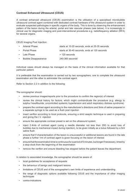
Contrast Enhanced <strong>Ultrasound</strong> (CEUS)<br />
A contrast enhanced ultrasound (CEUS) examination is the utilisation of a specialised microbubble<br />
ultrasound contrast agent combined with dedicated contrast hardware of the ultrasound system in order to<br />
evaluate suspected pathologies in specific organs of the body. This is done by observing the enhancement<br />
pattern of the lesion during the arterial, portal and late vascular phases (see below). It is increasingly in<br />
clinical use for diagnostic imaging and post-interventional procedures e.g. radiofrequency ablation (RFA)<br />
for several organs.<br />
CEUS Imaging Post Injection:<br />
• Arterial Phase<br />
• Portal Phase<br />
• Late Phase<br />
starts at 10-20 seconds, ends at 25-35 seconds<br />
starts at 30-45 seconds, ends at 120 seconds<br />
> 120 seconds<br />
1<br />
• Bubble Disappearance<br />
240-360 seconds<br />
Individual cases should always be managed on the basis of the clinical information available for that<br />
particular patient.<br />
It is preferable that the examination is carried out by two sonographers, one to complete the ultrasound<br />
examination and the other to administer the contrast agent.<br />
Refer to Section 2.2 in addition to the following:<br />
The sonographer should:<br />
• review previous images/reports prior to the procedure to confirm the region(s) of interest<br />
• review the clinical history for factors, which might contraindicate the procedure (e.g. allergy to<br />
sulphur hexaflouride, uncontrolled systemic hypertension and adult respiratory distress syndrome)<br />
• prepare the contrast agent according to the manufacturer’s directions and 5mls of saline prepared in<br />
a separate syringe to be used as a flush post injection<br />
• insert venflon according to local protocols, ensuring a strict aseptic technique is used in preparing<br />
and giving the I.V. injection<br />
• ensure the appropriate contrast preset is set on the ultrasound system<br />
• inject 2.4mls of contrast agent (using a needle diameter not less than 20G to avoid loss of<br />
bubbles due to mechanical impact during injection), to be given initially as a bolus followed by a 5ml<br />
saline flush<br />
• ensure that if characterisation of the lesion is unsuccessful or additional lesions are found in the late<br />
phase a further 1ml of contrast agent is given focusing on the lesion of interest<br />
• ensure that the examination time is continuous for a period of 5 minutes, but longer if necessary, timed by<br />
a stop-clock from the beginning of the examination<br />
• remove the venflon and ensure bleeding has stopped before the patient leaves the department.<br />
In relation to associated knowledge, the sonographer should be aware of:<br />
• local guidelines for acceptance of requests<br />
• the behaviour of benign and malignant lesions<br />
• limitations of CEUS and of the sonographer’s own limits of experience and understanding<br />
• the range of diagnostic options available following CEUS and the importance of other imaging<br />
techniques<br />
• contra-indications<br />
25 © UKAS


