Guidelines For Professional Working Standards Ultrasound Practice
Guidelines For Professional Working Standards Ultrasound Practice
Guidelines For Professional Working Standards Ultrasound Practice
Create successful ePaper yourself
Turn your PDF publications into a flip-book with our unique Google optimized e-Paper software.
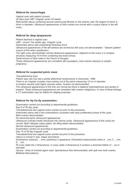
Referral for menorrhagia<br />
Vaginal scan with patient consent.<br />
25 days post LMP. Irregular cycle 4-6 weeks.<br />
Retroverted uterus containing several submucosal fibroids on the anterior wall, the largest of which is<br />
Xmm in diameter. <strong>Ultrasound</strong> appearances of both ovaries are normal with a corpus luteum in the left<br />
ovary.<br />
Referral for deep dyspareunia<br />
Patient declined a vaginal scan.<br />
LMP - unsure six weeks ago. Irregular cycle.<br />
Anteverted uterus with endometrial thickness Xmm.<br />
<strong>Ultrasound</strong> appearances of the left adnexa are normal but left ovary not demonstrated - absent (patient<br />
unsure of this when questioned.)<br />
The right ovary demonstrates normal ultrasound appearances. Adjacent to this ovary is a complex<br />
structure measuring YxYxYmm containing low level echoes.<br />
Small amount of fluid noted in the Pouch of Douglas.<br />
These ultrasound appearances are consistent with pyosalpinx, tubo-ovarian abscess or ectopic<br />
pregnancy.<br />
Referral for suspected pelvic mass<br />
Transabdominal scan.<br />
Patient says she has had a partial abdominal hysterectomy in December, 1986.<br />
There is an irregular complex mass arising out of the pelvis measuring 15 cm in diameter.<br />
It contains several solid highly vascular areas. Ovaries not demonstrated.<br />
The ultrasound appearances of the liver are normal but there is bilateral hydronephrosis and ascites is<br />
present. These ultrasound appearances are consistent with ovarian malignancy. In view of these findings<br />
a CT examination may be helpful for staging purposes.<br />
Referral for Hy-Co-Sy examination:<br />
Examination carried out according to departmental guidelines.<br />
Day 6 of 30 day cycle.<br />
Transabdominal and vaginal scans carried out prior to the procedure.<br />
Anteverted uterus with a thin endometrium consistent with early proliferative phase of the cycle.<br />
Both ovaries demonstrated.<br />
No abnormal pelvic ultrasound appearances.<br />
<strong>Ultrasound</strong> contrast media introduced into uterine cavity. <strong>Ultrasound</strong> appearances of the cavity are<br />
normal. Both Fallopian tubes patent. No filling defect demonstrated.<br />
Normal ultrasound appearances.<br />
Examination carried out according to departmental guidelines.<br />
Day 14 of 28 day irregular cycle.<br />
Transabdominal and vaginal scans carried out prior to the procedure.<br />
Uterus is normal in size, shape and texture.<br />
Endometrium measures ….mms, preliminary in appearance. Endometrial measurements where A …mm; C…..mm:<br />
E…..mm.<br />
Rt ovary (state the 3 dimensions); Lt ovary (state 3 dimensions) it contains a dominant follicle of …cm in<br />
diameter.<br />
Hycosy - Xmls of contrast agent used. Spontaneous flow demonstrated, with spill over both ovaries.<br />
Bilateral tubal patency.<br />
53 © UKAS


