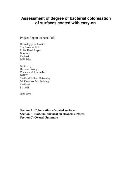
Assessment of degree of bacterial colonisation of ... - Addemar
Assessment of degree of bacterial colonisation of ... - Addemar
Assessment of degree of bacterial colonisation of ... - Addemar

You also want an ePaper? Increase the reach of your titles
YUMPU automatically turns print PDFs into web optimized ePapers that Google loves.
<strong>Assessment</strong> <strong>of</strong> <strong>degree</strong> <strong>of</strong> <strong>bacterial</strong> <strong>colonisation</strong><br />
<strong>of</strong> surfaces coated with easy-on.<br />
Project Report on behalf <strong>of</strong>:<br />
Urban Hygiene Limited<br />
Sky Business Park<br />
Robin Hood Airport<br />
Doncaster<br />
England<br />
DN9 3GA<br />
Written by<br />
Dr Jamie Young<br />
Commercial Researcher<br />
BMRC<br />
Sheffield Hallam University<br />
7th Floor Norfolk Building<br />
Sheffield<br />
S1 1WB<br />
June 2006<br />
Section A: Colonisation <strong>of</strong> coated surfaces<br />
Section B: Bacterial survival on cleaned surfaces<br />
Section C: Overall Summary
Section A: Colonisation <strong>of</strong> Coated surfaces<br />
Summary<br />
20 mm discs coated with easy-on durability coating were tested for their resistance to<br />
<strong>bacterial</strong> colonization over a period <strong>of</strong> 5 days. The growth <strong>of</strong> seven pathogenic<br />
<strong>bacterial</strong> species was monitored over time, with bacteria added to the discs in both<br />
growth medium and also in a simple salt-buffered solution. Easy-on coated discs were<br />
shown to prevent the colonization and growth <strong>of</strong> Staphylococcus aureus, Listeria<br />
monocytogenes, Salmonella enterica subs enterica and Pseudomonas aeruginosa after<br />
a period <strong>of</strong> 3 days. The survival <strong>of</strong> Klebsiella pneumoniae was also severely reduced.
Materials and Methods<br />
Bacterial Species<br />
Species<br />
Staphylococcus<br />
aureus subsp. aureus<br />
American Type<br />
Culture<br />
Collection<br />
(ATCC)<br />
identification<br />
number<br />
ATCC 12600<br />
Description<br />
Gram +ive, common skin<br />
organism<br />
Growth<br />
Media<br />
Nutrient<br />
Broth<br />
Escherichia coli ATCC 11775 Gram -ive, common gut<br />
organism, some strains are<br />
pathogenic<br />
Listeria<br />
monocytogenes<br />
ATCC 15313<br />
Gram +ive rod, 10% <strong>of</strong> human<br />
population are carriers, can<br />
cause severe infection in some<br />
patients<br />
Salmonella enterica<br />
subsp. enterica<br />
ATCC 43971 Gram -ive rod, common cause<br />
<strong>of</strong> salmonella food poisoning<br />
Bacillus cereus ATCC 14579 Gram +ive rod, implicated in<br />
food poisoning. Forms spores<br />
which are resistant to heat and<br />
some disinfectants<br />
Klebsiella<br />
pneumoniae subsp.<br />
pneumoniae<br />
Pseudomonas<br />
aeruginosa<br />
ATCC 13883<br />
ATCC 10145<br />
Gram -ive, common cause <strong>of</strong><br />
hospital acquired pneumonia<br />
Gram -ive, fourth most<br />
common cause <strong>of</strong> hospital<br />
acquired infections. Very<br />
simple growth requirements,<br />
and can easy colonise surfaces<br />
Nutrient<br />
Broth<br />
Brain<br />
Heart<br />
Infusion<br />
CASO<br />
broth<br />
Nutrient<br />
Broth<br />
Nutrient<br />
Broth<br />
Nutrient<br />
Broth<br />
All <strong>bacterial</strong> species used were supplied as vacuum dried cultures by DSMZ<br />
(Deutsche Sammlung von Mikroorganismen und Zellkulturen GmbH, Mascheroder<br />
Weg 1b, 38124 Braunschweig, Germany.)
Media<br />
Nutrient agar/broth, Caso agar/broth and brain heart infusion agar/broth were supplied<br />
by Sigma-Aldrich (UK)<br />
Nutrient Broth/Agar<br />
Peptone<br />
Meat extract<br />
Agar, if necessary<br />
Distilled water<br />
Adjust pH to 7.0.<br />
5.0 g<br />
3.0 g<br />
15.0 g<br />
1000.0 ml<br />
Caso Broth/Agar<br />
Peptone from casein 15.0 g<br />
Peptone from soymeal 5.0 g<br />
NaCl 5.0 g<br />
Agar, if necessary 15.0 g<br />
Distilled water<br />
1000.0 ml<br />
Adjust pH to 7.3.<br />
BHI Broth Agar<br />
Brain Heart Infusion<br />
Agar, if necessary<br />
Distilled water<br />
37.0 g<br />
15.0 g<br />
1000.0 ml<br />
Buffers and solutions<br />
Quarter strength Ringer’s solution<br />
Sodium chloride<br />
2.25 g/l<br />
Potassium chloride 0.105 g/l<br />
Calcium chloride 6H2O 0.12 g/l<br />
Sodium bicarbonate 0.05 g/l<br />
Distilled water<br />
to 1000.00 ml<br />
pH 7.0<br />
All Chemicals and reagents used in this experiment were supplied by Sigma-Aldrich<br />
(UK) unless otherwise stated
Methods<br />
Sterilisation <strong>of</strong> coated discs<br />
22mm discs coated with easy-on were placed in 90 % ethanol for 30 min. Following<br />
sterilisation discs were air-dried in a sterile flow hood.<br />
Growth <strong>of</strong> <strong>bacterial</strong> cultures<br />
Dried cultures were resuspended for 30 min in the appropriate media, and then added<br />
to culture flasks containing 250 ml <strong>of</strong> nutrient broth, Caso broth or BHI broth. Flasks<br />
were incubated overnight at 37 °C. Following incubation cultures were centrifuged<br />
and the medium removed. Cells were then re-suspended in quarter strength Ringer’s<br />
solution and centrifuged again. The supernatant was removed and cells suspended in<br />
either culture media or Ringer’s solution to give a working concentration <strong>of</strong> 10 7<br />
cells/ml. Cell numbers were determined using a haemocytometer and light<br />
microscopy.<br />
Addition <strong>of</strong> bacteria to discs<br />
200 µl <strong>of</strong> washed and resuspended cells were added to each coated disc. As a negative<br />
control 200 µl <strong>of</strong> either sterile growth media or sterile Ringer’s solution was added to<br />
separate discs. A set <strong>of</strong> discs was also used with no addition <strong>of</strong> either <strong>bacterial</strong> cells or<br />
media/Ringer’s solution. Discs were then placed in sterile Petri dishes and incubated<br />
at 22 °C.<br />
Sampling <strong>of</strong> coated discs<br />
Discs were sampled at Day 0, 1, 3 and 5, following initial addition <strong>of</strong> <strong>bacterial</strong><br />
cultures. Each disc was placed in a sterile Universal tube and 5ml <strong>of</strong> Ringer’s solution<br />
was added. Tubes were agitated for 10 min using a Griffin shaker to remove bacteria<br />
from the coated surface. A dilution series was created in Ringer’s solution. After this<br />
200 µl <strong>of</strong> the 10 -3 , 10 -4 and 10 -5 dilutions was plated out onto the appropriate agar<br />
plates. Plates were incubated overnight at 37 °C and cell counts taken the following<br />
morning.<br />
Each experiment was carried out on triplicate discs for reproducibility.<br />
Analysis<br />
The arithmetic mean was taken <strong>of</strong> each set <strong>of</strong> replicates and standard deviations<br />
calculated
Results<br />
Staphylococcus aureus<br />
Complete loss <strong>of</strong> culturable cells was noted at day 3 in bacteria suspended in Ringer’s<br />
solution, and by day 5 in bacteria suspended in growth media. Loss <strong>of</strong> <strong>bacterial</strong> cells<br />
appears to be quite rapid with a drop from 10 7 cfu/ml to 2x10 4 cfu/ml in Ringer’s<br />
solution and 7x10 4 cfu/ml in growth media by day 1 (Fig. 1)<br />
8<br />
7<br />
6<br />
log 10 cfu/ml<br />
5<br />
4<br />
3<br />
Ringer's solution<br />
Media<br />
2<br />
1<br />
0<br />
Day 0 Day1 Day3 Day5<br />
Figure 1. Cell concentration <strong>of</strong> Staphylococcus aureus added to coated discs over<br />
time. Error bars indicate standard deviation
Escherichia coli<br />
Viable E. coli cells were detected at day 5. Cell numbers did reduce over time, with<br />
no significant difference between media and Ringer’s solution. Final cell<br />
concentrations were 3.9x10 3 cfu/ml for Ringer’s solution and 2.5x10 3 cfu/ml in media<br />
(Fig 2).<br />
8<br />
7<br />
6<br />
log 10 cfu/ml<br />
5<br />
4<br />
3<br />
Ringer's solution<br />
Media<br />
2<br />
1<br />
0<br />
Day 0 Day1 Day3 Day5<br />
Figure 2. Cell concentration <strong>of</strong> Escherichia coli added to coated discs over time. Error<br />
bars indicate standard deviation.
Listeria monocytogenes<br />
Complete loss <strong>of</strong> cells in Ringer’s solution was noted by day 3, and by Day 5 in<br />
media. The drop in cell numbers was much more rapid in bacteria suspended in<br />
Ringer’s solution, falling to 2.5x10 3 cfu/ml by day 1. This compares to a <strong>bacterial</strong><br />
count <strong>of</strong> 7.9x10 4 cfu/ml in cells suspended in growth media (Fig. 3).<br />
8<br />
7<br />
log 10 cfu/ml<br />
6<br />
5<br />
4<br />
3<br />
Ringer's solution<br />
Media<br />
2<br />
1<br />
0<br />
Day 0 Day1 Day3 Day5<br />
Figure 3. Cell concentration <strong>of</strong> Listeria monocytogenes added to coated discs over<br />
time. Error bars indicate standard deviation.
Salmonella enterica subsp. enterica<br />
Total loss <strong>of</strong> <strong>bacterial</strong> cells was noted by day 3 in both cell suspensions. There<br />
appeared to be a faster loss <strong>of</strong> cell numbers when bacteria were resuspended in<br />
Ringer’s solution, compared to growth media, with cell numbers falling to 7.6x10 4<br />
cfu/ml and 1.8x10 5 cfu/ml respectively by day 1 (Fig. 4).<br />
8<br />
7<br />
log 10 cfu/ml<br />
6<br />
5<br />
4<br />
3<br />
Ringer's solution<br />
Media<br />
2<br />
1<br />
0<br />
Day 0 Day1 Day3 Day5<br />
Figure 4. Cell concentration <strong>of</strong> Salmonella enterica subsp. enterica added to coated<br />
discs over time. Error bars indicate standard deviation.
Bacillus cereus<br />
Bacterial cells were still viable at a high rate in both Ringer’s solution and Growth<br />
media. Cell numbers did fall but appeared to reach a stable level <strong>of</strong> 3.1x10 4 cfu/ml in<br />
Ringer’s solution and 1.2x10 4 cfu/ml in growth media by day 3. There was no<br />
significant difference between the cell concentrations in Ringer’s solution and media<br />
at day 5 (Fig. 5).<br />
8<br />
7<br />
6<br />
Ringer's solution<br />
Media<br />
log 10 cfu/ml<br />
5<br />
4<br />
3<br />
2<br />
1<br />
0<br />
Day 0 Day1 Day3 Day5<br />
Figure 5. Cell concentration <strong>of</strong> Bacillus cereus added to coated discs over time. Error<br />
bars indicate standard deviation.
Klebsiella pneumoniae<br />
A complete loss <strong>of</strong> cell numbers was noted when bacteria were resuspended in<br />
Ringer’s solution; however, cell numbers only fell to a level <strong>of</strong> 2.7x10 4 cfu/ml by<br />
day5 when bacteria were resuspended in media (Fig. 6).<br />
8<br />
7<br />
6<br />
Ringer's solution<br />
Media<br />
log 10 cfu/ml<br />
5<br />
4<br />
3<br />
2<br />
1<br />
0<br />
Day 0 Day1 Day3 Day5<br />
Figure 6. Cell concentration <strong>of</strong> Klebsiella pneumoniae added to coated discs over<br />
time. Error bars indicate standard deviation.
Pseudomonas aeruginosa<br />
Total loss <strong>of</strong> viable cells was noted at day 3 in cells suspended in Ringer’s solution<br />
and by day 5 in cells suspended in media. A slower drop in cell numbers occurred in<br />
bacteria in media, compared to bacteria in Ringer’s solution (Fig. 7).<br />
8<br />
log 10<br />
cfu/ml<br />
7<br />
6<br />
5<br />
4<br />
3<br />
Ringer's solution<br />
Media<br />
2<br />
1<br />
0<br />
Day 0 Day1 Day3 Day5<br />
Figure 7. Cell concentration <strong>of</strong> Pseudomonas aeruginosa added to coated discs over<br />
time. Error bars indicate standard deviation.<br />
Negative controls<br />
No <strong>bacterial</strong> growth was seen on any negative controls used in this experiment.
Conclusions<br />
• easy-on coating prevents the <strong>colonisation</strong> <strong>of</strong> Staphylococcus aureus, Listeria<br />
monocytogenes, Salmonella enterica subs enterica and Pseudomonas<br />
aeruginosa after a period <strong>of</strong> 3 days on treated surfaces.<br />
• easy-on coating significantly reduces the survival <strong>of</strong> Klebsiella pneumoniae in<br />
a buffered solution when added to the treated surface.<br />
• easy-on coating does not inhibit the <strong>colonisation</strong> <strong>of</strong> E. coli or Bacillus cereus.<br />
A drop in cell numbers however is seen.<br />
• Where loss <strong>of</strong> cell numbers is noted it is usually higher in cells resuspended in<br />
a simple salt-buffered solution.
Section B<br />
Bacterial survival on surfaces coated with easy-on durability<br />
coating after disinfection with Presept<br />
(Dichloroisocyanurate)<br />
Summary<br />
Wooden panels were coated with vinyl emulsion paint, acrylic paint or easy-on<br />
durability coating. Bacterial cultures were added and subsequently cleaned using a<br />
1000 ppm solution <strong>of</strong> Presept (Johnson and Johnson, US). Panels were incubated on<br />
agar plates for 1 and 3 days following cleaning. Results show that when panels coated<br />
with easy-on a simple cleaning procedure removes the bacteria. Panels coated with<br />
emulsion and acrylic paints show survival <strong>of</strong> bacteria after cleaning.
Materials and Methods<br />
Bacterial Strains<br />
The seven <strong>bacterial</strong> species used in Section A were used in this experiment.<br />
Preparation <strong>of</strong> Panels<br />
Wooden panels were coated with vinyl matt emulsion, vinyl matt emulsion and overcoated<br />
with easy-on coating, and with acrylic paint. Panels were cleaned thoroughly<br />
with 1000 ppm <strong>of</strong> Presept solution prior to addition <strong>of</strong> <strong>bacterial</strong> cultures<br />
Addition <strong>of</strong> Bacteria<br />
Cultures <strong>of</strong> bacteria were incubated overnight at 37 ºC and then diluted in culture<br />
media to achieve a <strong>bacterial</strong> concentration <strong>of</strong> 10 7 cells ml -1 . 100 l <strong>of</strong> <strong>bacterial</strong><br />
cultures were then added to each panel and incubated at room temperature for 30 min.<br />
Following incubation wooden panels were cleaned twice with sterile tissue soaked in<br />
1000 ppm Presept solution. A general cleaning action was simulated with the panels<br />
being wiped vertically twice in each cleaning step. Panels were then placed face down<br />
on nutrient agar plates for 30 minutes to allow transfer <strong>of</strong> bacteria to the agar surface.<br />
Panels were then removed and the plates incubated at 37 ºC overnight. Bacterial<br />
colonies were then counted. The process was then repeated with the bacteria<br />
incubated on the panels for 3 days prior to cleaning.
Results<br />
Panels coated with easy-on and subsequently cleaned with Presept solution showed no<br />
survival <strong>of</strong> <strong>bacterial</strong> species after 30 min and three days incubation.<br />
After 30 minutes incubation followed by cleaning, E.coli, L. monocytogenes, Staph.<br />
aureus and B. cereus all exhibited survival on both cleaned acrylic and emulsion<br />
coated panels (Fig 1). Salm. enterica, K. pneumoniae and Ps .aeruginosa only showed<br />
survival on acrylic coated surfaces. Results after 3 days incubation were identical,<br />
with the same survival patterns present.<br />
450<br />
400<br />
350<br />
c.f.u ml-1<br />
300<br />
250<br />
200<br />
Vinyl Emulsion<br />
Acrylic Paint<br />
Easy On Coating<br />
150<br />
100<br />
50<br />
0<br />
Negative control Escherichia coli Listeria monocytogenes<br />
800<br />
700<br />
c.f.u ml -1<br />
600<br />
500<br />
400<br />
300<br />
Vinyl Emulsion<br />
Acrylic Paint<br />
Easy On Coating<br />
200<br />
100<br />
0<br />
Negative control<br />
Staphylococcus aureus<br />
subs aureus<br />
Salmonella enterica subs<br />
enterica
1000<br />
900<br />
800<br />
c.f.u ml-1<br />
700<br />
600<br />
500<br />
400<br />
Vinyl Emulsion<br />
Acrylic Paint<br />
Easy On<br />
Coating<br />
300<br />
200<br />
100<br />
0<br />
Negative control Bacillus cereus Pseudomonas<br />
aeruginosa<br />
Klebsiella<br />
pneumoniae subs<br />
pneumoniae<br />
Figure 1. Survival <strong>of</strong> <strong>bacterial</strong> species on wooden panels with different coated<br />
surfaces, after 30 minutes <strong>of</strong> incubation followed by cleaning.<br />
Conclusions<br />
Following a basic cleaning regime, all bacteria were removed from surfaces coated<br />
with easy-on.<br />
Surfaces coated with acrylic paint allowed survival <strong>of</strong> all seven <strong>bacterial</strong> species<br />
tested after cleaning<br />
Surfaces coated with emulsion paint allowed survival <strong>of</strong> Bacillus cereus,<br />
Staphylococcus aureus, Escherichia coli and Listeria monocytogenes.
Section C<br />
Overall Summary<br />
• easy-on coating prevents the <strong>colonisation</strong> <strong>of</strong> several pathogenic <strong>bacterial</strong><br />
species including Staphylococcus aureus, Listeria monocytogenes, Salmonella<br />
enterica and Pseudomonas aeruginosa on treated surfaces.<br />
• easy-on coated surfaces are easier to clean than untreated surfaces with simple<br />
paint finishes. The durable coating allows for decontamination <strong>of</strong> surfaces<br />
using simple cleaning solutions with no damage to the surface<br />
• easy-on coated surfaces contaminated with bacteria can be thoroughly cleaned<br />
with a simple solution with a 100% <strong>bacterial</strong> removal rate. Staphylococcus<br />
aureus, Escherichia coli, Listeria monocytogenes, Salmonella enterica,<br />
Bacillus cereus, Klebsiella pneumoniae and Pseudomonas aeruginosa were all<br />
completely removed from coated surfaces after cleaning. This compares with<br />
surfaces with simple paint finishes, where bacteria remained on the surface<br />
after cleaning.